Clinical Research Paper
advertisement
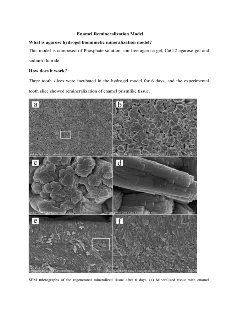
Enamel Remineralization Model What is agarose hydrogel biomimetic mineralization model? This model is composed of Phosphate solution, ion-free agarose gel, CaCl2 agarose gel and sodium fluoride. How does it work? Three tooth slices were incubated in the hydrogel model for 6 days, and the experimental tooth slice showed remineralization of enamel prismlike tissue. SEM micrographs of the regenerated mineralized tissue after 6 days. (a) Mineralized tissue with enamel prismlike crystals regenerated on the enamel. (b) Magnified micrograph of panel a to show the paralleled bundles. (c) Magnified micrograph of panel a to show the agglomerative hexagonal crystals. (d) Magnified micrograph of the rectangular area in panel a to show the side view of the crystal bundle. (e) Cross-sectional view of panel a to show that the regeneration layer was perpendicular to the underlying enamel. (f) Magnified micrograph of the rectangular area in panel e to show the interface between the regeneration tissue and the underlying enamel. Three-dimensional AFM tapping-mode images of the etched enamel and regenerated mineralized tissue. (a) Etched enamel. (b) Regenerated mineralized tissue after 2 days. (c) Regenerated mineralized tissue after 4 days. (d) Regenerated mineralized tissue after 6 days. Stage of material development Extracted human third molars were collected and three tooth slices were used for the experiment. The first tooth section with the nail varnish was the control groups, and the second tooth section with the nail varnish and 37% phosphoric acid, as well as the third tooth section only with 37% phosphoric acid were the experimental group. These three tooth sections were incubated in the hydrogel mineralization model for 2, 4, and 6 days for comparison. Stage of research The experiment was performed outside of human’s mouth and needs further studies by adding an organic matrix, such as enamel protein to the model to mimic the enamel matrix. Was it successful? Formation of regenerated mineralized tissue occurred after 6 days of incubation period with no difference in nanohardness between the regenerated and untreated enamel, which proved that hardness of natural enamel and regenerated enamel are similar. The experimental finding concluded that agarose hydrogel biomimietic mineralization model will help in regenerating enamel-like tissue for repairing enamel loss, especially for the patients with hypersensitivity due to abrasion, attrition and erosion. Furthermore, it could be used in clinical treatments and industrial application in the future, since the regenerated apatite crystals were formed after interaction of calcium, phosphate, and fluoride ions with the agarose hydrogel model. Reference Zhou, Y. Z.; Cao, Y.; Liu, W.; Chu, C. H.; Li, Q. L. ACS. Appl. Mater. Interfaces. (2013, December 19). Retrieved from http://pubs.acs.org/doi/full/10.1021/am4044823#showRef
