CRVO- Flame hemorrhages (red streaks) around BRVO
advertisement
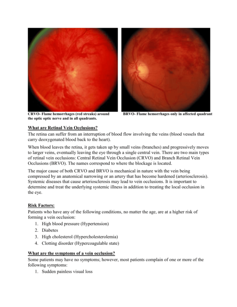
CRVO- Flame hemorrhages (red streaks) around the optic optic nerve and in all quadrants. BRVO- Flame hemorrhages only in affected quadrant What are Retinal Vein Occlusions? The retina can suffer from an interruption of blood flow involving the veins (blood vessels that carry deoxygenated blood back to the heart). When blood leaves the retina, it gets taken up by small veins (branches) and progressively moves to larger veins, eventually leaving the eye through a single central vein. There are two main types of retinal vein occlusions: Central Retinal Vein Occlusion (CRVO) and Branch Retinal Vein Occlusions (BRVO). The names correspond to where the blockage is located. The major cause of both CRVO and BRVO is mechanical in nature with the vein being compressed by an anatomical narrowing or an artery that has become hardened (arteriosclerosis). Systemic diseases that cause arteriosclerosis may lead to vein occlusions. It is important to determine and treat the underlying systemic illness in addition to treating the local occlusion in the eye. Risk Factors: Patients who have any of the following conditions, no matter the age, are at a higher risk of forming a vein occlusion: 1. High blood pressure (Hypertension) 2. Diabetes 3. High cholesterol (Hypercholesterolemia) 4. Clotting disorder (Hypercoagulable state) What are the symptoms of a vein occlusion? Some patients may have no symptoms; however, most patients complain of one or more of the following symptoms: 1. Sudden painless visual loss 2. General blurry vision 3. Blind spots in peripheral vision 4. Distorted vision CRVO- Intraretina hemorrhages in all quadrants CRVO-Fluorescein Angiography showing tortuosity of blood vessels in all quadrants How can the doctor determine the extent of my vein occlusion? The doctor will perform a dilated exam using a slit lamp to determine the extent of the occlusion and what effect it has on the center of your retina (the macula). To check the outer retina, the doctor will use an indirect ophthalmoscope. Since retinal swelling and edema are frequently present in a patient with a vein occlusion, the doctor may order several tests. What tests are performed? Testing is important because it helps the doctor to precisely document the vein occlusion, check for edema, and measure changes that occur. The three types of tests described below are performed in our clinic. Optical Coherence Tomography (OCT) is a high definition image of the retina taken by a scanning ophthalmoscope with a resolution of 5 microns. These images can determine the presence of swelling and edema by measuring the thickness of your retina. The doctor will use OCT images to objectively document the progress of the disease throughout the course of your treatment. Fundus Photography is an image taken by a digital fundus camera to document the vein occlusion in the retina. Fluorescein Angiography is a test that documents blood circulation in the retina using fluorescein dye which luminesces under blue light. Fluorescein is injected into a vein in your arm and digital fundus pictures are taken afterwards for 10 minutes. These pictures show the location and extent of blockage and the presence of leakage from blood vessels. Before treatment After treatment with intravitreal injections What treatments are available? Injections of anti-inflammatory medicines into the eye can be performed in our office to treat the vein occlusion. Sometimes focal laser to the retina is necessary. The doctor may recommend the treatments below: 1. Intravitreal injection of an anti-Vascular Endothelial Growth Factor (Avastin or Lucentis) 2. Intravitreal injection of a corticosteroid (Dexamethasone or Triamcinolone) 3. Intravitreal Injection of Ozurdex (a long lasting Dexamethasone tablet) 4. Focal Laser therapy 5. Pan-retinal photocoagulation therapy It is also very important that any underlying systemic conditions (hypertension, diabetes, hypercholesterolemia, hypercoagulable state) be evaluated and treated by your primary care doctor. What is my follow up care? Return visits with us are recommended to monitor your disease progress. It is important to detect changes in your condition and formulate treatment plans as needed. It is also important to inform your primary care doctor of your retinal vein occlusion, so he or she can evaluate and treat any underlying systemic illnesses.
