Milk Examination: Mastitis Diagnosis & Methods
advertisement
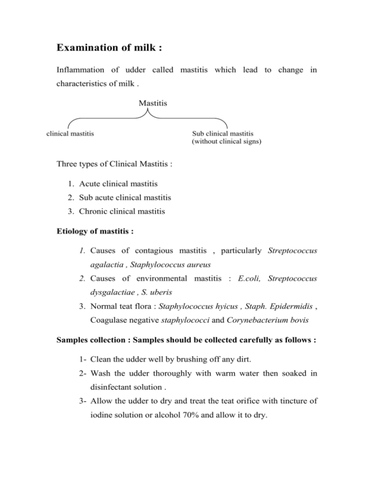
Examination of milk : Inflammation of udder called mastitis which lead to change in characteristics of milk . Mastitis clinical mastitis Sub clinical mastitis (without clinical signs) Three types of Clinical Mastitis : 1. Acute clinical mastitis 2. Sub acute clinical mastitis 3. Chronic clinical mastitis Etiology of mastitis : 1. Causes of contagious mastitis , particularly Streptococcus agalactia , Staphylococcus aureus 2. Causes of environmental mastitis : E.coli, Streptococcus dysgalactiae , S. uberis 3. Normal teat flora : Staphylococcus hyicus , Staph. Epidermidis , Coagulase negative staphylococci and Corynebacterium bovis Samples collection : Samples should be collected carefully as follows : 1- Clean the udder well by brushing off any dirt. 2- Wash the udder thoroughly with warm water then soaked in disinfectant solution . 3- Allow the udder to dry and treat the teat orifice with tincture of iodine solution or alcohol 70% and allow it to dry. 4- While the tincture of iodine is drying, label the tubes as to cow and quarter from which the sample will be taken . 5- The cap of the sterile tube is carefully removed and held between the fingers in such a manner that the inside of the cap is facing down ward. The tube should be held at a slight angle to prevent contamination of the sample by falling particles . 6- Immediately following collection, refrigerate the samples for transportation to the laboratory . Methods of examination of milk : including into A- Physical methods of diagnosis B- Chemical methods of diagnosis C- Culture methods of diagnosis A- Physical methods of diagnosis :(colour and consistence of udder,odor or smell of milk ). B- Chemical methods of diagnosis : determination specially sub-clinical mastitis including into( Card field test, pH test , Chloride test, White side test, Calfornia mastitis test , Catalase test , Somatic cell count , Microscopic examination of incubated milk ) 1- pH determination : Normal pH of milk (6.4-6.8) paper strips contian Bromthymol blue or Bromcresol purple . 2- Chloride test : The chloride test is dependent upon the determination of an abnormal quantity of chloride in the milk. Normal milk contains 0.08-0.14 % chloride .The test is conducted by adding 5ml of silver nitrate solution to 1ml of milk, followed by the addition of two drops of a potassium chromate solution and mixing by inversion of the tube .The appearance of yellow color indicator that more than 0.14% chlorides are present in the sample and brownish red color indicates that the sample contain less than that amount. Cows in either early or late lactation may give false positive reaction to the chloride test because of normal physiologic processes of the udder . 3- Catalase test : Most living cells (RBCs catalase help to release oxygen from peroxide hydrogen therefore action determination that amount catalase in WBCs milk) catalase isn’t found in milk normally except in the early and finally period lactation .2ml of milk incubated for 1-2 hours and then add drops from H2O2 and the positive test appearance bubble airs and negative test disappearance bubble airs . 4- White side test : Put 5 drops of milk on dark smooth surface on which the test can be conducted and then add 2 drops of sodium hydroxide 4% solution for 20-25 second. Milk from normal animals will have no change after the addition of the NaOH, but mastitis milk appear as the following : Nagative (-) The mixture is milky and opaque and entirely free of precipition .In such animals ,the leukocyte count is generally under 500,000 cells /cm3 . 1 + The background is less opaque but still somewhat milky, with large particles of coagulated material being present and thickly scattered throughout the area .A slight degree of clumping is observed .The leukocyte count is usually between 1 and 2 million . 2 ++ The background is more watery and large clump of coagulated material are present. If the stirring has been rapid ,fine threads or strings may be present .In such specimens the total leukocyte count is usually over 2 million. 3 +++ The background is very watery and whey like with large masses of coagulated material forming into strings and shreds. the total leukocyte count is usually several million . 5- California mastitis test : take 2ml of milk with 2ml CMT solution (2:2) mixing rotation movement and then for 10 second read results: Negative (-) the mixture remains liquid without precipitation 1+ distinct precipitate but no tendency toward gel formation 2++ the mixture thickens immediately, with mucous formation moving slowly on the paddle . 3+++distinct gel forms that tends to adhere to the bottom of the paddle and during swirling distinct central peak forms . 6- Direct microscopic total leukocytic count : taken drop of milk put in slide spreading and dryness staining by (Newman's-lampert stain) for 2 minute and dryness by filter paper and then washing by distill water and dryness after that count by microscopic and calculated in 30 fields = N/30 X400.000 N= number of cells 400.000= Zoom factor 7- Microscopic examination of incubated milk : taken precipitate milk which incubated 12-16 hours and putting in slide spread drop and left air for dry and then staining by gram stain diagnosis ) Take loopful for primary culture . After 24 hours at incubator then make gram stain . (rapid C- Bacterial Culture methods of diagnosis : Primary culture Culture Secondary culture Specimens (Milk) Primary culture (Nutrient agar ,Blood agar , Macconkey agar.) After 24h.incubated Gram stain (Genus of M.O.) Secondary culture Biochemical test (oxidase, catalase, indol test.. etc.) (Differential diagnosis for Species of M.O.)
