management_care__preeclampsia 2
advertisement

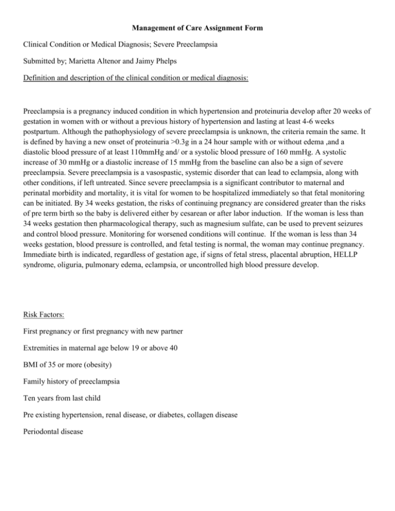
Management of Care Assignment Form Clinical Condition or Medical Diagnosis; Severe Preeclampsia Submitted by; Marietta Altenor and Jaimy Phelps Definition and description of the clinical condition or medical diagnosis: Preeclampsia is a pregnancy induced condition in which hypertension and proteinuria develop after 20 weeks of gestation in women with or without a previous history of hypertension and lasting at least 4-6 weeks postpartum. Although the pathophysiology of severe preeclampsia is unknown, the criteria remain the same. It is defined by having a new onset of proteinuria >0.3g in a 24 hour sample with or without edema ,and a diastolic blood pressure of at least 110mmHg and/ or a systolic blood pressure of 160 mmHg. A systolic increase of 30 mmHg or a diastolic increase of 15 mmHg from the baseline can also be a sign of severe preeclampsia. Severe preeclampsia is a vasospastic, systemic disorder that can lead to eclampsia, along with other conditions, if left untreated. Since severe preeclampsia is a significant contributor to maternal and perinatal morbidity and mortality, it is vital for women to be hospitalized immediately so that fetal monitoring can be initiated. By 34 weeks gestation, the risks of continuing pregnancy are considered greater than the risks of pre term birth so the baby is delivered either by cesarean or after labor induction. If the woman is less than 34 weeks gestation then pharmacological therapy, such as magnesium sulfate, can be used to prevent seizures and control blood pressure. Monitoring for worsened conditions will continue. If the woman is less than 34 weeks gestation, blood pressure is controlled, and fetal testing is normal, the woman may continue pregnancy. Immediate birth is indicated, regardless of gestation age, if signs of fetal stress, placental abruption, HELLP syndrome, oliguria, pulmonary edema, eclampsia, or uncontrolled high blood pressure develop. Risk Factors: First pregnancy or first pregnancy with new partner Extremities in maternal age below 19 or above 40 BMI of 35 or more (obesity) Family history of preeclampsia Ten years from last child Pre existing hypertension, renal disease, or diabetes, collagen disease Periodontal disease Nursing Diagnosis #1: Risk for injury (maternal) Risk factors: CNS irritability (seizures) Goals or Expected Outcomes: Patient will show diminished signs of seizures Deep tendon reflexes will be less than 2+ Patient shows absence of clonus Focused Nursing Assessment: OBJECTIVE DATA 1. Patients urine output is decreased with less than 30ml/hour SUBJECTIVE DATA 1. Patients complains of blurred vision 2. Patient is vomiting frequently 2. Patient complains of persistent headache 3. Patients blood pressure is 140/90 or more, or the systolic is increased by 30mmHg and/or diastolic increased by 15mmHg from their baseline 3. Patients states she is experiencing upper abdominal pain 4. Patients urine shows high levels of protein with a value >0.3g within a 24 hour period 4. Patient states her feet look extremely swollen 5. Patient shows deep tendon reflexes greater than 2+ when bicep and patellar reflexes were assessed 5. Patients states she feels “like something bad is going to happen” 6. Assessment for clonus is positive 6. Interventions (you can include both nursing interventions and medical management, example medications that you would expect to be administering: INTERVENTIONS 1.Establish a baseline for blood pressure, clonus, deep tendon reflexes, fetal heart rate, and level of consciousness 2. Monitor maternal vital signs every 15 minutes, level of consciousness, fetal heart rate less than 110beats/min, deep tendon reflexes, urine output less than 30ml/hr, proteinuria, IV flow rate, serum levels of magnesium sulfate between 4-7mEq/L RATIONALE 1. To use as a basis for evaluating effectiveness of treatment 2. To assess for and prevent magnesium sulfate toxicity, slurred speech, lethargy, drowsiness, fetal distress, oliguria, depressed respirations, sudden drop in blood pressure, decreased kidney function, and hyperreflexia 3. Administer IV magnesium sulfate per physicians orders, 4g IV bolus then 2g every hour through IV infusion pump 3. Administered to decrease hyperreflexia and minimize risk of seizure activity 4. Have calcium gluconate on the unit 4. To be available if needed as an antidote for magnesium sulfate toxicity 5. Maintain a quiet, darkened environment 5. To avoid stimuli that may precipitate seizure activity 6. Place in seizure precautions with raised and padded side rails 6. To prevent injury during seizure activity Nursing Diagnosis #2: Risk for injury (fetal) Risk factor: uteroplacental insufficiency Goals or Expected Outcomes: Fetus will remain free of injuries Fetus will maintain normal, baseline heart rate Fetus will maintain adequate oxygenation Fetus will show no signs of late decelerations Focused Nursing Assessment: OBJECTIVE DATA 1. Fetus displays abnormal heart rate of 110beats/min or less with continuous EFM SUBJECTIVE DATA 1. Patient states she feels less fetal movement than the babies normal trend 2. Non stress test shows insufficient accelerations over 40 minutes leading to a nonreactive result 2. Patient states she feels her belly has not shown much growth 3. Contraction stress test shows late decelerations occurring with at least 50% of contractions 3. 4. Ultrasound shows oligohydramnios 4. 5. Doppler blood flow analysis shows absent or reversed flow during diastole 5. 6. Amniocentesis shows L/S ratio less than 2:1 and a negative PG 6. Interventions (you can include both nursing interventions and medical management, example medications that you would expect to be administering: INTERVENTIONS 1. Establish baseline data for fetal heart rate with EFM, should be maintained between 110160beats/min RATIONALE 1. To assess for signs of fetal distress 2. Perform non stress test to monitor for fetal reactivity, for reactive NST fetus should show at least 2 movements/accelerations in 20 minutes 2. To determine the well being of the fetus 3. Perform a contraction stress test to assess the fetus’ response to stress, test should be negative showing no late decelerations with a minimum of 3 contractions over 10 minutes 3. To determine the presence of late decelerations in a continued pattern for more than 30 minutes which increases the risk of the negative effects of fetal hypoxia, acidosis, and asphyxia 4. Review ultrasound results to determine amniotic fluid volume, normal AFI is between 10 and 25cms 4. To determine the existence of oliohydramnios, an AFI of less than 5cms, which would indicate renal insufficiency in the fetus 5. Review doppler blood flow analysis, blood should flow freely with no signs of decreased, reversed, or absent flow during diastole 5. To assess for blood flow through the uterine artery, severely restricted uterine artery blood flow is indicated by absent or reversed flow during diastole, abnormal results indicate uteroplacental insufficiency 6.Review results of amniocentesis, should show an L/S ratio of 2:l or greater with a positive PG 6. To determine fetal lung maturity Nursing Diagnosis #3: Excess fluid volume related to high blood pressure and decreased kidney function Goals or Expected Outcomes: Extremities and dependent areas free of edema BUN and creatinine levels are maintained within normal limits Maintain a safe blood pressure, pulse, and body temperature Maintain adequate urine output at least 30 mL per hour Focused Nursing Assessment: OBJECTIVE DATA 1. Increased generalized edema that takes longer than 30 seconds to disappear SUBJECTIVE DATA 1. Patient states increased feeling of thirst 2. Decreased urine output less than 30ml/hr 2. Patient complains of cold, moist skin 3. Increased protein in the urine (greater than 0.3g) and elevated levels of creatinine in the blood (greater than 1.2mg/dl) 3. Patient states feet look swollen 4. Patient gained more than 4lbs in a week 4. Patient states “I am having difficulty breathing” 5. 5. 6. 6. Interventions (you can include both nursing interventions and medical management, example medications that you would expect to be administering: INTERVENTIONS 1. Monitor patient for increased edema, elevated serum creatinine, and dyspnea RATIONALE 1. To detect increased fluid volume due to decreased kidney function 2. Monitor intake and output, watch for trends reflecting decreased urine output from foley catheter in relation to fluid intake 2. To assess for adequate output of at least 30ml/hr 3. Monitor daily weight for increases; use the same scale and type of clothing and weigh at the same time each day, preferably in the morning 3. Changes in body weight reflect changes in body fluid volume 4. Listen to lung sounds for crackles and monitor respirations for effort 4. To assess for pulmonary edema which results from excessive shifting of fluid from the vascular space into the pulmonary interstitial space and alveoli resulting in dyspnea 5. Implement fluid restriction, as ordered 5. Fluid restriction may decrease intravascular fluid volume 6. Administer IV magnesium sulfate per physicians orders, 4g IV bolus then 2g every hour through the IV infusion pump 6. To relax vasospasms and increase renal perfusion Nursing Diagnosis #4: Risk for ineffective cerebral tissue perfusion Risk factors: cerebral vasospasms and cerebral edema Goals or Expected Outcomes: State absence of headache Demonstrate appropriate orientation to person, place, time, and situation Demonstrate stable vital signs and absence of signs in ICP Shows diminished signs of seizures Focused Nursing Assessment: OBJECTIVE DATA 1. Patient is disoriented to place and time SUBJECTIVE DATA 1. Patient complains of a headache 2. Patient is grimacing while holding her head 2. Patients complains of pressure in her head 3. Patient speech is slurred 3. Patient complains of blurry vision 4. Patient is restless 4. Patient complains of dizziness 5. Changes in vital signs from baseline, increase in blood pressure and decrease in heart rate and respirations 5. 6. 6. Interventions (you can include both nursing interventions and medical management, example medications that you would expect to be administering: INTERVENTIONS RATIONALE 1. Monitor and record the neurological status every 30 minutes and compare it to the standard or normal state 1. Assesses trends in LOC and potential for increased intracranial pressure, decreased mental status is suggestive of decreased cerebral perfusion 2. Assess for changes in vision such as blindness or visual field disturbances 2. Visual impairments can be indicative of decreased cerebral perfusion 3. Maintain bed rest by creating a peaceful environment and limiting activities and visitors 3. To decrease overstimulation which can lead to increased ICP causing further confusion and possible seizure activity 4. Monitor vital signs, especially blood pressure, pulse rate, and respirations, every 30 minutes to determine deviations from the baseline 4. Fluctuations in pressure may occur because of cerebral pressure, changes in heart rate, especially bradycardia can occur because of brain damage, irregularities in respirations can suggest an increase in ICP 5. Changes in cognitive function and speech are indicative of location and degree of cerebral damage and may indicate ICP 5. Assess higher functions including speech, if patient is alert 6. Administer magnesium sulfate, 4g IV bolus then 2g every hour through IV infusion pump 6. To decrease vasospasms and increase renal perfusion which can lead to a decrease in cerebral edema Diagnostic Studies or Lab Results that should be reviewed by the nurse: 1. Platelet count- preeclampsia may lead to an abnormally low number of platelets which can prevent normal clotting during bleeding 2. Liver enzymes- to determine how well the liver is functioning 3. Serum creatinine- an elevation would indicate a decrease in kidney function 4. BUN- an elevation would indicate a decrease in kidney function 5. Hematocrit- a high level means the body tissues are absorbing more blood plasma and concentrating the blood 6. Electrolytes- the amounts of electrolytes in the body can change if a decrease in kidney function occurs or if fluid leaks out of the blood vessels and into the surrounding tissue 7. Magnesium levels- should be monitored closely with the infusion of magnesium sulfate to prevent toxicity 8. Fibrinogen- high levels contribute to the hypercoagulability seen in preeclampsia 9. Prothrombin time- a measure of the time it takes for blood to clot which could be increased with preeclampsia 10. Uric acid- an increase would indicate a decrease in kidney function References Ackley, B. J., &Ladwig, G. B. (2011). Nursing diagnosis handbook. (9 ed., pp. 619-624 397401 521-523). St. Louis: Elsevier Inc. Ameh, C., Ekechi, C., &Turkr, J. (2012). Monitoring severe pre-eclampsia and eclampsia treatment in resources poor countries: skilled birth attendant perception of a new treatment and monitoring chart (LIVKAN Chart). Maternal & Child Health Journal, 16(5), 941-946. doi:10.1007/s10995-011-0832-7 Jakobson,T., Clausen, F., Rode, L., Dziegiel, M.,& Tabor, A. (2013) Identifying mild and severe preeclampsia in asymptomatic pregnant women by level of cell-free fetal DNA. Transfusion,53(9), 1956-1964.doi: 10.1111/trf.12073 Lowdermilk, D., Perry, S. E., Cashion, K., & Alden, K. R. (2012). Maternity and women's health care. (10 ed., pp. 656-663). St. Louis: Elsevier Inc. Shaker, O., & Shehata,H. (2011). Early prediction of preeclampsia in high –risk women.Journal Of Women’s Health , 20(4), 539-544.doi: 10.1089/jwh.2010.2378


