A fluorescence and Atomic force microscopy Study of the interaction
A fluorescence and Atomic force microscopy Study of the interaction of Bacteria with AMPs
Supervisors: Peter Fojan, Leonid Gurevich
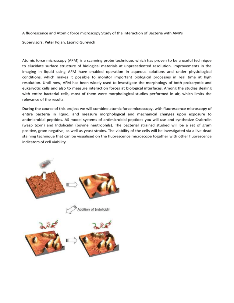
Atomic force microscopy (AFM) is a scanning probe technique, which has proven to be a useful technique to elucidate surface structure of biological materials at unprecedented resolution. Improvements in the imaging in liquid using AFM have enabled operation in aqueous solutions and under physiological conditions, which makes it possible to monitor important biological processes in real time at high resolution. Until now, AFM has been widely used to investigate the morphology of both prokaryotic and eukaryotic cells and also to measure interaction forces at biological interfaces. Among the studies dealing with entire bacterial cells, most of them were morphological studies performed in air, which limits the relevance of the results.
During the course of this project we will combine atomic force microscopy, with fluorescence microscopy of entire bacteria in liquid, and measure morphological and mechanical changes upon exposure to antimicrobial peptides. AS model systems of antimicrobial peptides you will use and synthesize Crabrolin
(wasp toxin) and Indolicidin (bovine neutrophils). The bacterial strainsd studied will be a set of gram positive, gram negative, as well as yeast strains. The viability of the cells will be investigated via a live dead staining technique that can be visualised on the fluorescence microscope together with other fluorescence indicators of cell viability.
