adenomatoid odontogenic tumour of maxilla: a case report
advertisement
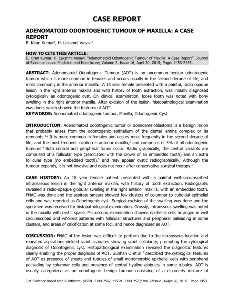
CASE REPORT ADENOMATOID ODONTOGENIC TUMOUR OF MAXILLA: A CASE REPORT E. Kiran Kumar1, H. Lakshmi Vasavi2 HOW TO CITE THIS ARTICLE: E. Kiran Kumar, H. Lakshmi Vasavi. ”Adenomatoid Odontogenic Tumour of Maxilla: A Case Report”. Journal of Evidence based Medicine and Healthcare; Volume 2, Issue 16, April 20, 2015; Page: 2453-2455. ABSTRACT: Adenomatoid Odontogenic Tumour (AOT) is an uncommon benign odontogenic tumour which is more common in females and occurs usually in the second decade of life, and most commonly in the anterior maxilla.1 A 18 year female presented with a painful, radio opaque lesion in the right anterior maxilla and with history of tooth extraction, was initially diagnosed cytologically as odontogenic cyst. On clinical examination, loose tooth was noted with bony swelling in the right anterior maxilla. After excision of the lesion, histopathological examination was done, which showed the features of AOT. KEYWORDS: Adenomatoid odontogenic tumour, Maxilla, Odontogenic Cyst. INTRODUCTION: Adenomatoid odontogenic tumor or adenoameloblastoma is a benign lesion that probably arises from the odontogenic epithelium of the dental lamina complex or its remnants.1,2 It is more common in females and occurs most frequently in the second decade of life, and the most frequent location is anterior maxilla,3 and comprises of 3% of all odontogenic tumours.4 Both central and peripheral forms occur. Radio graphically, the central variants are comprised of a follicular type (associated with the crown of an embedded tooth) and an extra follicular type (no embedded tooth),5 and may appear cystic radiographically. Although the tumour expands, it is not invasive and does not recur after conservative surgical therapy.6 CASE HISTORY: An 18 year female patient presented with a painful well-circumscribed intraosseous lesion in the right anterior maxilla, with history of tooth extraction. Radiographs revealed a radio-opaque globular swelling in the right anterior maxilla, with an embedded tooth. FNAC was done and the aspirate smears showed few clusters of columnar to cuboidal epithelial cells and was reported as Odontogenic cyst. Surgical excision of the swelling was done and the specimen was received for histopathological examination. Grossly, intraosseus swelling was noted in the maxilla with cystic space. Microscopic examination showed epithelial cells arranged in well circumscribed and whorled patterns with follicular structures and peripheral palisading in some clusters, and areas of calcification at some foci, and hence diagnosed as AOT. DISCUSSION: FNAC of the lesion was difficult to perform due to the intraosseus location and repeated aspirations yielded scant aspirates showing scant cellularity, prompting the cytological diagnosis of Odontogenic cyst. Histopathological examination revealed the diagnostic features clearly enabling the proper diagnosis of AOT. Gunhan O et al 7 described the cytological features of AOT as presence of sheets and tubules of small monomorphic epithelial cells with peripheral palisading by columnar cells and presence of central hyaline globules in some tubules. AOT is usually categorized as an odontogenic benign tumour consisting of a disorderly mixture of J of Evidence Based Med & Hlthcare, pISSN- 2349-2562, eISSN- 2349-2570/ Vol. 2/Issue 16/Apr 20, 2015 Page 2453 CASE REPORT odontogenic epithelium and odontogenic ectomesenchyme with calcification.8 hence it is considered as a benign slow growing hamartomatous lesion, rather than a true neoplasm.9 CONCLUSION: Cytological diagnosis of AOT is possible but sometimes difficult due to the intraosseous nature of the lesion and due to presence of cystic areas in some cases, but proper localization of the solid areas of the lesion, using appropriate needle and expertise can enhance the cell yield and help in the cytological diagnosis of this entity, and to differentiate it from odontogenic cyst. REFERENCES: 1. Juan Rosai. Mandible and maxilla. In: Rosai and Ackerman’s Surgical Pathology, Vol. I. 10th ed. Mosby Elsevier; 2011. 265-82. 2. Ivan Damjanov, James Linder. Mouth, teeth and pharynx. In Anderson’s Pathology, Vol. II, 10th ed. Mosby Elsevier: 2009. 1563-98. 3. Pindborg J, Kramer IRH, Torloni H. Histological typing of odontogenic tumours, jaw cysts and allied lesions. Geneva: World health organization; 35-36; 1972. 4. Virupakshappa D, Rajashekhara BS, Manjunatha BS, Das N. Adenomatoid odontogenic tumour in a 20-year-old woman. BMJ Case Rep. 2014 Apr 15:2014. Pii: bcr2013010436.doi: 10.1136/bcr-2013-010436. 5. Lee JS, Yoon SJ, Kang BC, Kim OJ, Kim YH. Adenomatoid odontogenic tumour associated with unerupted first primary molar. Pediatr Dent. 2012 Nov-Dec; 34(7): 493-5. 6. Vasudevan K, Kumar S, Vijayasamundeeswari, Vigneswari S. Adenomatoid odontogenic tumour. An uncommon tumour. Contemp Clin Dent. 2012 Apr: 3(2):2457.doi:10.4103/0976-237X.96837. 7. Gunhan O, Dogan N, Celasun B, Sengun O, Onder T, Finci R: Fine needle aspiration cytology of oral cavity and jaw bone lesions: A report of 102 cases. Acta Cytol; 37: 135-41; 1993 8. Sato D, Matsuzaka K, Yama M, Kakizawa T, Inoue T. Adenomatoid odontogenic tumor arising from the mandibular molar region: a case report and review of the literature. Bull Tokyo Dent Coll.2004 Nov; 45(4): 223-27. 9. Prakasam M, Tiwari S, Satpathy M, Banda VR. Adenomatoid odontogenic tumour. BMJ Case Rep. 2013 Jun 27:2013.pii:bcr2013010212. doi: 10.1136/bcr-2013-010212. J of Evidence Based Med & Hlthcare, pISSN- 2349-2562, eISSN- 2349-2570/ Vol. 2/Issue 16/Apr 20, 2015 Page 2454 CASE REPORT Fig. 1: Radiograph of OAT showing a radio-opaque globular intraosseus swelling in the right maxilla with an embedded tooth Fig. 2: Histological picture of OAT showing epithelial cells arranged in wellcircumscribed, whorled patterns with follicular structures and occasional palisading patterns and focal areas of calcification (H & E x100) AUTHORS: 1. E. Kiran Kumar 2. H. Lakshmi Vasavi PARTICULARS OF CONTRIBUTORS: 1. Associate Professor, Department of Pathology, Rajiv Gandhi Institute of Medical Sciences, Srikakulam, Andhra Pradesh. 2. Assistant Professor, Department of Pathology, Rajiv Gandhi Institute of Medical Sciences, Srikakulam, Andhra Pradesh. NAME ADDRESS EMAIL ID OF THE CORRESPONDING AUTHOR: Dr. E. Kiran Kumar, Plot No. 70, Near Sun School, Radhakrishna Nagar, Srikakulam, Andhra Pradesh. E-mail: meetkiran5@rediffmail.com Date Date Date Date of of of of Submission: 09/04/2015. Peer Review: 10/04/2015. Acceptance: 13/04/2015. Publishing: 20/04/2015. J of Evidence Based Med & Hlthcare, pISSN- 2349-2562, eISSN- 2349-2570/ Vol. 2/Issue 16/Apr 20, 2015 Page 2455

