Unicystic Ameloblastoma
advertisement
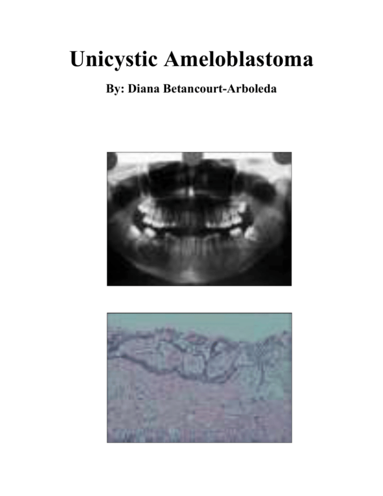
Unicystic Ameloblastoma By: Diana Betancourt-Arboleda Ameloblastoma is a true neoplasm of odontogenic epithelial origin. It is slow-growing, persistent and locally aggressive neoplasm accounting for 10% out of 30% of all odontogenic tumors.1 Unicystic ameloblastoma (UA) refers to those cystic lesions that show clinical, radiographic, or distinct features of a mandibular cyst, but on histologic examination show a typical ameloblastomatous epithelium lining part of the cyst cavity, with or without luminal and/or tumor growth. Its peak incidence is in the 3rd to 4th decades of life with no sexual or racial preference. It is most commonly seen, asymptomatically, in the posterior mandible. Most patients commonly present with swelling and facial asymmetry, pain being an occasional presenting symptom. Mucosal ulceration is rare, but may be caused by continued growth of the tumor. Small lesions may be identified on routine radiographic screening examinations. As the tumor enlarges, it forms a hard swelling and later may cause thinning of the cortical bone resulting in an “egg shell” crackling which can be elicited. Other effects include tooth mobility, displacement of teeth, resorption of roots, paraesthesia if the inferior alveolar canal is involved, and failure of eruption of teeth produced by the tumor.2 On a radiograph, ameloblastoma usually present as a well defined, multilocular radiolucency with scalloped border typically described as “honeycomb” or “soap bubble” appearance.1 However, UA typically presents as a unilocular radiolucency containing an impacted tooth. They typically infiltrate through the medullary bone, therefore the radiographic margins are not accurate indicators of the extent of involvement. Histologically, the criteria for diagnosing a lesion as UA is the presentation of a single cystic sac lined by odontogenic (ameloblastomatous) epithelium often seen only in focal areas. Also, UA can be differentiated from odontogenic cysts because they have a higher rate of recurrence. While chemotherapy, radiation therapy, curettage and liquid nitrogen have been effective in some cases of ameloblastoma, surgery or enucleation remains is the most common treatment for this tumor. Due to the invasive nature of the growth, excision of normal tissue near the tumor margin is often required. Some have referred the disease to basal cell carcinoma (a skin cancer) in its tendency to spread to adjacent bony and sometimes soft tissues without metastasizing. While not a cancer that actually invades adjacent tissues, ameloblastoma is suspected to spread to adjacent areas of the jaw bone via marrow space. Thus, wide surgical margins that are clear of disease are required for a good prognosis. This is very much like surgical treatment of cancer. Often, treatment requires excision of entire portions of the jaw. Unicystic ameloblastoma is a tumor of odontogenic origin. It is not a rare neoplasm and most often the lesion is asympthomatic. However, large lesions may cause painless swelling of the jaws. Ameloblastoma is tentatively diagnosed through radiographic examination and must be confirmed by histological examination, such as a biopsy. Radiographically, it appears as a radiolucency in the bone of varying size and can feature a single, well-demarcated lesion whereas it often demonstrates as a multiloculated "soap bubble" appearance. The role of the dental hygienist is crucial to help identify these lesions. Knowing histopathologic variations of odontogenic tumors and also knowledge about differential diagnosis and clinical and radiographic features can lead to appropriate diagnosis and treatment planning. 1. Zainab Chaudhary, Vandana Sangwan, U. S. Pal, and Pankaj Sharma. Unicystic ameloblastoma: A diagnostic dilemma. Natl J Maxillofac Surg. 2011 Jan-Jun. 2. Rakesh S Ramesh, Suraj Manjunath, Tanveer H Ustad, Saira Pais, and K Shivakumar. Unicystic ameloblastoma of the mandible. Heand and Neck Oncol, 2010 Jan.

