Imaging fluid displacement at the pore scale
advertisement

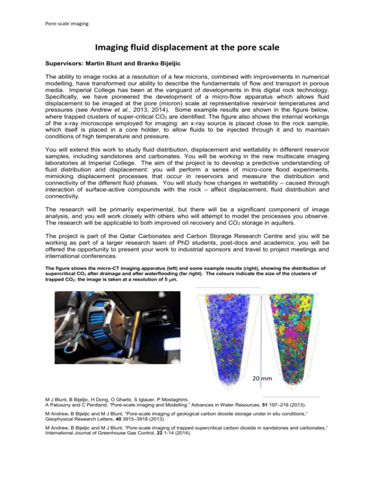
Pore-scale imaging Imaging fluid displacement at the pore scale Supervisors: Martin Blunt and Branko Bijeljic The ability to image rocks at a resolution of a few microns, combined with improvements in numerical modelling, have transformed our ability to describe the fundamentals of flow and transport in porous media. Imperial College has been at the vanguard of developments in this digital rock technology. Specifically, we have pioneered the development of a micro-flow apparatus which allows fluid displacement to be imaged at the pore (micron) scale at representative reservoir temperatures and pressures (see Andrew et al., 2013, 2014). Some example results are shown in the figure below, where trapped clusters of super-critical CO2 are identified. The figure also shows the internal workings of the x-ray microscope employed for imaging: an x-ray source is placed close to the rock sample, which itself is placed in a core holder, to allow fluids to be injected through it and to maintain conditions of high temperature and pressure. You will extend this work to study fluid distribution, displacement and wettability in different reservoir samples, including sandstones and carbonates. You will be working in the new multiscale imaging laboratories at Imperial College. The aim of the project is to develop a predictive understanding of fluid distribution and displacement: you will perform a series of micro-core flood experiments, mimicking displacement processes that occur in reservoirs and measure the distribution and connectivity of the different fluid phases. You will study how changes in wettability – caused through interaction of surface-active compounds with the rock – affect displacement, fluid distribution and connectivity. The research will be primarily experimental, but there will be a significant component of image analysis, and you will work closely with others who will attempt to model the processes you observe. The research will be applicable to both improved oil recovery and CO2 storage in aquifers. The project is part of the Qatar Carbonates and Carbon Storage Research Centre and you will be working as part of a larger research team of PhD students, post-docs and academics; you will be offered the opportunity to present your work to industrial sponsors and travel to project meetings and international conferences. The figure shows the micro-CT imaging apparatus (left) and some example results (right), showing the distribution of supercritical CO2 after drainage and after waterflooding (far right). The colours indicate the size of the clusters of trapped CO2: the image is taken at a resolution of 5 m. 20 mm M J Blunt, B Bijeljic, H Dong, O Gharbi, S Iglauer, P Mostaghimi, A Paluszny and C Pentland, “Pore-scale imaging and Modelling.” Advances in Water Resources, 51 197–216 (2013). M Andrew, B Bijeljic and M J Blunt, “Pore-scale imaging of geological carbon dioxide storage under in situ conditions,” Geophysical Research Letters, 40 3915–3918 (2013). M Andrew, B Bijeljic and M J Blunt, “Pore-scale imaging of trapped supercritical carbon dioxide in sandstones and carbonates,” International Journal of Greenhouse Gas Control, 22 1-14 (2014).


