TITLE 1. Provide as accurate and concise a description of the
TITLE
ABSTRACT
INTRODUCTION
BACKROUND
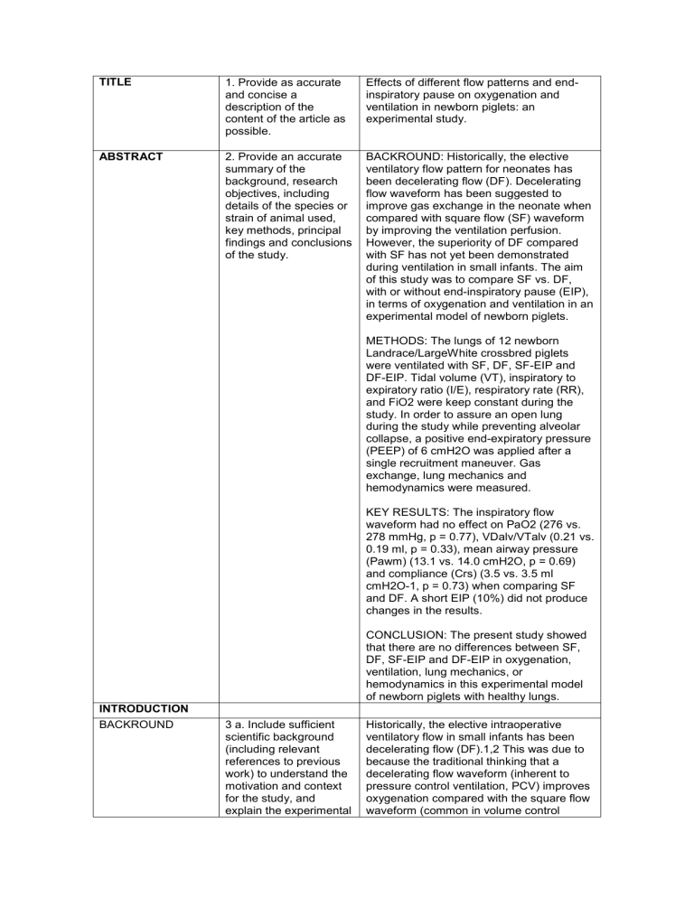
1. Provide as accurate and concise a description of the content of the article as possible.
Effects of different flow patterns and endinspiratory pause on oxygenation and ventilation in newborn piglets: an experimental study.
2. Provide an accurate summary of the background, research objectives, including details of the species or strain of animal used, key methods, principal findings and conclusions of the study.
3 a. Include sufficient scientific background
(including relevant references to previous work) to understand the motivation and context for the study, and explain the experimental
BACKROUND: Historically, the elective ventilatory flow pattern for neonates has been decelerating flow (DF). Decelerating flow waveform has been suggested to improve gas exchange in the neonate when compared with square flow (SF) waveform by improving the ventilation perfusion.
However, the superiority of DF compared with SF has not yet been demonstrated during ventilation in small infants. The aim of this study was to compare SF vs. DF, with or without end-inspiratory pause (EIP), in terms of oxygenation and ventilation in an experimental model of newborn piglets.
METHODS: The lungs of 12 newborn
Landrace/LargeWhite crossbred piglets were ventilated with SF, DF, SF-EIP and
DF-EIP. Tidal volume (VT), inspiratory to expiratory ratio (I/E), respiratory rate (RR), and FiO2 were keep constant during the study. In order to assure an open lung during the study while preventing alveolar collapse, a positive end-expiratory pressure
(PEEP) of 6 cmH2O was applied after a single recruitment maneuver. Gas exchange, lung mechanics and hemodynamics were measured.
KEY RESULTS: The inspiratory flow waveform had no effect on PaO2 (276 vs.
278 mmHg, p = 0.77), VDalv/VTalv (0.21 vs.
0.19 ml, p = 0.33), mean airway pressure
(Pawm) (13.1 vs. 14.0 cmH2O, p = 0.69) and compliance (Crs) (3.5 vs. 3.5 ml cmH2O-1, p = 0.73) when comparing SF and DF. A short EIP (10%) did not produce changes in the results.
CONCLUSION: The present study showed that there are no differences between SF,
DF, SF-EIP and DF-EIP in oxygenation, ventilation, lung mechanics, or hemodynamics in this experimental model of newborn piglets with healthy lungs.
Historically, the elective intraoperative ventilatory flow in small infants has been decelerating flow (DF).1,2 This was due to because the traditional thinking that a decelerating flow waveform (inherent to pressure control ventilation, PCV) improves oxygenation compared with the square flow waveform (common in volume control
OBJECTIVES
METHODS
ETHICAL
STATEMENT
STUDY DESIGN approach and rationale. b. Explain how and why the animal species and model being used can address the scientific objectives and, where appropriate, the study’s relevance to human biology.
4. Clearly describe the primary and any secondary objectives of the study, or specific hypotheses being tested.
5. Indicate the nature of the ethical review permissions, relevant licences (e.g. Animal
[Scientific Procedures]
Act 1986), and national or institutional guidelines for the care and use of animals, that cover the research.
6. For each experiment, give brief details of the study design including: a. The number of experimental and control groups. b. Any steps taken to minimise the effects of subjective bias when allocating animals to ventilation, VCV) related to a better intrapulmonary gas distribution. However, the superiority of decelerating flow compared with square flow has not yet been demonstrated during ventilation in small infants. Even in adults, the superiority has been questioned in clinical studies showing contradictory results.3-10
Most studies3-6 used VCV and PCV modes when comparing square vs. decelerating flow waveforms in terms of oxygenation and ventilation. However different VT between both modes could affect gas exchange. In order to prevent the changes in VT that can occur on PCV, we used VCV with decelerating flow as a surrogate of PCV because they show identical airway pressures and flow and volume waveforms under passive conditions when the same
VT is administered.
There are no reported studies comparing square flow and decelerating flow in small infants with healthy lungs. Based on recent data, we hypothesized that in the nonatelectasic healthy lungs of small infants there are no differences in intrapulmonary gas distribution between squared and decelerating flow
.
The primary outcome was to elucidate the differences in oxygenation between square and decelerating flow during ventilation in an experimental model of newborn piglets with healthy lungs at the same VT. A secondary outcome was a difference in ventilation and respiratory mechanics between the two flow waveforms
An experimental, prospective, controlled study was conducted. Approval was granted by the Ethical Committee for Experimental
Research at the University of Valencia,
Spain (Chairperson, Prof Dr. M. Real).
To prevent bias while applying the four ventilatory modes (DF, SF, DF-EIP, SF-EIP) to the same animal, a sequence of all modes, starting and ending with the same mode, was designed (e.g., DF, SF, DF-EIP,
SF-EIP, DF). The 12 possible combinations were determined, and the combinations were randomly applied to 12 animals.
After an initial hemodynamic stabilization, all animals were ventilated for 30 minutes
EXPERIMENTAL
PROCEDURES
EXPERIMENTAL
ANIMALS treatment (e.g. randomisation procedure) and when assessing results (e.g. if done, describe who was blinded and when). c. The experimental unit
(e.g. a single animal, group or cage of animals). d. A time-line diagram or flow chart can be useful to illustrate how complex study designs were carried out.
7. For each experiment and each experimental group, including controls, provide precise details of all procedures carried out. For example: a. How (e.g. drug formulation and dose, site and route of administration, anaesthesia and analgesia used
[including monitoring], surgical procedure, method of euthanasia).
Provide details of any specialist equipment used, including supplier(s). b. When (e.g. time of day). c. Where (e.g. home cage, laboratory, water maze). d. Why (e.g. rationale for choice of specific anaesthetic, route of administration, drug dose used).
8 a. Provide details of the animals used, including species, strain, sex, developmental stage (e.g. mean or median age plus age range) and weight (e.g. mean or median weight plus weight range). b. Provide further relevant information such as the source of ani mals, international with each mode. The VT, FiO2, PEEP, RR, and I/E relationships were kept constant during the study. To minimize error and variability, the mean value of the last 10 minutes of each mode was calculated for each variable of the ventilatory parameters.
These values were considered representative of the effect of the mode
(end-values) and were also taken as the baseline values for the following mode.19
Mean values of the hemodynamic and arterial blood gas analysis (BGA) parameters were calculated from 4 measurements made during the last 10 min.
Anesthesia management
Animals were premedicated with an intramuscular bolus of ketamine (1 mg Kg-
1), medetomidine (0.06 mg Kg-1), and azaperone (0.06 mg Kg-1). Midazolam (1 mg Kg-1) and fentanyl (0.03 mg kg-1) were then administered in order to induce anesthesia. Endotracheal intubation was performed with a 3-mm internal diameter cuffed tube to prevent changes in VT due to air-leakage as clinically recommended.11
During the study period, anesthesia was maintained with propofol (8 mg kg-1 min-1), remifentanil (0.15 µg kg-1 min-1), and cisatracurium (0.1 mg kg-1 h-1). Body temperature was maintained at 35- 36º C with a heat blanket.
Instrumentation
A 3-Fr thermodilution catheter (PV2013L07-
A, Pulsion Medical Systems AG, München,
Germany) was inserted by a cut down to the right femoral artery for cardiac output monitoring. A 4-Fr double-lumen catheter
(AK-14412, Arrow International, Inc, USA) was inserted by a cut down into the right or left internal jugular vein for drugs and fluid administration, and for transpulmonary thermodilution.
Landrace/Large White crossbred female piglets weighing 2.9-3.1 kg and 7 days of age were used in the study. At the end of the experimental protocol, the animals were euthanized with an overdose of potassium chloride under deep anesthesia.
HOUSING AND
HUSBANDRY strain nomenclature, genetic modification status (e.g. knock-out or transgenic), genotype, health/immune status, drug or test naïve, previous procedures, etc.
9. Provide details of: a. Housing (type of facility e.g. specific pathogen free [SPF]; type of cage or housing; bedding material; number of cage companions; tank shape and material etc. for fish). b. Husbandry conditions
(e.g. breeding programme, light/dark cycle, temperature, quality of water etc for fish, type of food, access to food and water, environmental enrichment). c. Welfare-related assessments and interventions that were carried out prior to, during, or after the experiment.
Mechanical ventilation (MV)
For mechanical ventilation, a Galileo gold
(Hamilton, Bonaduz, Switzerland) was used in the pediatric mode. The ventilator allows square and decelerating flow, and endinspiratory pause can be adjusted for a constant I/E relationship in VCV. The following ventilatory modes were compared in the study (Figure 1, table 1):
• Mode SF: Square flow, no endinspiratory pause.
• Mode DF: Decelerating flow, no end-inspiratory pause.
Also, by adjusting a short end-inspiratory pause, another two modes were possible.
The 10% end-inspiratory pause is based on our routine clinical practice, and due to a lack of evidence for the best short endinspiratory pause duration in healthy lungs:
• Mode SF-EIP: Square flow with an end-inspiratory pause of 0.06 s (10% TI).
• Mode DF-EIP: Decelerating flow with an end-inspiratory pause of 0.06 s
(10% Ti).
After induction of anesthesia, mechanical ventilation was initiated in volume-controlled ventilation (VCV) mode with a constant inspiratory flow (square wave) and the protective12,13 tidal volume (VT) set at 10 mL kg-1, inspiratory to expiratory ratio (I/E) at 1/2, respiratory rate (RR) of 30 breaths/min, and FiO2 of 0.5.
In order to assure a fully open lung during the study while preventing any alveolar collapse, a single recruitment maneuver
(RM) was performed before starting the experimental ventilatory protocol. This consisted of the application of 40 cmH2O of
CPAP for 10 seconds as described elsewhere,14 and adjusting for a positive end-expiratory pressure (PEEP) of 6 cmH2O thereafter. Response to RM and adequacy of the PEEP was confirmed by checking for a normal alveolar-arterial oxygen gradient (279 ± 20 mmHg) in the baseline control of the first ventilatory mode.15,16 No further RMs were performed, and the PEEP-level was kept constant during the whole experiment. In order to prevent de-recruitment, the sequence of changes in ventilatory modes
was performed without disconnecting the breathing circuit.
Respiratory monitoring
Volumetric capnography was recorded continuously using a NICO capnograph
(Respironics, Wallingford, CT, USA) connected to a laptop running DataColl software (Respironics, Wallingford, CT,
USA). The mainstream capnograph sensor
(single-patient airway adapter, neonatal:
6312-00) was placed between the endotracheal tube and the “Y” piece of the breathing circuit. Expired volume and CO2 data were downloaded into a custom
MatLab program (Mathworks, Natick, MA,
USA) that constructed breath-by-breath volumetric capnograms for offline analysis.
The VTCO2,br is the amount of CO2 eliminated during one breath, which is obtained by integration of expired airway flow and PCO2. PEtCO2 is the partial pressure of CO2 at the end of expiration.
Airway dead space (VDaw) was measured as the inflection point of phase II of the volumetric capnogram. Physiological dead space (VDphys) to tidal volume ratio
(VD/VT) was calculated using the Bohr –
Enghoff formula:
VD/VT=PaCO2 –PE´CO2/ PaCO2 where PE´CO2 is the mixed PCO2 of an expiration. VDphys was then calculated by multiplying VD/VT and tidal volume.
Alveolar dead space (VDalv) was obtained by subtracting VDaw from VDphys, which was then normalized by the alveolar tidal volume (VDalv/VTalv).
Peak inspiratory pressure (PIP) and mean airway pressure (Pawm) were determined through the NICO monitor with the pressure transducer placed between the endotracheal tube and the “Y” piece of the breathing circuit. Crs was automatically calculated as VT/(Pplat – PEEP).
Values of pH, PaO2, PaCO2, and bicarbonate were obtained from arterial blood gas analysis (i-STAT Analyzer, Abbott
Laboratories, East Windsor, NJ, USA).
Hemodynamic monitoring and management
A PiCCO monitor (Pulsion Medical System
AG, Munchen, Germany) was used for hemodynamic monitoring. Recent experimental studies using the PiCCO monitor in pigs have not reported any technical limitations17,18.
The cardiac index (CI) was obtained through transpulmonary thermodilution with the PiCCO monitor using the mean values of three 5-mL iced saline injections prior to each set of measurements. Mean arterial pressure (MAP) and heart rate (HR) were
SAMPLE SIZE
ALLOCATING
ANIMALS TO
EXPERIMENTAL
GROUPS
EXPERIMENTAL
OUTCOMES
STATISTICAL
METHODS
10. a. Specify the total number of animals used in each experiment, and the number of animals in each experimental group. b. Explain how the number of animals was arrived at. Provide details of any sample size calculation used. c. Indicate the number of independent replications of each experiment, if relevant
11.
a. Give full details of how animals were allocated to experimental groups, including randomisation or matching if done. b. Describe the order in which the animals in the different experimental groups were treated and assessed.
12. Clearly define the primary and secondary experimental outcomes assessed (e.g. cell death, molecular markers, behavioural changes).
13. a. Provide details of the statistical methods used for each analysis. b. Specify the unit of analysis for each recorded continuously by arterial pulse wave analysis. All hemodynamic parameters were obtained with an atmospheric pressure calibration measured at the mid-thoracic level while the animals were in a supine position. Throughout the study, the animals received a continuous intravenous infusion of crystalloids (Lactate
Ringer solution, 4-6 mL kg-1 h-1). a.
This was a crossover study including 12 animals in only one group. b.
The number of animals was base don previous studies.
There were not randomization in different groups.
After an initial hemodynamic stabilization, all animals were ventilated for 30 minutes with each mode. The VT, FiO2, PEEP, RR, and I/E relationships were kept constant during the study. To minimize error and variability, the mean value of the last 10 minutes of each mode was calculated for each variable of the ventilatory parameters.
These values were considered representative of the effect of the mode
(end-values) and were also taken as the baseline values for the following mode.19
Mean values of the hemodynamic and arterial blood gas analysis (BGA) parameters were calculated from 4 measurements made during the last 10 min.
Based on data from a previous study in healthy infants after cardiac surgery7, it was estimated that a total of 12 animals would be needed to detect at least a 25-mmHg difference in oxygenation between flows,
RESULTS
BASELINE DATA
NUMBERS
ANALYSED
OUTCOMES AND
ESTIMATION dataset (e.g. single animal, group of animals, single neuron). c. Describe any methods used to assess whether the data met the assumptions of the statistical approach.
14. For each experimental group, report relevant characteristics and health status of animals
(e.g. weight, microbiological status, and drug or test naïve) prior to treatment or testing. (This information can often be tabulated).
15.
a. Report the number of animals in each group included in each analysis. Report absolute numbers (e.g.
10/20, not 50%2). b. If any animals or data were not included in the analysis, explain why.
16. Report the results for each analysis carried out, with a measure of precision (e.g. standard error or confidence interval).
ADVERSE EVENTS 17. a. Give details of all important adverse with a 5% significance level, and 80% power. All data were entered into the statistical package, SPSS version 15.0
(SPSS, Chicago, IL, USA). The Friedman test was performed for homogeneity. To compare primary outcomes, differences between the end-values and baseline measurements in each ventilatory mode, or the differences in the end-values between the four ventilatory modes, a statistical analysis using the Wilcoxon signed-rank test was performed.20 To identify differences in the end-values between specific ventilatory modes (secondary outcome), the Bonferroni correction criteria was used to fit a type 1 risk to the chosen significance level (α = 0,05). All values are reported as the mean ± standard deviation
(SD).
12 animals in only one group.
All data was included
Table 2 shows mean ± SD of the oxygenation and ventilation parameters. No significant differences were found in any of the parameters measured when comparing end-values vs baseline in each ventilatory mode, with or without EIP (Table 2). No significant differences were found when the end-values of the four ventilatory modes were compared (Table 2). Table 3 shows the mean ± SD of the respiratory mechanic parameters. No significant differences were found in PIP, Pawm, and Crs when the endvalues of the four ventilatory modes were compared. The mean end-values of CI and
MAP were not significantly different between the four ventilatory modes (Table
4).
Any adverse event occur during the experimentation.
DISCUSSION
INTERPRETATION/
SCIENTIFIC
IMPLICATIONS events in each experimental group. b. Describe any modifications to the experimental protocols made to reduce adverse events.
18. a. Interpret the results, taking into account the study objectives and hypotheses, current theory and other relevant studies in the literature.
There were not modifications of the initial experimental protocol.
This study shows no differences in oxygenation (PaO2), ventilation
(VDalv/VTalv, PaCO2), lung mechanics
(Crs), or hemodynamics (CI) between the square and the decelerating inspiratory flow waveform in this experimental setting of newborn piglets with healthy lungs. The same results were obtained when a short end-inspiratory pause of 10% was added to the inspiratory time during both square and decelerating flow.
To the best of our knowledge, until now the optimal flow pattern and the effects of an end-inspiratory pause in terms of oxygenation and ventilation during ventilation in an animal model of small lungs has not been thoroughly investigated.
Some studies3-7,9,10,21-27 compared square (common in VCV) vs. decelerating flow (inherent to PCV) in terms of gas exchange, lung mechanics, and hemodynamics. Most of these studies, clinical and experimental, were performed in adults with heterogeneous lungs (with ALI), applied different methodologies, used different ventilator modes to compare flows
(PCV for decelerating flow and VCV for square flow), and produced contradictory results.
Our results in an experimental model of small healthy lungs are similar to those obtained in other settings in healthy lungs.
Kocis7 found no differences in PaO2,
PaCO2, MAP, and cardiac output (CO) between square and decelerating flow during postoperative cardiac surgery in infants with healthy lungs weighing over 5.5 kg when a level of 2-3 cmH2O of PEEP was set in all patients. It is true that our initial
PaO2 was much higher than the PaO2 in the infants included in Kocis study. The reason may be justified by methodological differences. Our initial PaO2 was after a recruitment maneuver with a PEEP level that prevents alveolar collapse, and the initial PaO2 in the Kocis study was after cardiac surgery where there was probably a lung collapse, no recruitment maneuvers, and lower PEEP levels. Smith8 compared 4 flow patterns and found no significant differences in PaO2, PaCO2 and VD/VT,
MAP, and CO.
Contrary to our results, some studies found differences between square and decelerating flow when applied in adults with lung injury. Al-Saady et al.3 and Davis et al.4 compared square vs. decelerating flow and showed that decelerating flow improved PaO2, VD/VT, and Crs. The authors justified these results because the higher mean airway pressure (Pawm) produced by the decelerating flow favored alveolar recruitment and gas redistribution and diffusion. Higher Pawm generates more alveolar recruitment, which improves the ventilation/perfusion relationship. This effect is based on a mathematical model25 and could be especially important in lungs with a high alveolar time constant3,26 accounting for better gas exchange. However, this effect is not apparent in a homogeneous lung.27
During mechanical ventilation, the infant respiratory system is prone to alveolar collapse,28-30 leading to a heterogeneous lung. In our study, in order to assure an homogeneous open lung (confirmed by a normal alveolar-arterial oxygen gradient), while preventing alveolar re-collapse, an alveolar recruitment maneuver (ARM)14 was performed in all animals and 6 cmH2O of PEEP was applied afterwards (based on previous experimental studies).31-33 In this situation, the possible benefits of alveolar recruitment secondary to higher Pawm with decelerating flow would disappear.9,10
However, the ARM and the supraphysiological PEEP levels used in this study for keeping the lung open are not common in clinical practice because of the supposed increased risk of barotrauma in small infants due to over-distension.
Recently, however, it was shown that higher airway pressures than those used to recruit healthy lung are needed to produce barotrauma in small lungs without chest wall
28.
The results of previous studies9,10,23 confirm our hypothesis. Markström10 showed no differences between square vs. decelerating flow in 13 pigs with ALI in
PaO2, functional residual capacity (FRC) and Crs despite the differences observed in
Pawm. As in our study, they performed an
ARM at the start of the study and they compared both flows with different PEEP levels. However, the Markström study showed that PaCO2 was significantly lower with PCV, demonstrating better alveolar ventilation. Recent studies in adults using imaging techniques confirm our results. No
b. Comment on the study limitations including any potential sources of bias, any limitations of the animal model, and the imprecision associated with the results2. c. Describe any implications of your experimental methods or findings for the replacement, refinement or reduction (the 3Rs) of the use of animals in research. differences in intrapulmonary gas distribution with CT-scanning was found between square and decelerating flow.9,23
In relation to alveolar ventilation and dead space, some studies in injured patients found a lower PaCO2 and VD/VT with decelerating flow.3,4,9,10,24 These results in lungs with different alveolar time constants could be justified by the theory of the mean distribution time (MDT),34 which establishes that CO2 elimination is enhanced when the time available for gas distribution and diffusion within the respiratory zone increases (as occurs with decelerating flow because of higher initial peak flow). However, recently endinspiratory flow (EIF) was shown to be a determinant of CO2 elimination. A high EIF enhances CO2 elimination, but an EIF of 0, as occurs with decelerating flow, worsens
CO2 elimination and could balance the positive effects of decelerating flow in
MDT.35 This could be especially important in healthy lungs35 and may explain our findings and those obtained by Smith et al.8
In relation to respiratory mechanics, several studies described higher PIP and lower
Pawm with SF compared with DF.
Differences could be significant in injured lungs with low compliance where PIP is lower with DF than with SF. As the flow decreases, the resistive pressure decreases, but the elastic pressure increases as the lungs fill.3 However, several studies have not found such differences in situations of normal or high compliance.7,9 In contrast, in patients with high resistance as in our study because the use of small-sized endotracheal tubes, the pressures are initially highest using DF with the fastest flow and could remain elevated throughout the respiratory cycle.36
Effects of the end inspiratory pause (EIP)
Until now, the effects of end-inspiratory pause (EIP) on the V/Q relationship during mechanical ventilation of small infant were unknown. In order to ascertain if the effect of flow waveform on oxygenation and ventilation was influenced by EIP but predominantly prevented the effects of EIP at the same time, a short (10% of the Ti) was added in both flows without modifications in the VT and the I/E relationship. We show that a 10% EIP did not produce differences in oxygenation
(PaO2) and ventilation (PaCO2,
VDalv/VTalv) in this experimental model of newborn piglets. The results in oxygenation are in concordance with previous studies37,38 and are justified because EIP
does not decrease shunt as it does not recruit alveoli. Different from previous studies (in healthy and injured lungs),34,35,37-39 our results did not show differences in ventilation. The EIP could improve ventilation because (1) it favors gas redistribution in the lungs with different alveolar time constants, improving the V/Q relationship and therefore improving VD/VT.
This effect may not appear in homogeneous lungs without different alveolar time constants as in our model of normal lungs; and (2) the EIP increases the MDT. We did not observe an improvement in ventilation
(PaCO2) with constant inspiratory time.
When I/E relationship is constant, previous studies in healthy lungs33,41 showed that higher EIP (20%) is required to improve ventilation, especially with high RR.
As we observed in our results, hemodynamics remained constant throughout the experimentation with no clinical differences between flows. These results are consistent with previous studies.4,7,9,24 Therefore, the effects of different flow patterns in oxygenation and ventilation are not explained by changes in pulmonary perfusion.
Historically, the elective intraoperative ventilatory mode in very small infants has been pressure control ventilation for two basic reasons. First, the very real limitation of older anesthesia machines to guarantee a constant VT during volume control ventilation, and second, because of the traditional thinking that DF (inherent to pressure control) improves oxygenation compared with SF (common in volume control). The results obtained in our study together with the new anesthesia machines that accurately ensure very low VT42 could make volume control the elective intraoperative mode.
Several limitations of this study need to be mentioned. Firstly, this is an experimental study which may limit the application of the findings to the clinical setting. Secondly, this study examined a small number of animals and therefore was not powered to expose small differences in some of the variables measured. Thirdly, currently there is no general indication for routinely applying
ARM and supraclinical levels of PEEP during intraoperative ventilatory management in infants. By not applying these techniques, lung homogeneity is not guaranteed, and decelerating flow may favor redistribution of gas and a better V/Q relationship. Fourth, The crossover
GENERALISABILITY/
TRANSLATION
FUNDINGS
CONCLUSION
19. Comment on whether, and how, the findings of this study are likely to translate to other species or systems, including any relevance to human biology.
20. List all funding sources (including grant number) and the role of the funder(s) in the study. methodology used could mask differences in the end values of the modes if the effects of the previous ventilatory mode spilled over into the next ventilatory mode. Fifth, a possible limitation were the effects of an unblinded study, however biases was minimized with a standarized protocol in management, monitoring, measurements and data colection. Finally, the clinical monitors used in the study are not completely validated for use in newborn piglets; however, they were applied throughout the study, and their inherent percent error was considered to be similar for the four conditions assessed.
The results of the study confirms that volumen controlled ventilation could be an alternative to pressure control ventilation during intraoperative mechanical ventilation in newborns.
There were not external fundings. The study was founded by the Clinical Research
Institute (INCLIVA) of the Hospital Clínico
Universitario of Valencia
In conclusion, the present study showed that there are no differences between square and decelerating flow, with or without EIP, in oxygenation, ventilation, lung mechanics, or hemodynamics in this experimental setting in an animal model of newborn piglets with healthy lungs.
However, further studies are needed to elucidate whether or not different flowwaveforms may have a direct effect when ventilating small lungs with acute lung injury.
