Shay/Wright retroviral protocol 2006
advertisement
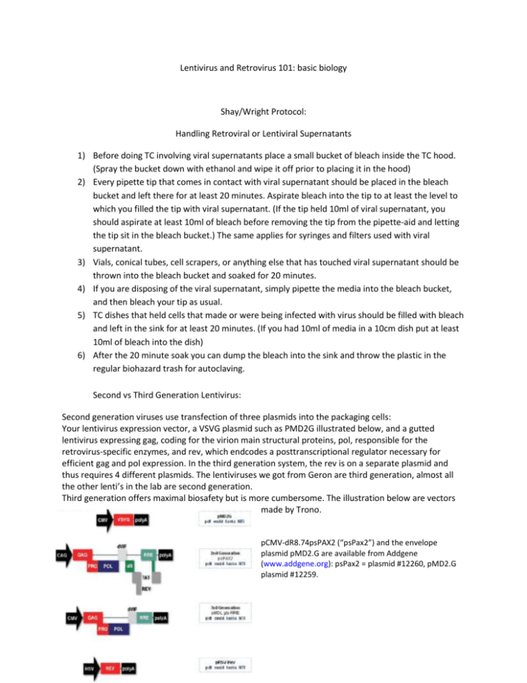
Lentivirus and Retrovirus 101: basic biology Shay/Wright Protocol: Handling Retroviral or Lentiviral Supernatants 1) Before doing TC involving viral supernatants place a small bucket of bleach inside the TC hood. (Spray the bucket down with ethanol and wipe it off prior to placing it in the hood) 2) Every pipette tip that comes in contact with viral supernatant should be placed in the bleach bucket and left there for at least 20 minutes. Aspirate bleach into the tip to at least the level to which you filled the tip with viral supernatant. (If the tip held 10ml of viral supernatant, you should aspirate at least 10ml of bleach before removing the tip from the pipette-aid and letting the tip sit in the bleach bucket.) The same applies for syringes and filters used with viral supernatant. 3) Vials, conical tubes, cell scrapers, or anything else that has touched viral supernatant should be thrown into the bleach bucket and soaked for 20 minutes. 4) If you are disposing of the viral supernatant, simply pipette the media into the bleach bucket, and then bleach your tip as usual. 5) TC dishes that held cells that made or were being infected with virus should be filled with bleach and left in the sink for at least 20 minutes. (If you had 10ml of media in a 10cm dish put at least 10ml of bleach into the dish) 6) After the 20 minute soak you can dump the bleach into the sink and throw the plastic in the regular biohazard trash for autoclaving. Second vs Third Generation Lentivirus: Second generation viruses use transfection of three plasmids into the packaging cells: Your lentivirus expression vector, a VSVG plasmid such as PMD2G illustrated below, and a gutted lentivirus expressing gag, coding for the virion main structural proteins, pol, responsible for the retrovirus-specific enzymes, and rev, which endcodes a posttranscriptional regulator necessary for efficient gag and pol expression. In the third generation system, the rev is on a separate plasmid and thus requires 4 different plasmids. The lentiviruses we got from Geron are third generation, almost all the other lenti’s in the lab are second generation. Third generation offers maximal biosafety but is more cumbersome. The illustration below are vectors made by Trono. pCMV-dR8.74psPAX2 (“psPax2”) and the envelope plasmid pMD2.G are available from Addgene (www.addgene.org): psPax2 = plasmid #12260, pMD2.G plasmid #12259. Lentiviruses and retrovirus are both large and are partially retained in 0.22 um filters, thus use 0.45 um filters to remove any contaminating cells. According to the TRC consortium, viral harvest growth media containing 10% serum + 1.1g/mL supplemental BSA is equivalent to viral harvest media containing 30% serum – both produce viral stocks with similar high titer (2x higher than in 10% serum). The BSA-supplemented media is more cost effective, easier to mix in standard 500mL media bottles, and may be preferred when transfecting cells that are sensitive to serum. The half-life at 37C for both lenti- and retroviruses is about 4-6 hours, so one typically collects supernatants twice a day—e.g., first thing in the morning, and last thing at the end of the day. Matt Porteus showed that including caffeine (to block DNA damage signaling and checkpoint activities) during lentiviral production increased titers 2-4-fold. 2,4, or 8 mM caffeine is added the morning after transfection (17 hours) and supernatant collected (in the presence of caffeine each time) at 48 hours and mornings/evenings thereafter. According to JY’s tests, 2mM caffeine was the best compromise between toxicity and increased titer. He made a 40 mM stock (20x) in Medium X, filter sterilized, and added it ~12 hours after transfection. No tests have been done on retroviral titers. Ecotropic refers to a virus that has the surface env protein that will bind to mouse but not human cells, while amphotropic refers to an envelope protein that will bind to both. An amphotropic packaging cell line expresses lots of the amphotropic env, and is resistant to infection by a retrovirus expressing the amphotropic env since all of its receptors are already tied up by the ligand it is already making. Retroviruses sometimes get rearranged following transfection into packaging cells, producing some non-infectious particles. These can be minimized by first transfecting the retrovirus proviral plasmid into an ecotropic packaging line, and then using the supernatant to infect an amphotropic packaging line. Only competent particles can infect the amphotropic line, and this thus eliminates all of the rearrangements so that one ends up with higher titers. We have both transient and stable systems for the production of retroviral supernatants. In general, a retrovirus that is for a special purpose and isn’t going to be used over and over is most conveniently just packaged in the transient line PhoenixA (A= amphotropic). Routinely used viruses are more conveniently produced in stable packaging lines. For this, first transfect into the transient packaging line PhoenixE (E=ecotropic), and then take the supernatant and use it to infect the stable amphotropic packaging line PA317. After selection using the appropriate drug, the stable line is easily expanded to produce as much supernatant as one wants and can be frozen down and later thawed for additional supernatant production. PE501 is the name of our stable ecotropic retroviral packaging line, but we rarely use it since the transient PhoenixE takes less time. IMPORTANT: retroviral packaging lines are immortal but they LOOSE expression of their packaging factors over time. We have reselected for retention of the factors (a mess, since diphtheria toxin resistance is one of the selection markers) and designated the reselected cells PD 0. BE SURE to keep track of PDs after thawing the cells and don’t keep them in culture for more than 60-90 PDs. Refreeze low passage cells after thawing them. The VSVG plasmid expressing the ligand for lentiviral packaging is toxic to cells, so stable packaging lines for lentiviral production aren’t readily available (some attempts to use an inducible VSVG have been done). Hela cells are somewhat resistant to retroviral infection, perhaps because they are infected with HPV. The env protein from HPV may be binding the receptor and making it unavailable. Several approaches around this include: 1) co-transfecting the proviral DNA into Phoenix A along with a VSVG env plasmid (from the lenti systems). VSVG uses a different cellular receptor, and thus gets in; 2) treating Hela transiently with tunicamycin, to block processing of potential endogenous env proteins. We actually haven’t tried this, and someone should at some point based on J.Virol 1992.66.78-42. They pretreated CHO cells with 0.1-0.3 ug/ml tunicamycin for 19 hours prior to infection with viral supernatants. Retroviruses only infect dividing cells, and a good retroviral supernatant can infect 10%-50% of the cells. Infecting the cells several times is OK. Lentiviruses can integrate into non-dividing cells, 11 12 18 23 27 32 MOI 0 0.1 0.2 0.3 0.4 0.5 0.6 0.7 0.8 0.9 1 3 30 Number of viral insertions into genome 0 1 2 3 100 0 0 0 90 9 0 0 82 16 2 0 74 22 3 0 67 27 5 1 61 30 8 1 55 33 10 2 50 35 12 3 45 36 14 4 41 37 16 5 37 37 18 6 5 15 22 22 0 0 0 0 % GFP negative %GFP+ MOI 100 0.0001 0.0001 90 10 0.1 82 18 0.2 74 26 0.3 67 33 0.4 61 39 0.5 55 45 0.6 50 50 0.7 45 55 0.8 41 59 0.9 37 63 1 5 95 3 0 100 30 >3 0 0 0 0 0 0 0 1 1 1 2 35 100 >25 0 0 0 0 0 0 0 0 0 0 0 0 84 MOI vs GFP 70 60 % GFP+ cells % with >1 50 40 30 20 10 0 0.1 0.2 0.3 0.4 0.5 0.6 0.7 0.8 0.9 MOI 1 and much higher titers can be produced. The tables below indicate the relationship between the number of cells that are GFP+ after infection with a GFP-lentivirus and the number of integrations per cell. The multiplicity-of-infection (MOI) refers to the number of infectious particles per cell. If one wants only a single integrant per cell, using an MOI such that <30% of the cells turn green is the best approach. For many situations, a high MOI is fine. Shay/Wright retroviral protocol 2006 Retroviral Protocols Discard pipets, dishes into 50% bleach Treat retroviral supernatants with respect but not panic: the half-life of the virus at 37 degrees is about 4 hours. hTERT is not an oncogene–it does not confer growth advantages by itself. pBabepuro hTERT-PA317 cells should be resistant to 3 ug/ml puromycin. We do not normally maintain them in puro. Wear gloves etc. To harvest supernatants: Grow packaging cells to near confluence or confluence. Replace medium with “minimal” amount of fresh medium to adequately cover the dish (e.g., 12-15 ml for a 15 cm dish with 150 square cm of surface area). Harvest medium after 6-8 hours or after overnight, and replace with fresh medium for the next harvest. Harvesting can be repeated twice/day, often for 3-4 days, until the cell layer detaches or the medium gets acidic too quickly. The more confluent the cells, the higher the viral titers will be. Supernatants should be filtered through a 0.45 micron sterile filter, and can be used fresh or frozen and stored for up to about six months at -80 degrees. Freezing causes as loss of about 50% of infectivity, but since the infectivity is usually much more than needed for most tissue culture applications it doesn’t matter. Convenient method: Harvest medium with a 12 ml syringe (no need to use a needle-just shake off the clinging drop back into the dish) and attach the syringe to a 0.45 micron filter, filter it into a 15 ml tube etc. To infect cells: Make viral supernatants 4 micrograms/ml with polybrene (some cells are sensitive and need 2 ug/ml polybrene). If needed, volumes can be increased (if need to infect multiple dishes, or large dishes etc.) by diluting the viral supernatant 1:1 with polybrene-containing fresh medium. Replace medium of growing cells (retroviruses only infect dividing cells, thus cells can’t be confluent) with enough viral supernatant to cover. Leave on at least four hours: anything more than 8 hours is probably a waste of time. If needed, one can infect first thing in the morning, then again overnight at the end of the day. One infection is usually enough, but can do 2-3 times if a cell type proves hard to infect. After a 36-48 hour expression period, put the cells under selection (puro, neo, etc.). Sample Retroviral Production Protocol—de Lange lab: Required Solutions Polybrene Stock is 10,000x in medium stored at 4C (40 mg/ml) and user stock is 100x. Final concentration is 4 μg/ml. Medium for Phoenix cells: DMEM, 10% FBS, 1% Non-essential amino acids, 1% Pen-Strep, 1% Glutamate Store medium at 4C, warm to 37C before use. 2.5 M CaCl2 Filter sterilize and store aliquots at -20C 2x HBS 50 mM HEPES pH 7.05 10 mM KCl 12 mM Dextrose 280 mM NaCl 1.5 mM Na2HPO4 (FW 141.96) The final pH of the solution should be 7.05 +/- 0.05. Filter through a 0.2 μm filter, aliquot, and store at -20°C. Try to avoid multiple freeze/thaw cycles. To thaw, warm to room temperature and invert or vortex the tube to achieve uniform mixing. Although it is unclear why this occurs, the ability of the 2x HBS solution to produce working CaPO4 precipitates deteriorates after 6 months to one year, even when the 2x HBS solution is stored at -20°C. Bleach All materials that have been in contact with the retrovirus must be bleached thoroughly before discarding in the biohazard waste. Virus production Day 0 Plate 2.5 x 10⁶ Phoenix packaging cells in 9 ml medium/10 cm dish in the afternoon. Note: It is very important to have good single cells suspensions (trypsinize well) and to evenly distribute the cells. Day 1 Transfect cells with 20 g DNA (±24 hrs after plating), using CaPO4 precipitation. Note: At the moment of transfection the cell density should be ± 40-50% such that the cells will be about 90% confluent at Day 3 and completely confluent at Day 4. 1. In a 2 ml eppendorf tube mix: 50 ul of 2.5 M CaCl2 20 ug DNA (Qiagen prep purified) MQ up to total of 500 ul 2. While vortexing the tube, slowly add 500 l 2x HBS drop by drop. Note: if more of the same transfection: mix in 15 ml tube (4 max). 3. Add the 1 ml mix drop by drop to the cells in medium and evenly distribute by swirling the plate. 4. Place cells back in incubator. Day 2 Change medium 5-20 hours after transfection (precipitate is very fine). Late in the afternoon or in evening replace medium with 9 ml of fresh medium. Note: Phoenix cells detach easily, be careful with all medium changes. Day 3 Collect first supernatant (T1) late in the afternoon (this is 48 hrs after transfection and not later than 24 hrs after changing medium): 1. Remove virus-containing medium and set aside for a moment to supply the packaging cells with 9 mls of fresh medium (1-2 plates at a time). 2. Filter the virus-containing medium through a 0.45 um filter and immediately use for infection. Day 4 Collect second supernatant (T2) in the morning. Collect third supernatant (T3) in the evening (8 hrs after T2). Lentiviral Packaging Protocols Below are a whole series of different protocols. Things to consider in using/designing your own protocol: including caffeine can increase titers 2-4x; some protocols claim higher titers in the absence of antibiotics; some protocols claim higher titers using 30% serum or 10% serum + 1000 mg/ml BSA (Note: JY claimed higher titers with lowering serum to 5%); for large scale production (e.g., many 15 cm plates for libraries) consider using CaPO4 ppt transfection protocols. I don’t know if anyone has tested combining caffeine, antibiotic free, and high protein. Polyjet protocol from Guido Stadler 2011 Transfection of 293 cells with PolyJet (Signagen # SL10068B) seed 800k (6 well) / 5M (10cm) 24 h before transfection in gelatin-coated dishes (gelatin prevents 293 cells from detaching when they become over-confluent during virus-harvest; gelatin is not needed for routine culture of 293 cells. Myoblast group routinely coats plates with gelatin, it’s easy) change medium to 1/5 ml fresh growth medium 30-60 min before transfection dilute 1/5 ug DNA into 50/250 ul medium X, vortex gently and spin down Lentivirus production: in 6 well dishes: 0.75 ug target plasmid, 0.75 ug helper plasmids (equimolar ratio of helper plasmids is recommended: ~0.49 ug psPAX2, ~0.26 ug pMD2G) dilute 3/15 ul poly jet into 50/250 ul medium X, vortex gently and spin down add diluted poly jet to the diluted DNA (not the other way round!), vortex and spin down, incubate 15 min (not longer than 20) at RT add the mixture drop-wise onto the medium in the dish, mix by swirling the plate change medium 12-18 h after transfection (5 h for sensitive cells) use 2mM caffeine in medium for virus production caffeine stock solution: dissolve 40mM in medium X, filter (MW=194.19 g/mol; 7.77 g/l (388.4 mg/50ml) = 40mM) Effectene protocol from Ugur Eskiocak~2010 Transient Transfection of 293FT cells using Effectene for virus production (6 well format) Protocol by Ugur Eskiocak 1. The day of transfection, dilute 1 ug DNA (0.5 ug vector + 0.2 ug PMD2G + 0.3 ug PsPAX2) in 100 ul (total volume) Buffer EC. 2. Add 8 ul Enhancer and mix by pipetting up and down. Incubate at room temperature for 2-5 min. 3. Add 8 ul Effectene to the DNA-Enhancer mixture. Mix by pipetting up and down. Incubate the samples for 5-10 min at room temperature to allow transfection-complex formation. 4. While complex formation takes place, wash, trypsinize and collect cells in a 15ml centrifuge tube. Count the cell numbers. 5. Add 600 ul growth medium (can contain serum and antibiotics) to the tube containing the transfection complexes. Mix by pipetting up and down, and immediately add the transfection complexes drop-wise onto well. 6. Plate 2M cells/well on the top of transfection mix in the 6 well. 7. Incubate the cells with the transfection complexes under their normal growth conditions and collect viral soups 24, 48, and 72 hours after transfection. Effectene protocol from Ryan Laranger for Myoblasts ~2010 MAKING "hCPK4 VIRUS" FOR TRANSDUCTION (in 293FT in MediaX 10%S) PMD26 = 285ng/ul PSPAX2 = 345ng/ul pUE3-lenti-hCDK4 = 250ng/ul l.MIX IN A TOTAL Of lug of DNA contained in 100ul of Buffer EC: 0.5 ug of~hCDK4 vector (2 ul) 0.2 ug of PMD2G (1 ul) 0.3 ug of PsPAX2 (1 ul) 2. • Add 8 ul of Enhancer and mix by pipetting up and down. • Incubate at room temp for 2-5 minutes 3.• Add 15 ul of Effectene to the DNA-Enhancer mixture. • Mix by pipetting up and down. • Incubate for 5-10 f inutes at RT 4.Wash, trypsinize and collect cells in a 15ml falcon tube. Count the cell and dilute them to l million/ml 5. Add 600 ul of growth medium to the tube containing the transfection complexes. Pipette up and down and immediately add the transfection complexes drop-wise onto wells 6.•Plate2 million cells per well on top of the transfection mix (6 well tray) 7.• Incubate cells with the transfection complexes and collect viral sups (replace l.5ml of media; freeze down 750ul and use 750ul to transduce). / Lentiviral Production Protocol adapted from Marcu lab, obtained from JY Kim~2009 -The day before transfection: I. Plate out __ 293FT cells per 10cm plate or __ (up to 6M) cells per I5cm plate in either 10 or 25ml X media, respectively. -The day of transfection: 1. Check to make sure cells are at least 25% confluent before continuing. 2. Combine the appropriate amount of plasmid DNAs in a 15ml conical tube: For 10cm plates: 5ug DNA 3-4ug psPAX2. (structural vector) 1-2ug pMDG2 (envelope vector) 10ugTOTAL For I5cm plates: I5ug DNA 7.5ug psPAX2 (structural vector) 7.5ug pMD2.G (envelope vector) 30ugTOTAL (rest of the protocol is for 15cm plates) 3. Add filtered water to a final volume of 878ul. ) 4. Add 122u12M CaCl2, (stock in -20C walk-in freezer) and mix. 5. Add 1000ul of2xHBS (stock in -20C walk-in freezer). 6. Use a 1 ml pipette tip and an automated pipettor to generate bubbles for l0s at the bottom of the tube and l0s going up and down the mixed solution. 7. Let mixture sit for 15m. 8. Remove appropriate amount of media from 293FT cells so that only 18ml is left. 9. Add all of mixture to the cells gently and evenly. Swirl to distribute evenly and put back in incubator OIN. -The day after transfection: 1. Check transfection efficiency for fluorescent marker if possible. 2. Change to the media for the cell type for which the virus is being produced. -The next day: 1. Harvest viral supematant and filter through a 0.45Um filter. (can collect viral supernatant up to 3 days after transfection) 2xHBS: 50mM HEPES 10mM KCl 12mM Dextrose 280mM NaCl 1.5mM Na2HPO4 Resuspend in 900 ml H2O, adjust pH to 7.04 (with NaOH if using HEPES free acid, with HCl if using HEPES sodium salt). Raise volume to 1000 ml and carefully re-adjust final pH to 7.05 exactly (IMPORTANT: pH needs to be very precise at pH 7.04 – 7.05). Filter through 0.22 uM and store in aliquots at -20°C (should be good for up to 6 months). The RNAi Consortium Protocol 1/18/07 Section II: Lentiviral Production Introduction: This section contains protocols for the production of lentivirus stocks from hairpin-pLKO.1 plasmids in 6 cm plates. Lentiviral production consists of the following steps: Day 0 Seed 293T packaging cells Day 1 (pm) Transfect packaging cells with 3 lentivirus plasmids (hairpin-pLKO.1 vector, packaging plasmid, envelope plasmid) Day 2 (am) 18 hours post-transfection: Remove media; replace with fresh high-BSA or high-serum media Day 3 (am) 24 hours after media change: Harvest virus; replace with fresh high-BSA or high-serum media Day 4 (am) 24 hours after harvest 1: Harvest virus; discard packaging cells These procedures should be carried out in accordance with biosafety requirements of the host institution Part 1: Lentiviral Production in 6 cm plates I. Materials Transfection-quality plasmid DNA for: - hairpin-pLKO.1 vector (TRC library plasmid – see Section I) - 2nd generation packaging plasmid containing gag, pol and rev genes (e.g. pCMV-dR8.91 or pCMVdR8.74psPAX2)* - envelope plasmid (e.g. VSV-G expressing plasmid, pMD2.G)* * recommended: use endotoxin-free plasmid isolation kits (Qiagen) TransIT-LT1 transfection reagent (Mirus Bio, MIR 2300/5/6) alternative: FuGENE 6 (Roche, #1 814 443 or #1 988 387) OPTI-MEM serum-free media (Invitrogen, #31985-070) 293T packaging cells (recommended: passage number < 10) Cell seeding media: Low-antibiotic 293T growth media (DMEM + 10% iFBS + 0.1x Pen/Strep) 500 mL DMEM (Dulbecco's Modification of Eagle's Medium; e.g. Mediatech #10-013-CV) 50 mL iFBS (heat-inactivated Fetal Bovine Serum; e.g. HyClone #SH30071.03) 0.5 mL 100x Pen/Strep (10,000 IU/mL penicillin, 10,000 μg/mL streptomycin; e.g. Mediatech #30-002-CI) Viral harvest media: High-BSA 293T growth media (DMEM + 10% iFBS + 1.1g/100mL BSA + 1x Pen/Strep) 500 mL DMEM (Dulbecco's Modification of Eagle's Medium; e.g. Mediatech #10-013-CV) 50 mL iFBS (heat-inactivated Fetal Bovine Serum; e.g. HyClone # SH30071.03) 32 mL 20g/100mL BSA stock (microbiology-grade Bovine Serum Albumin; VWR #14230-738) 5 mL 100x Pen/Strep (10,000 IU/mL penicillin, 10,000 μg/mL streptomycin; e.g. Mediatech #30-002-CI) alternative viral harvest media: High-serum 293T growth media (DMEM + 30% iFBS + 1x Pen/Strep) 500 mL DMEM (Dulbecco's Modification of Eagle's Medium; e.g. Mediatech #10-013-CV) 200 mL iFBS (heat-inactivated Fetal Bovine Serum; e.g. HyClone # SH30071.03) 5 mL 100x Pen/Strep (10,000 IU/mL penicillin, 10,000 μg/mL streptomycin; e.g. Mediatech #30-002-CI) 6 cm tissue culture plates Polypropylene storage tubes II. Instructions 1. Seed 293T packaging cells at 1.3-1.5x10⁵ cells/mL (6 mL per plate) in low-antibiotic growth media (DMEM + 10% iFBS + 0.1x Pen/Strep) in 6 cm tissue culture plates. 2. Incubate cells for 24 hours (37 °C, 5% CO2), or until the following afternoon. After ~24 hours, the cells should be ~70% confluent. 3. Transfect packaging cells: a. Prepare a mixture of the 3 transfection plasmids: Reagent per 6 cm plate packaging plasmid (e.g. pCMV-dR8.91 or pCMV-R8.74psPAX2) 900 ng envelope plasmid (e.g. VSV-G/pMD2.G) 100 ng hairpin-pLKO.1 vector 1 μg OPTI-MEM to total volume 10 to 30 μL* * The volume of OPTI-MEM per well can be adjusted for optimal handling. b. Dilute TransIT-LT1 transfection reagent in OPTI-MEM. Add the TransIT-LT1 reagent dropwise and mix by swirling the tip or gently flicking the tube (do not mix by pipetting or vortexing). Incubate 5 minutes at room temperature. Reagent per 6 cm plate TransIT-LT1 6 μL OPTI-MEM to total volume 90 μL c. Add the 3 plasmid mix dropwise to the diluted TransIT-LT1 reagent and mix by swirling the tip or gently flicking the tube. d. Incubate the transfection mix for 20 - 30 minutes at room temperature. e. Carefully transfer the transfection mix to the packaging cells (in low-antibiotic growth media). The packaging cells can be sensitive to perturbation - take care not to dislodge the cells from the plate. The total volume of transfection mix should be 100 to 125 μL per plate. 4. Incubate cells for 18 hours (37 °C, 5% CO2), or until the following morning. 5. Change media to remove the transfection reagent and replace with 6 mL high-BSA growth media or high serum growth media for viral harvests. 6. Incubate cells for 24 hours (37 °C, 5% CO2). 7. Harvest media containing lentivirus at ~40 hours post-transfection. Transfer media to a polypropylene storage tube. Replace with 6 mL high-BSA growth media or high serum growth media for viral harvests. 8. Repeat viral harvesting every 12-24 hours and replace with 6 mL high-BSA growth media or high serum growth media for viral harvests. Viral titer tends to decrease in later harvests; we typically collect a total of 2-3 time points. After the final harvest, discard the packaging cells. The viral harvests may be pooled as desired. 9. Spin the media containing virus at 1250 rpm for 5 minutes to pellet any packaging cells that were collected during harvesting. Transfer the supernatant to a sterile polypropylene storage tube. 10. Virus may be stored at 4 °C for short periods (hours to days), but should be frozen at -20 °C or -80 °C for long-term storage. To reduce the number of freeze/thaw cycles, aliquot large-scale virus preps to smaller storage tubes prior to long-term storage. RESOURCES pINDUCER lentivirus elicits inducible cDNA and shRNA expression. Meerbrey K L et al. PNAS 2011;108:3665-3670 National Academy of Sciences Openbiosystems 62K shRNA lentiviral library in pGIPz MGC 16K cDNA cloneset in pSport6 (a gateway compatible vector) and its lentiviral gateway transfer recipient 711 Map pSSI 14207 pDONR LTR-CMVattP1-ccdB-CAT-attP2-SV40-Hygro-LTR. Summary of Gateway system wrt transferring MGC library. The MGC library is an "Expression Clone" (i.e., it has attB sites), and thus can only be transferred to what is called a "Donor Vector" containing attP sites. This "donor" designation is just semantic according to their nomenclature—it could be a retro/lenti proviral plasmid with the attP-toxic ccdB-attP sites following a CMV promoter. (Nb:Bsp1407 and BsrGI cute t/gtaca at position 9 in attP1&2,Rsa1 is gt/ac) The CcdB protein interferes with E. coli DNA gyrase (Bernard and Couturier, 1992), thereby inhibiting growth of most E. coli strains (e.g. OmniMAX™ 2-T1R, DH5�™, TOP10). Because of the lethal effects of the CcdB protein, all Gateway® vectors containingthe ccdB gene must be propagated in an E. coli strain that is resistant to CcdB effects. We recommend using the ccdB Survival T1R E. coli strain which is resistant to CcdB effects (Bernard and Couturier, 1992; Bernard et al., 1993; Miki et al., 1992). One Shot® ccdB Survival T1R Chemically Competent E. coli are available from Invitrogen (Catalog no. C7510-03) for transformation. See page 18 for the genotype of ccdB Survival T1R E. coli. The recombination can occur between DNAs of any topology (i.e. supercoiled, linear, or relaxed), although efficiency varies. For optimal efficiency, perform the BP recombination reaction using: �� Linear attB substrates (see the next page for guidelines to linearize attB expression clones �� Supercoiled attP-containing donor vector Note: Supercoiled or relaxed attB substrates may be used, but will react less efficiently than linear attB substrates. (thus nomenclature is BP clonase to recombine between B and P sites, LR clonase to recombine between L and R sites) For your convenience, we suggest using the following nomenclature to catalog your Gateway® vectors and clones. (thus CMVpSport6 would be "pEXPCMVpSprot6", and need a pDONR-type as recipient) Below is the map of the lenti backbone we made so that one can directly transfer MGC clones to a lentivirus. The map automatically labeled the att sites as multiple forms, but this vector has only attP1 and attP2 sites. Use BP Clonase (rather than LR Clonase) to do the transfer. Protocol for inducible cDNA expression gateway cloning from Lu Zhang 2011: 1. Design primers flanking cDNA ORF sequence with attB sequence at the ends 2. Perform PCR (If using the cDNA in pCMV-sports6 vector, don’t do PCR. First linearize the vector with a restriction enzyme that cuts outside the attB sites but not within your cDNA. Streak the cut DNA on amp plates to verify adequate cutting and the lack of colonies. Use 50ng for BP reaction) 3. Incubate attB-flanked PCR product(50fmol) with donor vector(pDONR™221 Kan,150ng) overnight using BP Clonase protocol, treat with proteinase K for 10min and do transformation 4. Pick colonies and sequence(typically 80% correct) 5. Incubate entry clone(150ng or double or more) with destination vector (pINDUCER20 or 21, 150ng) for 1 hour using suggested amount on LR Clonase protocol , treat with proteinase K for 10 min and do transformation 6. Pick colonies and sequence(about 50% are correct)
