The balance between life and death in plasma cells di Caterina
advertisement
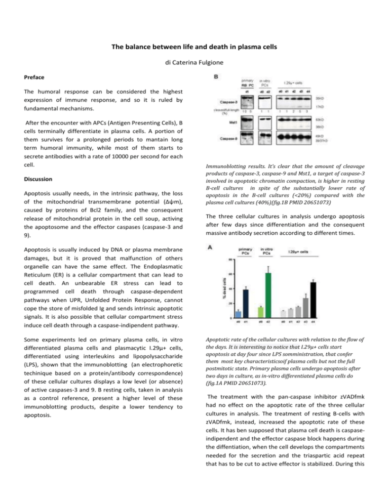
The balance between life and death in plasma cells di Caterina Fulgione Preface The humoral response can be considered the highest expression of immune response, and so it is ruled by fundamental mechanisms. After the encounter with APCs (Antigen Presenting Cells), B cells terminally differentiate in plasma cells. A portion of them survives for a prolonged periods to mantain long term humoral immunity, while most of them starts to secrete antibodies with a rate of 10000 per second for each cell. Discussion Apoptosis usually needs, in the intrinsic pathway, the loss of the mitochondrial transmembrane potential (Δψm), caused by proteins of Bcl2 family, and the consequent release of mitochondrial protein in the cell soup, activing the apoptosome and the effector caspases (caspase-3 and 9). Immunoblotting results. It’s clear that the amount of cleavage products of caspase-3, caspase-9 and Mst1, a target of caspase-3 involved in apoptotic chromatin compaction, is higher in resting B-cell cultures in spite of the substantially lower rate of apoptosis in the B-cell cultures (<20%) compared with the plasma cell cultures (40%)(fig.1B PMID 20651073) The three cellular cultures in analysis undergo apoptosis after few days since differentiation and the consequent massive antibody secretion according to different times. Apoptosis is usually induced by DNA or plasma membrane damages, but it is proved that malfunction of others organelle can have the same effect. The Endoplasmatic Reticulum (ER) is a cellular compartment that can lead to cell death. An unbearable ER stress can lead to programmed cell death through caspase-dependent pathways when UPR, Unfolded Protein Response, cannot cope the store of misfolded Ig and sends intrinsic apoptotic signals. It is also possible that cellular compartment stress induce cell death through a caspase-indipendent pathway. Some experiments led on primary plasma cells, in vitro differentiated plasma cells and plasmacytic I.29µ+ cells, differentiated using interleukins and lipopolysaccharide (LPS), shown that the immunoblotting (an electrophoretic techinique based on a protein/antibody correspondence) of these cellular cultures displays a low level (or absence) of active caspases-3 and 9. B resting cells, taken in analysis as a control reference, present a higher level of these immunoblotting products, despite a lower tendency to apoptosis. Apoptotic rate of the cellular cultures with relation to the flow of the days. It is interesting to notice that I.29µ+ cells start apoptosis at day four since LPS somministration, that confer them most key characteristicsoif plasma cells but not the full postmitotic state. Primary plasma cells undergo apoptosis after two days in culture, as in-vitro differentiated plasma cells do (fig.1A PMID 20651073). The treatment with the pan-caspase inhibitor zVADfmk had no effect on the apoptotic rate of the three cellular cultures in analysis. The treatment of resting B-cells with zVADfmk, instead, increased the apoptotic rate of these cells. It has ben supposed that plasma cell death is caspaseindipendent and the effector caspase block happens during the diffentiation, when the cell develops the compartments needed for the secretion and the triaspartic acid repeat that has to be cut to active effector is stabilized. During this development of the secretory pathway , the proteosome does not improve its capacity, but, intriguingly, the capacity of the proteasome to degrade misfolded proteins decreases during plasma cell differentiation. That is a surprinsing detection, because it would be expected that, if the secretion and the relative amount of misfolded Ig increase, an enlarged degradation capacity would be requested. That find has been considered part of a balance between the necessity of an efficient secretive capacity and the need of a strictly controlled humoral response. The rise of ER stress does not induce apoptosis with the celerity of the caspase-dipendent pathway, but it also cause a situation that does not allow the cell to keep living. The cell reach a level of ER stress, that can be tunamycininduced, after which the ER dependent caspase-4 (12 in mice), responsible of nuclear apoptosis, is expressed, and two Bcl2 proteins, Bim e Bax, induce a non reversible change of Δψm. At this point, apoptosis happens according to mechanisms caspase-indipendent that are not clearly understood. The same experiments had been carried out on nonlymphoid cells, as MEFs (fibroblast embryonic murine). A MEF cellular culture was double knock out (DKO) for caspase-3 and caspase-9 (the MEFs where deprived of both alleles for effector caspases), while another one was heterozygote. The results of DKO culture were the same of plasmacytic differentiated I.29µ+, while the heterozygote culture underwent apoptosis through caspase-dipendent pathways. The caspase-indipendent apoptotic mechanisms can be considered common to others cellular types, too. Conclusion The delay caused by the caspase-block, which is mostly due to the triaspartic acid stabilization on the cleavage site of caspases 3 and 9, allows the cell to keep secreting in a condition that would have usually been critical, and maintaining an high rate of Ig secretion. On the other side, the restricted proteosome capacity does not allow to dispose of misfolded Ig, determining an unbearable long term cellular environment. All these device are useful to induce cell death after the accomplishment of its function. The short-lived plasma cell life span is regulated to permit an effective humoral immune response, that has to face a danger for the system and be strictly defined in time. A further knowledge of short-lived plasma cell apoptosis has clinical implication: mice lacking the proapoptotic protein Bim show a tendency to autoimmune kidneys disease, and both murine and human myeloma have a block of the caspase-dipendent apoptotic pathways, that could be reactivated as a treatment for that disease. Bibliography Auner, Beham-Schmid, Dillon e Sabbattini “The live span of short lived plasma cells is partly determined by a block on activation of apoptotic caspases acting in combination with endoplasmatic reticulum stress”,2010 PMID 20651073 Ferri, K.F.,G.Kroener “Organelle-specific initiation of cell death pathways”, 2001 PMID 11715037 Pelletier N, Casamayor-Pallejà M, De Luca K, Mondière P, Saltel F, Jurdic P, Bella C, Genestier L, Defrance T. ”The endoplasmic reticulum is a key component of the plasma cell death pathway”, 2006 PMID:16424160 B. Alberts et al. “Biologia molecolare della cellula”, 5^ ed. Zanichelli

