anatomy 8 veins 2
advertisement
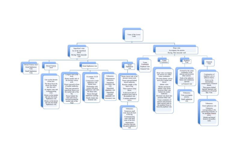
Veins of the Lower Limb Deep veins Accompany the arteries Having Thin muscular wall Superficial veins Lie in the superficial fascia Having Thick muscular wall Dorsal Venous Arch Great Saphenous Vein Small Saphenous Vein Dorsal Venous Arch Lies on the dorsum of the foot. The blood from the whole foot drains into this arch via digital veins and communicating veins. Drains on the medial side into the Great Saphenous vein Drains on the lateral side into the Small Saphenous vein Small Saphenous vein Great Saphenous vein Longest vein in the body. Drains medial side of the dorsum venous arch. Passes upward in front of medial malleolus. Then runs upward in superficial fascia over the medial side of the leg. Passes behind the knee and curves forward around the medial side of the thigh It contains 10-20 valves. Connected to small saphenous vein behind knee. Perforating veins connect the great saphenous vein with the deep veins. Passes through saphenous opening. Finally drains into femoral vein Tributaries: Subcutaneous tributaries Superficial circumflex iliac vein Superficial epigastric vein Superficial external pudendal vein Accessory vein Drains lateral side of the dorsum venous arch. Passes upward behind lateral malleolus. Reaches middle of the back of leg. Then it pierces deep fascia. In the lower part of popliteal fossa ends in the popliteal vein. Contains numerous valves. Tributaries: Numerous small veins from the back of the leg. Communicating veins with deep veins of the foot. Anastomotic branches that join the great saphenous vein. Venae Comitantes Popliteal Vein Femoral Vein Venae Comitantes Deep veins accompany the arteries are called venae comitantes. The deep plantar venous arch gives medial and lateral plantar veins. Medial and lateral plantar veins forms posterior tibial vein behind the medial malleolus. Peroneal vein drain into posterior tibial vein. Venae comitantes of anterior and posterior tibial arteries unite in the popliteal fossa to form the popliteal vein. Popliteal Vein Formed by the venae comitantes of the anterior and posterior tibial arteries. Ends at the adductor hiatus and continuous upward as Femoral vein Tributaries: Veins accompany arteries. Small saphenous vein. Femoral Vein Continuation of popliteal vein at adductor hiatus. Enters in the intermediate compartment of femoral sheath. Then passes behind inguinal ligament and continuous as External Iliac Vein. Tributaries: Great saphenous vein Tributaries corresponding to the perforating branches of the profunda femoris artery. Medial and lateral femoral circumflex veins.
