Mass Spectrometry - Background In mass spectrometry, a substance
advertisement
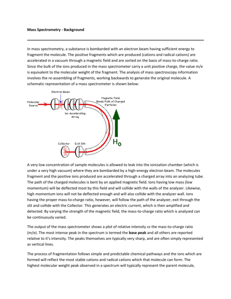
Mass Spectrometry - Background In mass spectrometry, a substance is bombarded with an electron beam having sufficient energy to fragment the molecule. The positive fragments which are produced (cations and radical cations) are accelerated in a vacuum through a magnetic field and are sorted on the basis of mass-to-charge ratio. Since the bulk of the ions produced in the mass spectrometer carry a unit positive charge, the value m/e is equivalent to the molecular weight of the fragment. The analysis of mass spectroscopy information involves the re-assembling of fragments, working backwards to generate the original molecule. A schematic representation of a mass spectrometer is shown below: A very low concentration of sample molecules is allowed to leak into the ionization chamber (which is under a very high vacuum) where they are bombarded by a high-energy electron beam. The molecules fragment and the positive ions produced are accelerated through a charged array into an analyzing tube. The path of the charged molecules is bent by an applied magnetic field. Ions having low mass (low momentum) will be deflected most by this field and will collide with the walls of the analyzer. Likewise, high momentum ions will not be deflected enough and will also collide with the analyzer wall. Ions having the proper mass-to-charge ratio, however, will follow the path of the analyzer, exit through the slit and collide with the Collector. This generates an electric current, which is then amplified and detected. By varying the strength of the magnetic field, the mass-to-charge ratio which is analyzed can be continuously varied. The output of the mass spectrometer shows a plot of relative intensity vs the mass-to-charge ratio (m/e). The most intense peak in the spectrum is termed the base peak and all others are reported relative to it's intensity. The peaks themselves are typically very sharp, and are often simply represented as vertical lines. The process of fragmentation follows simple and predictable chemical pathways and the ions which are formed will reflect the most stable cations and radical cations which that molecule can form. The highest molecular weight peak observed in a spectrum will typically represent the parent molecule, minus an electron, and is termed the molecular ion (M+). Generally, small peaks are also observed above the calculated molecular weight due to the natural isotopic abundance of 13C, 2H, etc. Many molecules with especially labile protons do not display molecular ions; an example of this is alcohols, where the highest molecular weight peak occurs at m/e one less than the molecular ion (m-1). Fragments can be identified by their mass-to-charge ratio, but it is often more informative to identify them by the mass which has been lost. That is, loss of a methyl group will generate a peak at m-15; loss of an ethyl, m-29, etc. The mass spectrum of toluene (methyl benzene) is shown below. The spectrum displays a strong molecular ion at m/e = 92, small m+1 and m+2 peaks, a base peak at m/e = 91 and an assortment of minor peaks m/e = 65 and below. The molecular ion, again, represents loss of an electron and the peaks above the molecular ion are due to isotopic abundance. The base peak in toluene is due to loss of a hydrogen atom to form the relatively stable benzyl cation. This is thought to undergo rearrangement to form the very stable tropylium cation, and this strong peak at m/e = 91 is a hallmark of compounds containing a benzyl unit. The minor peak at m/e = 65 represents loss of neutral acetylene from the tropylium ion and the minor peaks below this arise from more complex fragmentation. Mass Spectrometry - Functional Groups Alkanes: Simple alkanes tend to undergo fragmentation by the initial loss of a methyl group to form a (m-15) species. This carbocation can then undergo stepwise cleavage down the alkyl chain, expelling neutral two-carbon units (ethene). Branched hydrocarbons form more stable secondary and tertiary carbocations, and these peaks will tend to dominate the mass spectrum. Aromatic Hydrocarbons: The fragmentation of the aromatic nucleus is somewhat complex, generating a series of peaks having m/e = 77, 65, 63, etc. While these peaks are difficult to describe in simple terms, they do form a pattern (the "aromatic cluster") that becomes recognizable with experience. If the molecule contains a benzyl unit, the major cleavage will be to generate the benzyl carbocation, which rearranges to form the tropylium ion. Expulsion of acetylene (ethyne) from this generates a characteristic m/e = 65 peak. Aldehydes and Ketones: The predominate cleavage in aldehydes and ketones is loss of one of the sidechains to generate the substituted oxonium ion. This is an extremely favorable cleavage and this ion often represents the base peak in the spectrum. The methyl derivative (CH3C O+) is commonly referred to as the "acylium ion". Another common fragmentation observed in carbonyl compounds (and in nitriles, etc.) involves the expulsion of neutral ethene via a process known as the McLafferty rearrangement, following the general mechanism shown below. Esters, Acids and Amides: As with aldehydes and ketones, the major cleavage observed for these compounds involves expulsion of the "X" group, as shown below, to form the substituted oxonium ion. For carboxylic acids and unsubstituted amides, characteristic peaks at m/e = 45 and 44 are also often observed. Alcohols -alkyl groups (or hydrogens) to form the oxonium ions shown below. For primary alcohols, this generates a peak at m/e = 31; secondary alcohols generate peaks with m/e = 45, 59, 73, etc., according to substitution. Ethers: Following the trend of alcohols, ethers will fragment, often by loss of an alkyl radical, to form a substituted oxonium ion, as shown below for diethyl ether. Halides: Organic halides fragment with simple expulsion of the halogen, as shown below. The molecular ions of chlorine and bromine-containing compounds will show multiple peaks due to the fact that each of these exists as two isotopes in relatively high abundance. Thus for chlorine, the 35Cl/37Cl ratio is roughly 3.08:1 and for bromine, the 79Br/81Br ratio is 1.02:1. The molecular ion of a chlorine-containing compound will have two peaks, separated by two mass units, in the ratio 3:1, and a brominecontaining compound will have two peaks, again separated by two mass units, having approximately equal intensities. Common Mass Spectrum Fragments The lists given above are by no means exhaustive and represents only the simplest and most common fragments seen in the mass spectrum. Now, You Try!! For the following 4 spectrum, explain the reason for each of the peaks and match the spectrum to one of the 4 possible unknowns. 1) 2) 3) 4) Possible unknowns: 1) ethyl 3-oxy-3-phenylpropanoate (ethyl benzoylacetate) or 2) Bromomethylbenzene or 3) 1-phenyl-2-propanone 4) or or 2-propenyl ethanoate (allyl acetate)
