Microscope Lab - jl041.k12.sd.us
advertisement
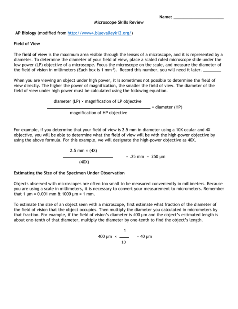
Name: ____________________ Microscope Skills Review AP Biology (modified from http://www4.bluevalleyk12.org/) Field of View The field of view is the maximum area visible through the lenses of a microscope, and it is represented by a diameter. To determine the diameter of your field of view, place a scaled ruled microscope slide under the low power (LP) objective of a microscope. Focus the microscope on the scale, and measure the diameter of the field of vision in millimeters (Each box is 1 mm 2). Record this number, you will need it later. ________ When you are viewing an object under high power, it is sometimes not possible to determine the field of view directly. The higher the power of magnification, the smaller the field of view. The diameter of the field of view under high power must be calculated using the following equation. diameter (LP) × magnification of LP objective = diameter (HP) magnification of HP objective For example, if you determine that your field of view is 2.5 mm in diameter using a 10Χ ocular and 4Χ objective, you will be able to determine what the field of view will be with the high-power objective by using the above formula. For this example, we will designate the high-power objective as 40Χ. 2.5 mm × (4X) = .25 mm = 250 μm (40X) Estimating the Size of the Specimen Under Observation Objects observed with microscopes are often too small to be measured conveniently in millimeters. Because you are using a scale in millimeters, it is necessary to convert your measurement to micrometers. Remember that 1 μm = 0.001 mm & 1000 μm = 1 mm. To estimate the size of an object seen with a microscope, first estimate what fraction of the diameter of the field of vision that the object occupies. Then multiply the diameter you calculated in micrometers by that fraction. For example, if the field of vision’s diameter is 400 μm and the object’s estimated length is about one-tenth of that diameter, multiply the diameter by one-tenth to find the object’s length. 1 400 μm × = 40 μm 10 Question: 1. A student determines that the field of view with a 10X ocular and a 4X objective is 2.1 mm in diameter. What is the diameter of the field of view with the same ocular and a 40X objective? 2. The illustration below is a sample view of the organisms you might see in a drop of lake water, using a 10X ocular and 10X objective. Three of these organisms are indicated by A, B, and C. Using the space below the diagram, describe each organism as completely as you can, including its shape and dimensions, the magnifications used, and the diameter of your field of view. Give all dimensions in micrometers (μm). Specimen A B C Shapes and Dimensions (μm) Magnification Diameter of Field of View (μm) Procedure for Examining Flower Petal 1. 2. 3. 4. 5. 6. Take a microscope from the storage area and place it about 10 cm from the edge of the desk. Carefully clean the ocular and objective lenses with lens paper. Place a drop of water in the center of a clean glass slide. Carefully place a coverslip over the drop of water and leaf. Place the slide on the stage of the microscope with the leaf directly over the opening in the stage. Using the low-power objective, locate the leaf under the microscope. Turn the coarse adjustment knob (the bigger one) until the leaf comes into focus. 7. Switch to the high-power objective. CAUTION: When turning the high-power objective, you should always look at the objective from the side of your microscope so that the objective does not hit or damage the slide. 8. Take two pictures of the flower petal. The first should be under a lower objective. Next, take a photo under the 40x objective and try to get as much detail as possible of the inside of a cell. Paste both photos below and, next to each, record the total magnification and estimate the size of a flower petal cell. Procedure for Examining Flower Pollen 1. 2. 3. 4. 5. 6. Take a microscope from the storage area and place it about 10 cm from the edge of the desk. Carefully clean the ocular and objective lenses with lens paper. Place a drop of water in the center of a clean glass slide. Carefully place a coverslip over the drop of water and leaf. Place the slide on the stage of the microscope with the pollen directly over the opening in the stage. Using the low-power objective, locate the pollen under the microscope. Turn the coarse adjustment knob (the bigger one) until the leaf comes into focus. 7. Switch to the high-power objective. CAUTION: When turning the high-power objective, you should always look at the objective from the side of your microscope so that the objective does not hit or damage the slide. 8. Take two pictures of the pollen. The first should be under a lower objective. Next, take a photo under the 40x objective and try to get as much detail as possible. Paste both photos below and, next to each, record the total magnification and estimate the size of a pollen grain. Take photos of the following specimens: Elodea leaf, paramecium and amoeba. In each photo, label any interior structures/organelles that might be visible and record total magnification, size of cell, and estimate of the size of any organelles.


