Microscope Lab (with Measurement Part)
advertisement
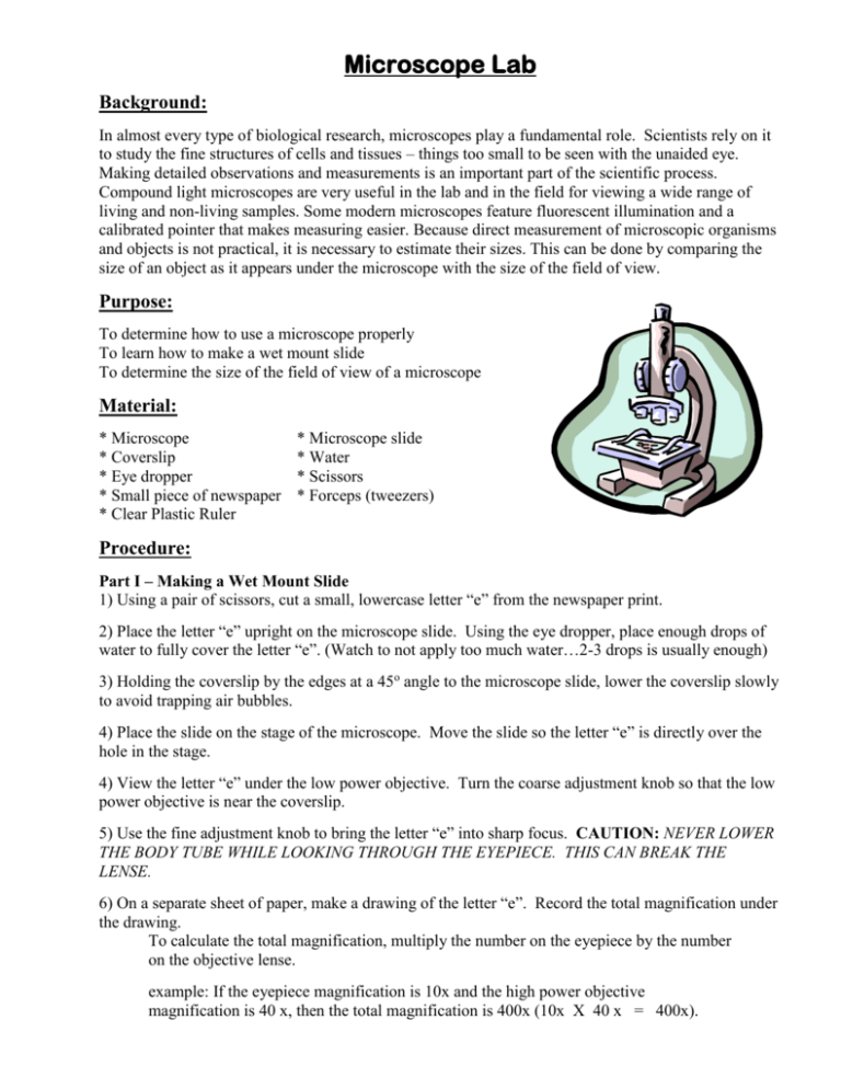
Microscope Lab Background: In almost every type of biological research, microscopes play a fundamental role. Scientists rely on it to study the fine structures of cells and tissues – things too small to be seen with the unaided eye. Making detailed observations and measurements is an important part of the scientific process. Compound light microscopes are very useful in the lab and in the field for viewing a wide range of living and non-living samples. Some modern microscopes feature fluorescent illumination and a calibrated pointer that makes measuring easier. Because direct measurement of microscopic organisms and objects is not practical, it is necessary to estimate their sizes. This can be done by comparing the size of an object as it appears under the microscope with the size of the field of view. Purpose: To determine how to use a microscope properly To learn how to make a wet mount slide To determine the size of the field of view of a microscope Material: * Microscope * Coverslip * Eye dropper * Small piece of newspaper * Clear Plastic Ruler * Microscope slide * Water * Scissors * Forceps (tweezers) Procedure: Part I – Making a Wet Mount Slide 1) Using a pair of scissors, cut a small, lowercase letter “e” from the newspaper print. 2) Place the letter “e” upright on the microscope slide. Using the eye dropper, place enough drops of water to fully cover the letter “e”. (Watch to not apply too much water…2-3 drops is usually enough) 3) Holding the coverslip by the edges at a 45o angle to the microscope slide, lower the coverslip slowly to avoid trapping air bubbles. 4) Place the slide on the stage of the microscope. Move the slide so the letter “e” is directly over the hole in the stage. 4) View the letter “e” under the low power objective. Turn the coarse adjustment knob so that the low power objective is near the coverslip. 5) Use the fine adjustment knob to bring the letter “e” into sharp focus. CAUTION: NEVER LOWER THE BODY TUBE WHILE LOOKING THROUGH THE EYEPIECE. THIS CAN BREAK THE LENSE. 6) On a separate sheet of paper, make a drawing of the letter “e”. Record the total magnification under the drawing. To calculate the total magnification, multiply the number on the eyepiece by the number on the objective lense. example: If the eyepiece magnification is 10x and the high power objective magnification is 40 x, then the total magnification is 400x (10x X 40 x = 400x). 7) Recognize spatial relationships: Move the slide to the left, to the right, toward you, and away from you. Note the direction in which the letter “e” appears to move. 8) Draw the letter “e” as it appears under medium and high power. Record the total magnification under the drawing. Part II – Measuring the Field of View 9) Measure the diameter of the field of view. Be sure the low-power objective is in place. Put a clear plastic ruler on the microscope stage. Using low power, focus on the millimeter marks of the ruler. Move the ruler so that one of the millimeter marks is at the left edge of the field of view, as shown below. Knowing the diameter of the field of view can help you estimate the actual size of objects seen through the microscope. Field of view with a diameter of 3.7 mm 10) Count the number of whole millimeters to the right edge of the field and estimate any fractions to determine the diameter of the field of view. Record the estimated diameter of the low-power field of the view in millimeters in Table 1. Express a fraction of a millimeter in decimal form. Table 1 Objectives Diameter of the Field of View Millimeters (mm) Area of the Field of View in square mm (mm2) Micrometers (μm) Low-power Medium-power High-power (calculated) 11) Move the medium-power objective into place and repeat this procedure for the medium-power field of view. Record the estimated diameter of the medium-power objective in millimeters in Table 1. 12) Since microscopic dimensions are very small, they are usually measured in micrometers (μm) rather than millimeters. There are 1,000 micrometers in a millimeter or a micrometer is 1/1,000 of a mm. Find the diameter of the objective lens in μm by using the following formula. # of millimeters (mm) X 1,000 = ________ micrometers (μm) Record the estimated diameter of each field of view in micrometers in Table 1. 13) Calculate the diameter of the high-power field of view using the diameter of the low-power field of view. The calculated value is more accurate than the estimated value and should be used whenever the diameter of the high-power field of view is required. Calculate the diameter of the high-power field of view in millimeters and micrometers with the following formula: Diameter of high-power field of view = Magnification of low-power objective X Diameter of low-power FOV Magnification of high-power objective For example, a microscope has a low-power objective with a magnification of 10X and a high-power objective with a magnification of 40X. If the low-power field of view (FOV) is 3 mm wide, the diameter of the high-power field of view is (10 X 3 mm)/40 = 0.75 mm. 14) Record the calculated diameter of the high-power field of view in millimeters and micrometers in Table 1. 15) Now that you know the diameters of the fields of vision in micrometers, you can estimate the sizes of objects under the microscope by comparing them with the diameter of the field of vision. For example, assume a field of vision diameter of 350 μm under high-power. A tiny shrimp takes up approximately one-half the field of vision under high-power. To find its size in micrometers, use the following formula: proportion of field of view X diameter of power objective = size of object 16) Calculate the area of the low-power, medium-power, and high-power fields of view. To determine the area of the field of view, use the formula: A = pi ( r2 ) where A is the area, the value of pi is 3.14, and r is the radius, or one-half the diameter of the field of view. For example, a field of view with a diameter of 4 mm would have a radius of 2 mm and an area of 3.14 X (2 mm)2 , or 12.56 mm2. Calculate the area for the low-power, medium-power, and highpower fields of view of your microscope, and record the area of each field of view in Table 1. Part III - Extension 17) View other objects, such as hair or thread, under the microscope. Make a drawing of what you see. Record the total magnification under the drawing. Discussion: 1) When you move the slide toward you, how does the letter “e” appear to move under the microscope? 2) Describe the appearance of the letter “e” under high power as compared to low power. 3) What is the purpose of the coverslip? 4) Why should the coverslip be held by the edges? 5) How do you calculate the total magnification by which an object was viewed under? 6) Why must a specimen be very thin to be viewed under the microscope? 7) Which objective lens provides the larger field of view? 8) Why are micrometers, rather than millimeters, used for microscopic measurements? 9) What is the importance of knowing the diameter and area of the field of view? 10) Calculate the estimated size of the tiny shrimp in Step 15 of the lab procedure. Show your work. Name ___________________________ Period ____________ Date ___________________ Drawings of the Letter “e” ________________________ total magnification _____________________ total magnification __________________ total magnification Table 1 Objectives Diameter of the Field of View Millimeters (mm) Area of the Field of View in square mm (mm2) Micrometers (μm) Low-power Medium-power High-power (calculated) Discussion: 1) __________________________________________________________________________ ____________________________________________________________________________ 2) __________________________________________________________________________ ____________________________________________________________________________ _____________________________________________________________________________ 3) ___________________________________________________________________________ _____________________________________________________________________________ 4) ___________________________________________________________________________ _____________________________________________________________________________ 5) ___________________________________________________________________________ _____________________________________________________________________________ 6) ___________________________________________________________________________ _____________________________________________________________________________ 7) ___________________________________________________________________________ _____________________________________________________________________________ 8) ___________________________________________________________________________ _____________________________________________________________________________ 9) ___________________________________________________________________________ _____________________________________________________________________________ 10) __________________________________________________________________________ _____________________________________________________________________________ Extension ____________________________ object being viewed __________________________ object being viewed ____________________________ total magnification __________________________ total magnification
