The RV: Anatomy, Physiology and Pathophysiology
advertisement
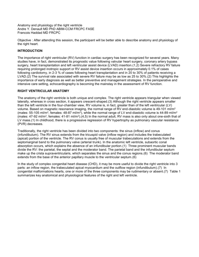
Anatomy and physiology of the right ventricle Andre Y. Denault MD PhD ABIM-CCM FRCPC FASE Francois Haddad MD FRCPC Objective : After attending this session, the participant will be better able to describe anatomy and physiology of the right heart. INTRODUCTION The importance of right ventricular (RV) function in cardiac surgery has been recognized for several years. Many studies have, in fact, demonstrated its prognostic value following valvular heart surgery, coronary artery bypass surgery, heart transplantation and left ventricular assist device (LVAD) insertion.(1,2) Severe refractory RV failure requiring prolonged inotropic support or RV assist device insertion occurs in approximately 0.1% of cases following cardiotomy, in 2-3 % of cases following heart transplantation and in 20 to 30% of patients receiving a LVAD.(2) The survival rate associated with severe RV failure may be as low as 25 to 30%.(2) This highlights the importance of early diagnosis as well as better preventive and management strategies. In the perioperative and intensive care setting, echocardiography is becoming the mainstay in the assessment of RV function. RIGHT VENTRICULAR ANATOMY The anatomy of the right ventricle is both unique and complex. The right ventricle appears triangular when viewed laterally, whereas in cross section, it appears crescent-shaped.(3) Although the right ventricle appears smaller than the left ventricle in the four-chamber view, RV volume is, in fact, greater than of the left ventricular (LV) volume. Based on magnetic resonance imaging, the normal range of RV end-diastolic volume is 49-101 ml/m2 (males: 55-105 ml/m2, females: 48-87 ml/m2), while the normal range of LV end-diastolic volume is 44-89 ml/m2 (males: 47-92 ml/m2, females: 41-81 ml/m2).(4,5) In the normal adult, RV mass is also only about one-sixth that of LV mass.(1) In childhood, there is a progressive regression of RV hypertrophy as pulmonary vascular resistance (PVR) decreases. Traditionally, the right ventricle has been divided into two components: the sinus (inflow) and conus (infundibulum). The RV sinus extends from the tricuspid valve (inflow region) and includes the trabeculated (apical) portion of the ventricle. The RV conus is usually free of muscular trabeculations and extends from the septomarginal band to the pulmonary valve (arterial trunk). In the anatomic left ventricle, subaortic conal absorption occurs, which explains the absence of an infundibular portion.(1) Three prominent muscular bands divide the RV: the parietal, the septal and the moderator band. The parietal band and the infundibular septum make up the crista supraventricularis, which separates the sinus and the conus regions.(6) The moderator band extends from the base of the anterior papillary muscle to the ventricular septum.(6) In the study of complex congenital heart disease (CHD), it may be more useful to divide the right ventricle into 3 parts: an inflow region, the trabeculated apical myocardium and the outflow region (infundibulum).(7) In congenital malformations hearts, one or more of the three components may be rudimentary or absent.(7) Table 1 summarizes key anatomical and physiological features of the right and left ventricle. Table 1. Characteristics of Right and Left Ventricle Characteristics Right ventricle Left ventricle Structure Inflow region, trabeculated myocardium, infundibulum No infundibulum Mitro-aortic continuity Shape From the side: triangular Cross-section: crescentic Elliptic Volume (end-diastolic) 49-101 ml/m2 44-89 ml/m2 Mass (g/m2) < 35 g/m2 ≈ 1/6 LV mass < 130 g/m2 (men) < 100 g/m2 (women) Ejection fraction 40-68 % > 45%a 57-74% > 50%a Ventricular elastance (mmHg/ml) 1.30 ± 0.84 5.48 ± 1.23 Ventricular compliance Higher compliance than LV 5.0 ± 0.52 x 10⁻² Adaptation to disease Better adaptation to volume overload states Better adaptation to pressure overload states Normal parameters of the right compared to the left ventricle. Range of normal values depends on method of acquisition.a Lower value of normal RV and LV ejection fraction used in clinical practice.(1,3,5,8-13) Adapted from Haddad et al.(14) RIGHT VENTRICULAR PHYSIOLOGY The primary function of the right ventricle is to receive systemic venous return and pump it into the pulmonary system. Under normal conditions the right ventricle, in contrast to the left ventricle, is coupled to a low pressure, highly distensible arterial system.(1,14) The right ventricle is normally connected in series with the left ventricle. In the absence of shunt physiology or significant valvular regurgitation, the stroke volume of the right ventricle will normally match that of the left. Because of the greater end-diastolic volume of the right ventricle, RV ejection fraction (RVEF) is lower than the left. The lower limit of normal RVEF ranges from 40-45% compared to 50-55% for LV ejection fraction (LVEF).(5,10,11,15) Several mechanisms contribute to RV ejection, the most important being the bellows-like inward movement of the free wall. Other important mechanisms include the contraction of the longitudinal fibers, shortening of the long axis, drawing the tricuspid annulus toward the apex, and the traction on the free RV wall at its points of attachment to the left ventricle as a result of LV contraction.(3) In contrast to the left ventricle, twisting and rotational movements do not contribute significantly to RV contraction.(16) Furthermore, the contraction of the right ventricle is also sequential, starting with the trabeculated myocardium and ending with the contraction of the infundibulum (normally separated by approximately 25 to 50 msec).(1,17) In order to better understand the complex relationship between RV contractility, preload and afterload, many investigators have studied the pressure-volume relationship of the right ventricle. One of their major findings was that the right ventricle follows a time-varying elastance model in which ventricular elastance is described by the relationship between systolic pressure and volume under variable loading conditions.(13,17,18) Many studies have shown that RV elastance may also be approximated by a linear relationship.(1) RV maximal elastance is considered by many investigators to be the most reliable index of RV contractility.(1) RV systolic elastance is lower than that of the left ventricle. This arrangement implies that the RV is far more sensitive to increases in afterload.(13,19) This can be illustrated in the acute setting where RV stroke volume decreases significantly following an increase in pulmonary arterial pressure.(1,14,20) The pulmonary circulation is an important determinant of RV afterload. The pulmonary vascular bed is a low pressure, low resistance system that is highly compliant. In the presence of normal pulmonary circulation, the right ventricle performs approximately one-fourth of the LV stroke work.(3) Several factors modulate PVR, including hypoxia, hypercarbia, cardiac output, pulmonary volume and pressure as well as specific molecular pathways, most prominent being the nitric oxide pathway (vasodilatation), the prostaglandin pathway (vasodilatation) and the endothelin pathway (vasoconstriction).(1,14,21) Pulmonary vessels constrict with hypoxia (Euler-Liljestrand reflex) and relax in the presence of hyperoxia.(22) In some instances, hypercarbia may also be a strong pulmonary vasoconstrictor. Lung volumes have a differential effect on intra- and extra-alveolar vessels which accounts for the unique Ushaped relationship between lung volume and PVR. PVR is minimal at functional residual capacity and increased at large and small lung volumes alike, this may be observed clinically when hyperinflation of the lungs greatly increases PVR.(22) Application of high levels of positive end-expiratory pressure (PEEP) may narrow the capillaries in the well-ventilated lung areas and divert flow to less well ventilated or non-ventilated areas, potentially leading to hypoxia. An increase in cardiac output distends open vessels and may recruit previously closed vessels decreasing PVR. Regional blood flow to the lungs is also influenced by gravity where pulmonary blood flow is greater in the dependant areas of the lung. Ventricular interdependence refers to the concept that through direct mechanical interactions: the size, shape and compliance of one ventricle may affect the size, shape and pressure-volume relationship of the other.(23) The main anatomical determinants for ventricular interdependence include the ventricular septum, the pericardium and continuity between myocardial fibers of the right and left ventricles. Ventricular interdependence may occur in both systole and diastole. Although always present, ventricular interdependence is most evident with changes in loading conditions such as those observed during respiration or sudden postural changes.(1,14,23) Ventricular interdependence also plays an important part in the pathophysiology of RV dysfunction. References 1. Dell'Italia LJ. Curr Probl Cardiol 1991;16:653-720. 2. Kaul TK, Fields BL. Cardiovasc Surg 2000;8:1-9. 3. Leng J. Right ventricle. In: Weyman AE, ed. Principle and practice of echocardiography, Lippincott Williams & Wilkins, 1994:901-21. 4. Mahler DA, Matthay RA, Snyder PE, et al. J Appl Physiol 1985;58:1818-22. 5. Lorenz CH, Walker ES, Morgan VL, et al. J Cardiovasc Magn Reson 1999;1:7-21. 6. Farb A, Burke AP, Virmani R. Cardiol Clin 1992;10:1-21. 7. Kirklin JW. Cardiac surgery. John Wiley and Sons ed. New-York: 1986. 8. Leyton RA, Sonnenblick EH. Methods Achiev Exp Pathol 1971;5:22-59. 9. Gaasch WH, Cole JS, Quinones MA, Alexander JK. Circulation 1975;51:317-23. 10. Hurwitz RA, Treves S, Kuruc A. Am Heart J 1984;107:726-32. 11. Pfisterer ME, Battler A, Zaret BL. Eur Heart J 1985;6:647-55. 12. Starling MR, Walsh RA, Dell'Italia LJ, et al. Circulation 1987;76:32-43. 13. Dell'Italia LJ, Walsh RA. Cardiovasc Res 1988;22:864-74. 14. Haddad F, Hunt SA, Rosenthal DN, Murphy DJ. Circulation 2008;117:1436-48. 15. Lang RM, Bierig M, Devereux RB, et al. J Am Soc Echocardiogr 2005;18:1440-63. 16. Petitjean C, Rougon N, Cluzel P. J Cardiovasc Magn Reson 2005;7:501-16. 17. Lee FA. Cardiol Clin 1992;10:59-67. 18. Brown KA, Ditchey RV. Circulation 1988;78:81-91. 19. Guyton AC, Lindsey AW, Gilluly JJ. Circ Res 1954;2:326-32. 20. MacNee W. Am J Respir Crit Care Med 1994;150:833-52. 21. McLaughlin VV, McGoon MD. Circulation 2006;114:1417-31. 22. Fischer LG, Van AH, Burkle H. Anesth Analg 2003;96:1603-16. 23. Santamore WP, Dell'Italia LJ. Prog Cardiovasc Dis 1998;40:289-308.


