Page 1 – Detecting Diffusion
Detecting Diffusion
Group Leader: ____________________
Materials Technician: _____________________
Recorder: ________________________
Materials Technician: ______________________
Problem:
How can you determine whether solutes are diffusing across a membrane?
Background
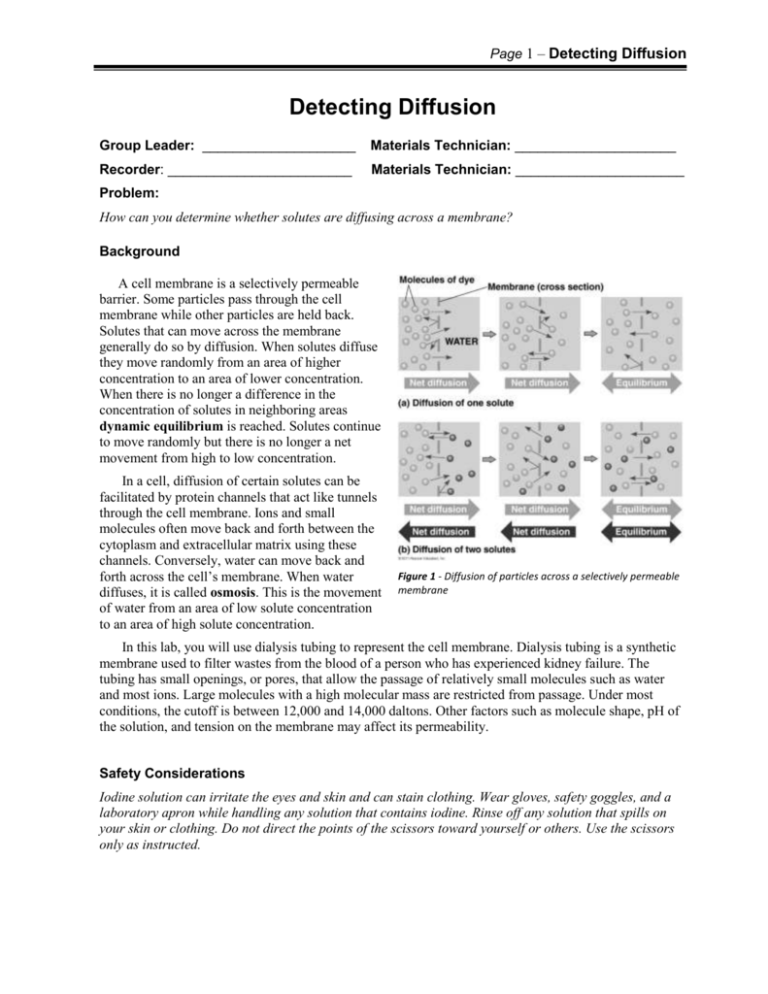
A cell membrane is a selectively permeable
barrier. Some particles pass through the cell
membrane while other particles are held back.
Solutes that can move across the membrane
generally do so by diffusion. When solutes diffuse
they move randomly from an area of higher
concentration to an area of lower concentration.
When there is no longer a difference in the
concentration of solutes in neighboring areas
dynamic equilibrium is reached. Solutes continue
to move randomly but there is no longer a net
movement from high to low concentration.
In a cell, diffusion of certain solutes can be
facilitated by protein channels that act like tunnels
through the cell membrane. Ions and small
molecules often move back and forth between the
cytoplasm and extracellular matrix using these
channels. Conversely, water can move back and
forth across the cell’s membrane. When water
diffuses, it is called osmosis. This is the movement
of water from an area of low solute concentration
to an area of high solute concentration.
Figure 1 - Diffusion of particles across a selectively permeable
membrane
In this lab, you will use dialysis tubing to represent the cell membrane. Dialysis tubing is a synthetic
membrane used to filter wastes from the blood of a person who has experienced kidney failure. The
tubing has small openings, or pores, that allow the passage of relatively small molecules such as water
and most ions. Large molecules with a high molecular mass are restricted from passage. Under most
conditions, the cutoff is between 12,000 and 14,000 daltons. Other factors such as molecule shape, pH of
the solution, and tension on the membrane may affect its permeability.
Safety Considerations
Iodine solution can irritate the eyes and skin and can stain clothing. Wear gloves, safety goggles, and a
laboratory apron while handling any solution that contains iodine. Rinse off any solution that spills on
your skin or clothing. Do not direct the points of the scissors toward yourself or others. Use the scissors
only as instructed.
Page 2 –Detecting Diffusion
Pre-Lab Questions
Read the entire laboratory, then answer these questions on a separate sheet of paper to be submitted
prior to beginning the laboratory.
1. Research the molecular sizes of glucose and starch molecules. Which is larger? Predict which, if
either, of these molecules would be able to travel through the pores in dialysis tubing?
2. Recall what occurs when starch solution and iodine solution are added together. How will you know
if these two substances come together in this laboratory?
3. Using your answer from #2 above, how will you know whether starch has diffused across the
membrane? How will you know whether iodine has diffused across the membrane?
4. How will you be able to tell whether glucose has diffused across the membrane?
Materials
dialysis tubing
Scissors
metric ruler
250-mL beakers
tubing clamps, string, or twist ties
10-mL graduated cylinder
15% glucose//1% starch solution
iodine solution
forceps
glucose test strips
Procedure
1. Obtain and wear goggles, a lab apron, and if you are sensitive to iodine, then you should
wear nitrile or latex gloves.
2. Cut a 15-cm length of dialysis tubing. Soak the tubing for one minute in a 250-mL beaker
filled with 50 mL of water.
3. Remove the tubing from the water. Fold up 1 cm of the tubing at one end. Use a tubing
clamp, twist tie, or piece of string to tightly seal the folded end.
4. Roll the unsealed end of the tubing between your fingers until it opens. Pour 3 mL of 15%
glucose/1% starch solution into the tubing.
5. Fold down 1 cm of the tubing at the open end Use a second tubing clamp, twist tie, or piece
of string to tightly seal this end.
6. Use tap water to gently but thoroughly rinse the outside of the tubing. Be sure to rinse the
clamps or twist ties as well. Do not squeeze the dialysis tubing.
Page 3 –Detecting Diffusion
7. Place the tubing in the 250-mL beaker. Fill the
beaker with enough water to completely submerge
the tubing.
8. Add 4 drops of iodine solution to the water in the
beaker.
Dialysis tubing with
15% glucose and 1%
starch solution
9. Record your initial observations. Use a glucose test
strip to measure the glucose concentration in the
beaker. Record your results in the data table.
10. Wait 10 minutes, and then record your final
observations.
11. Use a forceps to remove and dispose of the tubing as instructed by your teacher.
12. Use your data to answer the Analysis Questions.
Data & Results
Inside Tubing
Color
Initial
Final
Other observations:
Is starch Is iodine Is glucose
Color
present? present? present?
Outside Tubing (Beaker)
Is starch
present?
Is iodine
present?
Is glucose
present?
Page 4 –Detecting Diffusion
Analysis
After completing the laboratory answer these questions on a separate sheet of paper.
1. With respect to diffusion, infer what happened to the iodine, starch, and glucose between your initial
observations at the start of the experiment and your final observations at the end.
2. Were the predictions that you made for Pre-Lab question #1 accurate? Use what you know about the
structure of starch and glucose molecules to explain your results.
3. What substance other than iodine, starch, or glucose moved across the membrane? In which direction
did this substance move, and why?
4. Red blood cells are placed in water that has been distilled so that there are no solutes in the water. Are
the red blood cells likely to swell up or shrink? Why?
5. The function of the kidney organ in humans is to remove wastes and excess fluid from the blood. If a
person experiences kidney failure, they may require dialysis treatment to perform these functions.
Research kidney dialysis treatment and explain how dialysis tubing would be used for this treatment.
6. A student performing this lab observed the solution outside of the tubing turning black. What might
have happened?
7. Design an experiment to determine if the concentration of glucose inside the dialysis tubing has an
effect on its diffusion rate.
References:
"Diffusion." Unit 4 - Correia Life Science. Web. 11 Oct 2010. <http://www.bio.miami.edu/~cmallery/150/memb/c8.7x11.diffusion.jpg>
How Does Dialysis Work? Northwest Kidney Centers, 2014. Web. 13 Dec. 2014.
<http://www.nwkidney.org/dialysis/startingOut/basic/howDialysisWork.html>.
Kaplan Test Prep. Dialysis tubing in beaker. Learningpod. Learningpod, Inc., 18 Feb. 2013. Web. 13 Dec. 2014.
<https://www.learningpod.com/question/ap-biology-question-below-refer-to-the-figure-below-in-which-a-dialysistubing-bag-is-filled/e2b7ced6-5c3f-46b0-961c-39bca96dc6b1>.
Miller, Kenneth R., and Joseph S. Levine. Biology-Laboratory Manual A. Teacher ed. Boston: Pearson Prentice Hall, 2010. Print.

