From mouse to human: Are the biophysical properties and the
advertisement
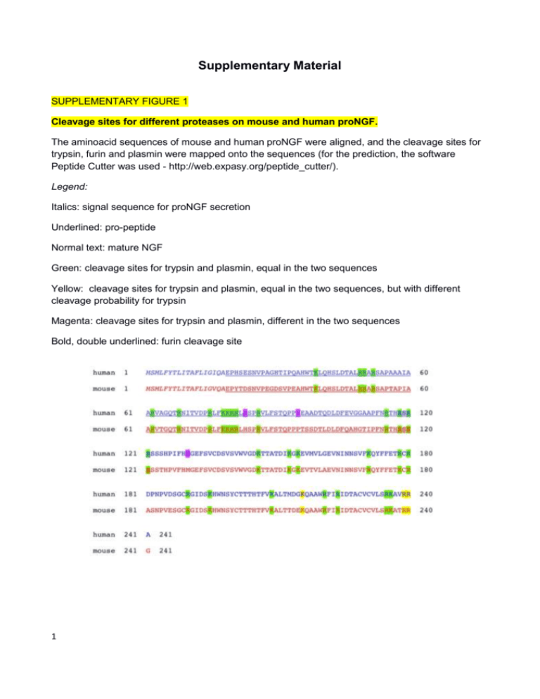
Supplementary Material SUPPLEMENTARY FIGURE 1 Cleavage sites for different proteases on mouse and human proNGF. The aminoacid sequences of mouse and human proNGF were aligned, and the cleavage sites for trypsin, furin and plasmin were mapped onto the sequences (for the prediction, the software Peptide Cutter was used - http://web.expasy.org/peptide_cutter/). Legend: Italics: signal sequence for proNGF secretion Underlined: pro-peptide Normal text: mature NGF Green: cleavage sites for trypsin and plasmin, equal in the two sequences Yellow: cleavage sites for trypsin and plasmin, equal in the two sequences, but with different cleavage probability for trypsin Magenta: cleavage sites for trypsin and plasmin, different in the two sequences Bold, double underlined: furin cleavage site 1 SUPPLEMENTARY FIGURE 2 Detailed Biacore kinetics for the binding to the anti-NGF MAB 256 R&D System Detailed SPR binding kinetics of the neurotrophins over the anti-NGF antibody R&D MAB 253. A mouse NGF, B - mouse proNGF, C - human NGF, D - human proNGF. Concentrations used, from top to bottom: 100, 50, 25, 6.3, 3.1, 1.6, 0.8, 0.4, 0.2, 0.1 nM. 2 SUPPLEMENTARY FIGURE 3 Detailed Biacore kinetics for the binding to the anti-NGF MAb D11 Detailed SPR binding kinetics of the neurotrophins over the anti-NGF antibody MAb D11. A mouse NGF, B - mouse proNGF, C - human NGF, D - human proNGF. Concentrations used, from top to bottom: 100, 50, 25, 6.3, 3.1, 1.6, 0.8, 0.4, 0.2, 0.1 nM. 3 SUPPLEMENTARY FIGURE 4 Detailed Biacore kinetics for the binding to the anti-proNGF MAb Millipore clone EP1318Y Detailed SPR binding kinetics of the neurotrophins over the anti-proNGF MAb Millipore clone EP1318Y. A - mouse proNGF, B - human proNGF. Concentrations used, from top to bottom: 100, 50, 25, 6.3, 3.1, 1.6, 0.8, 0.4, 0.2, 0.1 nM. 4 SUPPLEMENTARY TABLE 1 Summary of binding kinetics of the mouse and human NGF MAb anti-NGF R&D MAb anti-NGF αD11 mNGF hNGF ka (M-1s-1) 5.20 x 10⁶ 8.32 x 10⁶ kd (s-1) 1.03 x 10⁻³ 8.64 x 10⁻⁴ 2 20 7 ka (M-1s-1) 1.20 x 10⁷ 4.31 x 10⁶ kd (s-1) 2.07 x 10⁻¹⁵ 3.63 x 10⁻⁷ 2 0 0 SPR analysis: Summary of the derived kinetic binding constants of mNGF and hNGF towards the MAbs anti-NGF MAB 256 R&D and D11 . 5 SUPPLEMENTARY TABLE 2 Summary of binding kinetics of the mouse and human proNGF MAb anti-NGF R&D MAb anti-NGF αD11 MAb anti-proNGF Millipore mproNGF hproNGF ka (M-1s-1) 9.98 x 10⁵ 5.37 x 10⁴ kd (s-1) 1.81 x 10⁻³ 1.89 x 10⁻³ 2 30 6 ka (M-1s-1) 3.57 x 10⁶ 1.20 x 10⁶ kd (s-1) 14.60 x 10⁻³ 3.63 x 10⁻⁷ 2 13 8 ka (M-1s-1) 2.82 x 10⁴ 2.34 x 10⁵ kd (s-1) 4.84 x 10⁻⁴ 1.22 x 10⁻³ 2 9 18 SPR analysis: Summary of the derived kinetic binding constants of mproNGF and hproNGF towards the MAbs anti-NGF MAB 256 R&D and D11 and tha MAb anti proNGF Millipore clone EP1318Y. 6

