MRI Lumbar Spine
advertisement
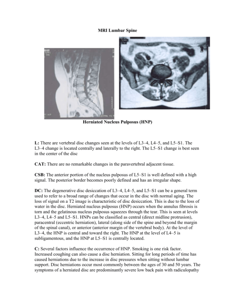
MRI Lumbar Spine Herniated Nucleus Pulposus (HNP) L: There are vertebral disc changes seen at the levels of L3–4, L4–5, and L5–S1. The L3–4 change is located centrally and laterally to the right. The L5–S1 change is best seen in the center of the disc CAT: There are no remarkable changes in the paravertebral adjacent tissue. CSB: The anterior portion of the nucleus pulposus of L5–S1 is well defined with a high signal. The posterior border becomes poorly defined and has an irregular shape. DC: The degenerative disc desiccation of L3–4, L4–5, and L5–S1 can be a general term used to refer to a broad range of changes that occur in the disc with normal aging. The loss of signal on a T2 image is characteristic of disc desiccation. This is due to the loss of water in the disc. Herniated nucleus pulposus (HNP) occurs when the annulus fibrosis is torn and the gelatinous nucleus pulposus squeezes through the tear. This is seen at levels L3–4, L4–5 and L5–S1. HNPs can be classified as central (direct midline protrusion), paracentral (eccentric herniation), lateral (along side of the spine and beyond the margin of the spinal canal), or anterior (anterior margin of the vertebral body). At the level of L3–4, the HNP is central and toward the right. The HNP at the level of L4–5 is subligamentous, and the HNP at L5–S1 is centrally located. C: Several factors influence the occurrence of HNP. Smoking is one risk factor. Increased coughing can also cause a disc herniation. Sitting for long periods of time has caused herniations due to the increase in disc pressures when sitting without lumbar support. Disc herniations occur most commonly between the ages of 30 and 50 years. The symptoms of a herniated disc are predominantly severe low back pain with radiculopathy into the buttocks, legs, and feet. The pain is generally made worse with coughing, straining, or laughing. Patients also complain of tingling or numbness in the legs and feet. Depending on the specific nerve involved, there may be associated numbness or weakness due to compression of that nerve. There are several tests that may help diagnose an HNP. Those include, x-ray, MRI, CT, myelogram, and EMG. For the majority of patients, a period of rest with anti-inflammatory medications followed by physical therapy is a sufficient treatment. For those who need further treatment, steroid injections or surgery may be the next options.

