Hany sayed ahmed Bauiomy_full paper
advertisement
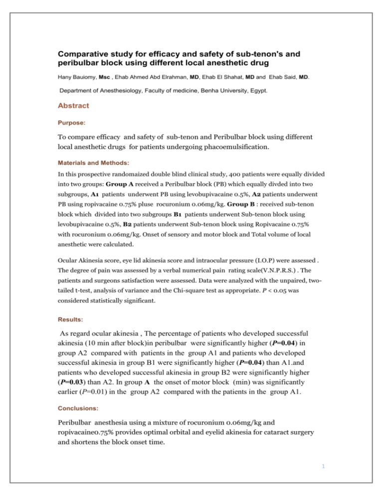
Comparative study for efficacy and safety of sub-tenon's and peribulbar block using different local anesthetic drug Hany Bauiomy, Msc , Ehab Ahmed Abd Elrahman, MD, Ehab El Shahat, MD and Ehab Said, MD. Department of Anesthesiology, Faculty of medicine, Benha University, Egypt. Abstract Purpose: To compare efficacy and safety of sub-tenon and Peribulbar block using different local anesthetic drugs for patients undergoing phacoemulsification. Materials and Methods: In this prospective randomaized double blind clinical study, 400 patients were equally divided into two groups: Group A received a Peribulbar block (PB) which equally divded into two subgroups, A1 patients underwent PB using levobupivacaine 0.5%, A2 patients underwent PB using ropivacaine 0.75% pluse rocuronium 0.06mg/kg. Group B : received sub-tenon block which divided into two subgroups B1 patients underwent Sub-tenon block using levobupivacaine 0.5%, B2 patients underwent Sub-tenon block using Ropivacaine 0.75% with rocuronium 0.06mg/kg. Onset of sensory and motor block and Total volume of local anesthetic were calculated. Ocular Akinesia score, eye lid akinesia score and intraocular pressure (I.O.P) were assessed . The degree of pain was assessed by a verbal numerical pain rating scale(V.N.P.R.S.) . The patients and surgeons satisfaction were assessed. Data were analyzed with the unpaired, twotailed t-test, analysis of variance and the Chi-square test as appropriate. P < 0.05 was considered statistically significant. Results: As regard ocular akinesia , The percentage of patients who developed successful akinesia (10 min after block)in peribulbar were significantly higher (P=0.04) in group A2 compared with patients in the group A1 and patients who developed successful akinesia in group B1 were significantly higher (P=0.04) than A1.and patients who developed successful akinesia in group B2 were significantly higher (P=0.03) than A2. In group A the onset of motor block (min) was significantly earlier (P=0.01) in the group A2 compared with the patients in the group A1. Conclusions: Peribulbar anesthesia using a mixture of rocuronium 0.06mg/kg and ropivacaine0.75% provides optimal orbital and eyelid akinesia for cataract surgery and shortens the block onset time. 1 Keywords: safety and efficacy, peribulbar ,sub-tenon,local anesthetic, Levobupivacaine vs. ropivacaine,rocuronium, INTRODUCTION Patient comfort, safety and low complication rates are the essentials of local anesthesia. The anesthetic requirements for ophthalmic surgery are dictated by the nature of the proposed surgery, the surgeon’s preference and the patient’s wishes.¹ Investigators have reported that racemic bupivacaine markedly reduced the stroke index, ejection fraction, acceleration index, and produced greater prolongation of the QT interval compared to levobupivacaine. Other studies have reported that levobupivacaine has a better profile compared to racemic bupivacaine in terms of neurotoxicity.² The reduced toxic profile of levobupivacaine is beneficial for achieving higher plasma concentrations and dose without signs of cardiovascular or systemic toxicity. 2 Furthermore, the success rate of cardiopulmonary resuscitation after toxic doses of levobupicaine is higher compared to bupivacaine intoxication.There is a relative paucity of studies describing the use of levobupivacaine 0.5% for the standard peribulbar block (PB) or for superficial extraconal anesthesia.³ The safety margin of ropivacaine is quite high, a higher volume is used in achieving the desired anaesthetic effect due to a lower potency than bupivacaine while performing different surgeries thus raising the concerns of systemic toxicity.4 Neuromuscular blocking drugs, such as vecuronium, and atracurium have also been added to the local anesthetic mixture and have been shown to improve the quality of peribulbar anesthesia. Atracurium, however, has histamine-releasing property and could result in undesirable local hyperemia. Rocuronium, on the other hand, is devoid of this adverse effect, has a faster onset of action, and its effects in a low dose on the quality of peribulbar anesthesia.5 The aim of this study was to compare efficacy and safety of sub-tenon's and peribulbar block using different local anaesthetic drugs for patients scheduled for phacoemulsification. METHODS After obtaining approval from the Institutional Ethics Committee and written informed consent from all the patients, 400 adult patients, scheduled for elective cataract surgery , were included in this prospective, double-blind, randomized study inclusion criteria included patients with axial length 22-28mm,Age ranged between 40-80years, American Society of Anesthesiologists (ASA) I-III.Methods of randomization: Closed envelope.These patients will randomly allocated into two equal groups: Group A : Patients of this group formed of 200 Patients divided into two subgroups:A1: formed of 100 patients underwent conventional peribulbar block using levobupivacaine 0.5%. A2: formed of 100 patients underwent conventional peribulbar block using Ropivacaine 0.75% pluse rocuronium 0.06mg/kg. 3 peribulbar anesthesia was given using a 25-G 16-mm short-bevel needle. The position of the injection site At the medial 2/3rd and lateral 1/3rd of the lower eyelid. After negative aspiration, 5-7 mL of the LA solution was slowly injected . Mechanical orbital compression was applied for 15 min in both groups, using a Honan balloon adjusted to 30 mmHg. Group B :Patients of this group formed of 200 Patients Will receive sub-tenon block divided into two subgroups B1: formed of 100 patients underwent Sub-tenon block using levobupivacaine 0.5%. B2: formed of 100 patients underwent Sub-tenon block using Ropivacaine 0.75% pluse rocuronium 0.06mg/kg. Sub-tenon block involve transconjunctival infiltration of local anesthetic agent directly to subtenons space, through a small incision done in a tent of conjunctiva raised about 5-7 mm from limbus with ophthalmic scissor. after instillation of local anesthetic drops in conjunctiva . A curved blunt subtenon cannula(19 G, 25mm) was used for local anesthetic injection. Exclusion criteria included a Patients on anti-glaucoma drugs, patients refusing LA, patients with a single eye, allergy to LA solutions, clotting abnormalities, history of sleep apnea, impaired mental status, and drug abuse. This study was conducted in benha university hospital between the period of October 2013 and May 2015. All operations were carried out by the same technique. Patients were admitted to the operating room fasted for 6-8 hrs. and unpremedicated. A peripheral i.v. catheter was inserted and standard monitoring was conducted and recorded, including heart rate (HR), noninvasive arterial blood pressure, electrocardiogram (5 leads), and peripheral oxygen saturation (SpaO2). A nasal cannula was applied and supplemental oxygen was given throughout the procedure at 3 L/min. The patients were randomly divided (using the block randomization method) to 1 of 2 groups according to the LA solution used to receive either 0.5% levobupivacaine (Chirocaine, Abbott Laboratories, Elverum, Norway) (levobupivacaine group, n=100) or 0.75% ropivacaine (Naropin, Astra-Zeneca, Zug, Switzerland) plus rocuronium 0.06mg/kg. (ropivacaine rocuronium group, n=100). 4 The studied LA solutions were prepared at the bedside before the injection and provided in patient specific, sealed packaging by a member of staff not otherwise involved in the study. All peribulbar and sub-tenon blocks were performed by the team experienced in these techniques who was blinded to the kind of LA solution used. The patients also were blinded to the LA solution used. Motor block was evaluated after 10 minute by the assessment of the akinesia in the 4 quadrants using a 3-point scoring system, which was categorized as follows: 0=akinesia, 1=partial akinesia, and 2=normal movement, with a maximal score of 8 for the 4 muscles. The block can only be considered successful, if the akinesia score was 3 or less.[6] In such event, if one or more of the components of ocular movement showed inadequate motor blockade (akinesia score >3) 10 min after block, supplementary anesthesia (3mL) was injected into the involved quadrant using the same length needle as for the primary block. After that, an additional assessment was performed 5 min later. Intraocular pressure (I.O.P) was measured preoperative and after local anesthetic injection 5, 10 and 15 min using a tonopen. Eye lid movement score will be categorized as full movement=2, flicker=1, and No movement=0. The incidence of any complications was routinely recorded. Sensory block was considered along with abolition of the corneal reflex next to instillation of drops of balanced salt solution on the conjunctiva and cornea. The total volume of LA solution injected (mL) was calculated. The onset of motor block and onset of sensory block (min) was calculated. At the end of the procedure, surgeons who were masked to the type of local anesthetic used were requested to rate their satisfaction with the block quality (0 = absolutely not satisfied to 10 = totally satisfied). Patients who were also masked to type of local anesthetic used were requested to rate the degree of pain on verbal numerical pain rating scale (VNPRS) (0 = no pain to 10 = most severe pain) at 1njection, 1and 2h postoperatively. If the intraoperative VNPRS for pain was >4, topical anesthetic drops(benzocaine) were added by the surgeon. The statistical analysis of our results was conducted using the computer program SPSS version 15.0 for Windows (SPSS, Chicago, IL, USA). Data were expressed as mean±SD or number (percentages). The 2-way repeated measures analysis of variance was used to compare the interval data, and Student's t test was used as the post hoc test to determine differences between and within groups. Fisher's exact test was used to compare nominal data or percentages. Bonferroni correction for repeated comparisons was applied if necessary. P<0.05 was considered significant. 5 RESULTS This study demonstrated comparable results as the patients were homogenous as regards age , sex, weight and A.S.A. classification . Table(1) As regards ocular akinesia The percentage of patients who developed successful akinesia (10 min after block)in peribulbar were significantly higher (P=0.04) in the group A2 (78%) compared with the patients in the group A1 (65%) and in sub tenon there was no significant difference between B1 and B2.Also patients who developed successful akinesia in group B1 were significantly higher (P=0.04) than A1.Also patients who developed successful akinesia in group B2 were significantly higher (P=0.03) than A2. Figure (1) In the group A, the mean volume of levobupivacaine administered was 7.52 mL, which was not significantly higher (P=0.64) than the volume of ropivacaine rocuronium group (7.05 mL), Also in group B the the mean volume of levobupivacaine administered was 4.42 mL, which was not significantly higher (P=0.81) than the volume of ropivacaine rocuronium group (4.4 mL).in group A the onset of motor block (min) was significantly earlier (P=0.01) in the ropivacaine rocuronium group (8.37±1.50) compared with the patients in the levobupivacaine group (8.90±1.61). The onset sensory block (min) was not significant (P=0.07) between A1 group (5.02±0.90) compared with the patients in the A2 group (5.28±1.1).in group B the onset of sensory and motor block did not show any significant difference between B1 and B2. Table(2) As regard I.O.P. measurement before and 5min,10min.and 15min. after injection there was no significant difference between all groups. Figure(2) As regard complication there was no significant difference between all groups. 6 Table (1) Demographic distribution of study groups and A.S.A classification. Group A1 Group A2 Group B1 Group B2 Age 57.43±6.33 57.39±6.48 56.95±6.44 55.31±6.68 Weight 79.42±6.40 80.97±6.97 80.39±6.17 81.41±5.81 Height 173.26±5.10 173.47±4.31 172.7±4.64 172.49±4.86 Male/Female 65/35 56/44 62/38 58/42 I 20 15 17 18 II 65 67 70 73 III 15 18 13 9 A.S.A. Data expressed as a mean and SD or numbers and percent ASA : American Society of Anesthiesiology. In the postoperative period, the patients in the group A1 and B1 achieved lower values of verbal numeric rating scale of pain compared with the patients in the group A2 and B2, respectively, when comparing patients of group A1 with B1 and patients of group A2 with B2 they did not show any significant difference among the period from injection to 2 h postoperatively fig(3) and (4). 7 Table 2.ocular akinesia score,eye lid movement score,V.N.P.R.S,onset of sensory and motor block,patient &surgeon satisfaction and total vol. of local anesthetic used in all study groups. Group A1 Group A2 Group B1 P value Group B2 A1&A2 B1&B2 A1&B1 A2& B2 0 65 78 78 89 0.04* 0.07 0.04* 0.03* I 22 17 19 10 0.37 0.24 0.59 0.14 II 13 5 3 1 0.04* 0.14 0.009* 0.09 0 63 77 60 75 0.03* 0.02* 0.66 0.74 I 22 18 30 19 0.47 0.07 0.19 0.85 II 15 5 10 6 0.01* 0.29 0.26 0.75 INJECTIO N 1HR. 3.39±1.39 3.61±1.12 3.40±1.09 3.57±0.90 0.21 0.23 0.95 0.78 1±0.63 1.18±0.53 1.02±0.77 1.16±0.52 0.03* 0.13 0.84 0.78 2HRS. 0.94±0.52 1.12±0.53 0.85±0.74 1.03±0.52 0.01* 0.04* 0.32 0.22 Onset of sensory block 5.02±0.90 5.28±1.12 3.3±0.46 3.42±0.49 0.07 0.07 0.001** 0.001 Onset of motor block 8.90±1.61 8.37±1.50 4.81±0.91 4.64±0.75 0.01* 0.15 0.001** 0.001** Patient satisfaction 6.97±1.07 7.31±1.26 8.1±1.02 8.44±1.20 0.04* 0.03* 0.001** 0.001** Surgeon satisfaction 7.02±1.02 7.31±1.11 8.06±1.25 8.44±1.23 0.05* 0.03* 0.001** 0.001** 7.52±7.52 7.05±7.05 4.42±0.58 4.4±0.61 0.64 0.81 0.001** 0.001** Ocular Akinesia grades Eye lid movement score V.N.P.R.S Total vol. of L.A. injected Data expressed as a mean and SD or numbers and percent. V.N.P.R.S : verbal nuemircal pain rating scale. p value denote statistically significant difference between groups p ˂ 0.05 is statistically significant and p ˂ 0.001 is statistically highly significant. 8 Figure 1: Akinesia score in all groups of study. Figure 2: comparing between all groups as regards I.O.P. . 9 Figure 3: comparing betweenA1 &A2 as regards V.N.P.R.S : Verbal Numerical Pain Rating scale . Figure 4: comparing between B1& B2 as regards V.N.P.R.S : Verbal Numerical Pain Rating scale 10 DISCUSSION Levobupivacaine and ropivacaine are pure S (_) isomers of the family of n-alkyl– substituted pipecholyl xylidines. Their physicochemical properties are relatively similar, but the issue of their clinical profile is to some extent controversial. Levobupivacaine is known to be more lipophilic and theoretically more potent than ropivacaine, but clinical investigations show conflicting findings in terms of anesthetic and analgesic characteristics.6 This study has demonstrated that the addition of rocuronium to ropivacaine for peribulbar block in cataract surgery provided more effective peribulbar anesthesia with more patients who developed successful block (10 min after block),fewer patients who required supplementary injection, and less volume of LA solution was used compared with levobupivacaine. Furthermore, the onset of motor block was significantly earlier in ropivacaine rocuronium group than levobupivacaine group, the onset of sensory block was not significant between both groups. In terms of efficacy of postoperative analgesia, levobupivacaine provided more effective postoperative analgesia. On the other side addition of rocuronium to ropivacaine for sub tenon block show more patient who developed successful block but was not significant when compared with levobupivacaine alone, this is may be attributed to technical issue. As far as we know, this is the foremost study to evaluate the efficacy and safety of different local anesthetic and concentrations of 0.5% levobupivacaine versus 0.75% ropivacaine plus rocuronium for peribulbar and sub-tenon block in cataract surgery. However, comparing unequipotent doses or concentrations of these LAs or their usage in different surgeries have been described in previous studies.7 Di Donato and colleagues8 who compared 0.5% levobupivacaine with 0.75% ropivacaine for peribulbar anesthesia in cataract surgery and found that the sensory and motor block offset times and akinesia scores (6 min after block) were higher in the levobupivacaine-treated group than in the ropivacaine-treated group. Also, Borazan and colleagues9 evaluated the efficacy of 0.75% levobupivacaine and 1% ropivacaine in peribulbar anesthesia for cataract surgery with phacoemulsification. They reported that the akinesia score was similar in both groups at 10 min after block, which could be attributed to the higher concentration of ropivacaine used, while the verbal pain score at 4 h postoperatively was significantly less in the patients received 0.75% levobupivacaine compared with the patients received 1% ropivacaine. It is worthy to note that this study was not a masked study as the same person served as surgeon and observer. 11 In previous studies designed for different clinical applications, Fournier and colleagues10 compared equipotent concentrations and dosage of levobupivacaine 0.5% and ropivacaine 0.5% to provide analgesia after sciatic nerve block. They reported that 0.5% levobupivacaine provided longer-lasting analgesia after foot and ankle surgery compared with the same dose of ropivacaine. The time for the first request of pain medication with levobupivacaine 0.5% was significantly longer than with ropivacaine. The call for postoperative rescue analgesia was higher in the ropivacaine group. Furthermore, Casati and colleagues11 reported that the patients receiving 0.5% ropivacaine for lumbar epidural anesthesia may have a higher incidence of inadequate intraoperative motor block compared with 0.5% levobupivacaine. On the other hand, previous studies designed for ocular blocks using different LA drugs, Aksu and colleagues12 compared 0.5% levobupivacaine, with 0.5% bupivacaine, and 2% lidocaine for retrobulbar anesthesia in vitreoretinal surgery. They reported that levobupivacaine provides longer motor and sensory block duration and higher surgeon and patient satisfaction than lidocaine. Also, Birt and Cummings13 evaluated the efficacy and safety of 0.75% levobupivacaine vs 0.75% bupivacaine for peribulbar anesthesia. They reported that levobupivacaine and bupivacaine are equally successful in achieving clinically satisfactory peribulbar anesthesia with few adverse effects. Aissaoui and colleagues have reported that the addition of rocuronium 0.06 mg/kg to a local anesthetic mixture of 2% lidocaine and 0.5% bupivacaine improves the akinesia scores.5 The exact mechanism through which the local administration of a non-depolarizing muscle relaxant improves orbital and eyelid akinesia is not known, but is believed to be due to local effects at the muscles motor end-plate. In this study, the dose of rocuronium chosen was less than one-tenth the dose administered intravenously for clinical neuromuscular blockade. This was planned so that in the unlikely event of inadvertent intravascular injection during block administration, the main adverse effects would be related to the local anesthetic mixture as opposed to rocuronium itself. The other potential adverse event of peribulbar anesthesia is central spread of the local anesthetic. There are currently no data on the potential effects of a small dose of rocuronium if injected intrathecally. In this study, patients were constantly monitored in the operating room to intervene in the event of the remote theoretical chance of muscle weakness developing due to central spread of anesthesia.14 12 When comparing levobupivacaine and ropivacaine, the differences in molarity must be taken into consideration due to differences in the molecular weight. Thus, taking molecular weights into account, levobupivacaine has 7%–8% more active molecules than ropivacaine.15 Pertaining to this study, it was proven that the onset of motor block was significantly earlier in ropivacaine rocuronium group than levobupivacaine group. and the onset of sensory block was not significant between both groups . This is clearly not only explained by the difference in molarity but also potentially by the difference in protein binding between levobupivacaine (95%) and ropivacaine (90%–92%). However, our results cannot be concluded to a generalized concept in clinical practice because there is a contention in the literature about the controversial results, with different results according to the site of deposition of LAs.6,7 The type of block may also have an influence on the potency ratio between these 2 drugs, because clinical equipotency has mainly been demonstrated in patients undergoing sciatic nerve block and epidural analgesia.10,11 We are concluding that, adding rocuronium to ropivacaine improve significantly onset and quality of motor block for cataract surgery compared to 0.05% levobupivacaine and still levobupivacaine provide more effective postoperative analgesia compared to 0.5% ropivacaine. In conclusion, peribulbar anesthesia using a mixture of rocuronium 0.06mg/kg and ropivacaine0.75% provides optimal orbital and eyelid akinesia for cataract surgery and shortens the block onset time. However, this does not significantly decrease the need for block supplementation with local anesthesia and that effects was not so obvious in sub-tenon group which may be attributed to sub-tenon technique which draw the local anesthetic directly to optic nerve and ciliary ganglia in contrast to peribulbar technique which depend mainly on diffusion. 16 13 . References 1-Narendra P Datti,Kanthamani Krishnappa,Guruprasad BS,Prashanth Krishnappa,Joyita Guha,Amol Bansal .2013: Efficacy and safety of subtenons and peribubar anaesthesia in manual small incision cataract surgery. 2-Riad W, Ahmed N.2012:Single injection ,peribulbar anesthesia with short needle combined with digital compression. Anesth Analg.;107:1751–3. 3- Pacella E, Collini S, Pacella F, Piraino DC, Santamaria V, De Blasi RA. Levobupivacaine vs.racemic bupivacaine in peribulbar anaesthesia: A randomized double blind study in ophthalmic surgery. Eur Rev Med Pharmacol Sci. 2010;14:539–44. [PubMed 4-Bajwa SJ,Bajwa S,Kaur J,Singh G,Arora V,Gupta S,et al. 2011:Dexmedetomidine and clonidine in epidural anesthesia:Acompartive evaluation.Indian J Anaesth. 55:116-21. 5-Aissaoui Y,Belyamani L,Kamili ND.2010: Effects of local low-dose rocuronium on the quality of peribulbar anesthesia for cataract surgery,Acta Anaesthesiol Belg.2010;61(2):51-4. 6. Casati A, Vinciguerra F, Santorsola R, Aldegheri G, Putzu M, Fanelli G. Sciatic nerve block with 0.5% levobupivacaine, 0.75% levobupivacaine or 0.75% ropivacaine: A double-blind, randomized comparison. Eur J Anesthesiol. 2005;22:452– 6. [PubMed] 7- S. Vamadevan, U.Paralkar and Y.Lam 2007: subtenon block –demonstration of the learning curve doi:10.1016/j.ram.2007.06.384. 8. Di Donato A, Fontana C, Lancia F, Celleno D. Efficacy and comparison of 0.5% levobupivacaine with 0.75% ropivacaine for peribulbar anaesthesia in cataract surgery. Eur J Anaesthesiol. 2006;23:487–90. [PubMed] 14 9. Borazan M, Karalezli A, Oto S, Algan C, Aydin Akova Y. Comparison of a bupivacaine 0.5% and lidocaine 2% mixture with levobupivacaine 0.75 and ropivacaine 1% in peribulbar anaesthesia for cataract surgery with phacoemulsification. Acta Ophthalmol Scand. 2007;85:844–7. [PubMed] 10. Fournier R, Faust A, Chassot O, Gamulin Z. Levobupivacaine 0.5% provides longer analgesia after sciatic nerve block using the labat approach than the same dose of ropivacaine in foot and ankle surgery. Anesth Analg.2010;110:1486–9. [PubMed] 11. Casati A, Santorsola R, Aldegheri G, Ravasi F, Fanelli G, Berti M, et al. Intraoperative epidural anesthesia and postoperative analgesia with levobupivacaine for major orthopedic surgery: A double-blind, randomized comparison of racemic bupivacaine and ropivacaine. J Clin Anesth. 2003;15:126– 31. [PubMed] 12. Aksu R, Bicer C, Ozkiris A, Akin A, Bayram A, Boyaci A. Comparison of 0.5% levobupivacaine, 0.5% bupivacaine, and 2% lidocaine for retrobulbar anesthesia in vitreoretinal surgery. Eur J Ophthalmol. 2009;19:280– 4. [PubMed] 13. Birt DJ, Cummings GC. The efficacy and safety of 0.75% levobupivacaine vs 0.75% bupivacaine for peribulbar anaesthesia. Eye. 2003;17:200–6. [PubMed] 14. Birt DJ, Cummings GC. The efficacy and safety of 0.75% levobupivacaine vs 0.75% bupivacaine for peribulbar anaesthesia. Eye. 2003;17:200–6. [PubMed] 15. McLeod GA, Columb MO. Moles, weights and potencies: Freedom of expression% Br J Anaesth. 2005;95:110– 1. [PubMed] 16. Zafirakis P, Voudouri A, Rowe S, Livir-Rallatos G, Livir-Rallatos C, Canakis C, et al. Topical versus sub-Tenon's anesthesia without sedation in cataract surgery. J Cataract Refract Surg. 2001;27:873–9.[PubMed] 15 . 16
