Muscles of The Shoulder
advertisement
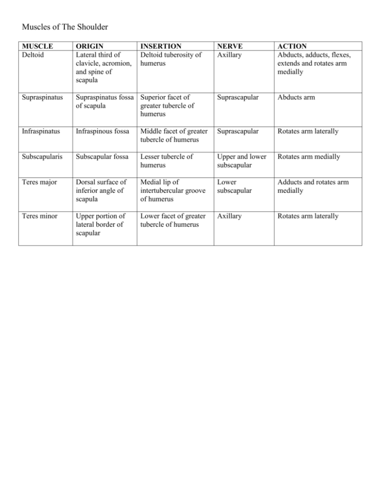
Muscles of The Shoulder MUSCLE Deltoid ORIGIN Lateral third of clavicle, acromion, and spine of scapula Supraspinatus INSERTION Deltoid tuberosity of humerus NERVE Axillary ACTION Abducts, adducts, flexes, extends and rotates arm medially Supraspinatus fossa Superior facet of of scapula greater tubercle of humerus Suprascapular Abducts arm Infraspinatus Infraspinous fossa Middle facet of greater tubercle of humerus Suprascapular Rotates arm laterally Subscapularis Subscapular fossa Lesser tubercle of humerus Upper and lower subscapular Rotates arm medially Teres major Dorsal surface of inferior angle of scapula Medial lip of intertubercular groove of humerus Lower subscapular Adducts and rotates arm medially Teres minor Upper portion of lateral border of scapular Lower facet of greater tubercle of humerus Axillary Rotates arm laterally Muscles of the Arm MUSCLE PROXIMAL Coracobrachialis Coracoid process DISTAL Middle third of medial surface of humerus NERVE ACTION Musculocutaneous Flexes and adducts arm Biceps brachii Long head, supraglenoid tubercle; short head, coracoid process Radial tuberosity of radius Musculocutaneous Flexes arm and forearm, supinates forearm Brachialis Lower anterior surface of humerus Coronoid process of ulna and ulnar tuberosity Musculocutaneous Flexes forearm Triceps Long head, infraglenoid tubercle, lateral head, superior to radial groove of humerus; medial head, inferior to radial groove Posterior surface of olecranon process of ulna Radial Extends forearm Aconeus Lateral epicondyle of humerus Olecranon and upper posterior surface of ulna Radial Extends forearm (Elbow joint stabilizer) Muscles of the Anterior Forearm MUSCLE Pronator teres PROXIMAL Medial epicondyle and coronoid process of ulna DISTAL Lateral radius NERVE Median ACTION Pronates forearm Flexor carpi radialis Medial epicondyle of humerus Bases of SECOND and sometimes third metacarpals Median Flexes forearm, flexes and abducts hand (ie flexes and abducts at the wrist) Palmaris longus Medial epicondyle of humerus Palmar aponeurosis (flexor retinaculum) Median Flexes hand and forearm Flexor carpi ulnaris Medial epicondyle, upper posterior border of ulna Pisiform, hook of hamate, and base of fifth metacarpal Ulnar Flexes and adducts hand, flexes forearm Flexor digitorum superficialis Medial epicondyle, humeroulnar head arises from the common flexor tendon and the medial border of coronoid process; radial head arises from the proximal anterior border of radius Base of middle phalanges, digits 2-5 Median w/ ulnar artery both pass bet 2 heads of origin Flexes proximal interphalangeal joints, flexes hand and forearm DEEP MUSCLES (all innervated by ANTERIOR INTEROSSEUS NERVE – a branch of the median nerve) Flexor Anteromedial Bases of distal Ulnar (medial Flexes distal digitorum surface of ulna, phalanges of digits 2-5 portion of FDP) interphalangeal joints and profundus interosseous and anterior hand membrane interosseous nerve Flexor pollicis Anterior surface of Bases of distal phalanx Anterior Flexes thumb longus radius, interosseous of thumb interosseous membrane nerve Pronator quadratus Anterior surface of distal ¼ of ulnar Anterior surface of distal ¼ of radius Anterior interosseous nerve Pronates forearm Muscle of Posterior Forearm (origin – humerus and COMMON EXTENSOR TENDON) MUSCLE PROXIMAL DISTAL Brachioradialis Lateral Lateral lower end of supracondylar radius (base of radial ridge of humerus styloid process) NERVE Radial ACTION Flexes forearm Extensor carpi radialis longus Lateral supracondylar ride of humerus Dorsum of base of second metacarpal Radial Extends and abducts hand Extensor carpi radialis brevis Lateral epicondyle of humerus Posterior base of third metacarpal Radial (deep) Extends fingers and abducts hands Extensor digitorum Lateral epicondyle of humerus Extensor expansion of digits 2-5 Posterior interosseous nerve Extends fingers and hand (2-5); extends digits and all three phalanxes Extensor digiti minimi Common extensor tendon and interosseous membrane Extensor expansion of digit 5 Post interosseous n. Extends little finger Extensor carpi ulnaris Lateral epicondyle and posterior surface of ulna Base of fifth metacarpal Posterior Interosseous nerve Extends and adducts hand DEEP MUSCLES (mostly innvervated by POSTERIOR INTEROSSEOUS NERVE) Supinator Lateral epicondyle, Lateral border of radius Radial Supinates forearm (deep) Abductor Interosseous Lateral surface of base Posterior Abducts thumb (and hand) pollicis longus membrane, of first metacarpal Interosseous n. posterior surfaces of radius and ulna Extensor pollicis longus Interosseous membrane and posterior surface of ulna Extensor Interosseous pollicis brevis membrane and posterior surface of radius Extensor indicis Posterior surface of ulna and interosseous membrane Aconeus Lateral epicondyle of humerus Base of distal phalanx of thumb Posterior interosseous n. Extends distal phalanx of thumb and abducts hand Base of proximal phalanx of thumb Posterior interosseous n. Extends proximal phalanx of thumb and abducts hand Extensor expansion of index finger (2nd digit) Posterior interosseous n. Extends index finger Olecranon and upper posterior surface of ulna Radial Extends forearm (assists triceps) Muscles of Hand MUSCLE PROXIMAL DISTAL THENAR COMPARTMENT (RECURRENT MEDIAN NERVE) Abductor pollicis Flexor retinaculum, Sesamoid bone, proximal brevis scaphoid, trapezium phalanx and extensor expansion Flexor pollicis brevis Flexor retinaculum, Sesamoid, proximal scaphoid, trapezium phalanx and extension expansion Opponens pollicis Flexor retinaculum First metacarpal and trapezium (lateral side) HYPOTHENAR COMPARTMENT Palmaris brevis Medial side of flexor retinaculum, palmar aponeurosis Abductor digiti mini Pisiform Flexor digiti minimi brevis Opponens digiti minimi hamate hamate MID-PALMAR COMPARTMENT Adductor pollicis (2 2nd metacarpal and heads) capitate. Transverse head origin. 3rd metacarpal. Lumbricals (4) Lateral side of tendons of flexor digitorum profundus DEEP MUSCLES OF HAND Dorsal interossei (4) 1st – 1st and 2nd metacarpals (adjacent sides of metacarpal bones) Palmar interossei (3) 1st origin - Medial side of 2nd metacarpal; 2nd - lateral sides of 4th metacarpal; 3rd – lateral side of 5th metacarpals. (medial side of second metacarpal; lateral sides of fourth and fifth metacarpals) NERVE ACTION Recurrent median nerve Abduction and flexion of proximal phalanx Superficial head by recurrent median nerve Recurrent median nerve Flexion of proximal phalanx Skin of medial side of palm Superfical branch of ulnar n. proximal phalanx of 5th digit (little finger) proximal phalanx of 5th digit 5th metacarpal Ulnar (deep branch) Draws the skin at the ulnar side of palm towards the middle of the palm. Deepens the hollow of the palm. (wrinkles skin on medial side of palm) Abduction of digit 5 Medial sesamoid bone and base of proximal phalanx, and extensor expansion of thumb Lateral side of extensor expansion of 2nd to 5th digits. Ulnar (deep branch) Adduction of thumb, aids in opposition Recurrent Median (two lateral lumbricals) and ulnar (deep) (two medial 3rd & 4th) Flex metacarpophalangeal joints and extend interphalangeal joints 1st – lateral side of prox phalanx of index finger; 2nd & 3rd – middle finger, 2nd lat side & 3rd medial side. 4th – medial side of ring finger (Lateral sides of bases of proximal phalanges; extensor expansion) 1st – medial side proximal phalanx digit 2, and extensor expansion; 2nd – lat side prox phalanx digit 4 and extensor expansion; 3rd – lat side prox. Phalanx digit 5. and extensor expansion; (proximal phalanges in same sides as their origins; extensor expansion) Ulnar (deep branch) Abduction of digits 2,3,4; Abduct fingers; flex metacarpophalangeal joints, extend interphalangeal joints; adducts fingers toward the middle fingers Ulnar (deep) Adduct fingers toward the middle finger (flex metacarpolangeal joints; extend interphalangeal joints) Ulnar (deep) Ulnar (deep) Rotation of metacarpal during opposition (opposes thumb to other digits) Flexion of proximal phalanx, digit 5 Draws 5th metacarpal anteriorly thus bringing digit 5 into contact with thumb during opposition. Only thumb can oppose other fingers. (Opposes little finger) MUSCLES OF THE BACK MUSCLE ORIGIN THE EXTRINSIC MUSCLES The Superficial Group Trapezius Occiptal region of skull; ligamentum nuchae, spinous processes of CV7-T12 INSERTION NERVE ACTION Spine of scapula, acromion, and lateral third of clavicle Subtrapezial plexus = accessory nerve CNXI, ventral rami of C3-C4 spinal nerves Dorsal scapular nerve (C5); spinal nerves C3 & C4; Dorsal scapular nerve (C5), with small contribut from C4 & Dorsal scapula artery Dorsal scapular nerve (C5), with small contribut from C4 & Dorsal scapula artery Thoracodorsal nerve (C6,C7 and some contribution by C8) and artery Superior fibers elevate scapula; middle fibers retract scapula; lower fibers depress scapula and lower the shoulder Intercostal nerves (C8-T3) As the muscle pulls toward its origin during inspiration, it elevates the first four (or five) ribs increasing the diameter of the thorax During inspiration, the muscle depresses the inferior ribs, preventing them from being pulled superiorly by the diaphragm. Levator scapulae Transverse processes of C1-C4 vertebrae Blood supply: transverse cervical artery Superior part of medial border of scapula Rhomboid minor Ligamentum nuchae and spinous processes of C7 & T1 vertebrae Medial border of scapula (usually at level of scapula spine) Rhomboid major Spinous processes of T2-T5 Medial border of scapula (inferior to scapula spine) Latissimus dorsi Aponeurosis from the Floor of intertubercular spinous processes of groove of humerus (ant inferior 6 vertebrae: surface) T5-T12, thoracolumbar fascia, sacral spines, iliac crest, lower 4 ribs: 9-12 The Intermediate Group (two muscles responsible for aiding in respiration Serratus Posterior Inferior part of Superior Borders of second Superior ligamentum nuchae to fourth (or fifth ribs) and spinous process of C7 and T1-T3 vertebrae Serratus Posterior Spinous processes of Inferior border of the Inferior last two thoracic T11inferior 3 or 4 ribs (ribs (9) L2) and first two 10-12) lumbar vertebrae INTRINSIC MUSCLES of the Back (the deep group) Splenius muscle Inferior half of the A. Splenius capitis:lateral (superficial layer ligamentum nuchae aspect of mastoid region of of intrinsic (CV4-CV6) and the the temporal bone to the muscles) spinous processes of later 1/3rd of the superior CV7, T1-T6 nuchal line of the occipital bone. B. Splenius cervicis: Posterior tubercles located on the transverse processes of C1-C4) Erector Spinae Illiocostalis (lateral Longissimus (intermediate muscle (superficial column): regions – column): regions – layer) illiocostalis, longissimus throacis, lumborum, thoracis cervicis, and capitis and cervices The Transversospinalis group (the deep layer of intrinsic muscles) Semispinalis (forms Multifidus Rotatores the largest muscle mass in posterior aspects of the neck Intercostal nerves (T9-T11) Elevates scapula and helps to tilt its glenoid cavity inferiorly by rotating the scapula Retract scapula and rotate it to depress the glenoid cavity Retract scapula and rotate it to depress the glenoid cavity Adducts, extends, and rotates arm medially Dorsal rami of cervical nerves Acting alone, laterally flex and rotate the head and neck to the same side. Acting together, they extend the neck back Spinalis( medial column): regions – spinalis thoroacis, cervicis, and capitis Major: Extend the vertebral column and bend it posteriorly. Minor: maintain posture and head movement Levator costorum (longus and brevis) The semispinalis, multifidus, rotators muscles lat flex vert column (alone) & assist w/ post ext when working together. Levator costorum assist w/ the process of inspiration by elevating the ribs. Muscles of Pectoral Region MUSCLE Pectoralis major ORIGIN Medial half of clavicle, manubrium, body of the sternum, upper six costal cartilages INSERTION Lateral tip of the intertubercular groove (the crest of the greater tubercle) of humerus NERVE ACTION Medial and lateral Adducts and medially pectoral nerves rotates arm; clavicular part – rotates arm medially and flex it; sternocostal part depresses arm and shoulder, its lower fibers can help extend arm when it is flexes Pectoralis minor External surfaces of second to fifth ribs Coracoid process Medial and lateral Depresses shoulder pectoral nerves Subclavius Junction of the first rib and its cartilage Lower surface of the clavicle Nerve to subclavius Depresses lateral portion of clavicle Serratus anterior External surfaces of ribs 1-8 Medial border of inferior angle of scapula Long thoracic nerve Rotates scapula upward, so that the inferior angle swings laterally and abducts the arm and elevates it above a horizontal position
