Skeletal System Notes: Bone Structure & Disorders
advertisement

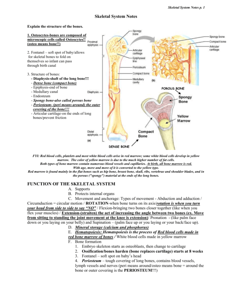
Skeletal System Notes p. 1 Skeletal System Notes Explain the structure of the bones. 1. Osteocytes-bones are composed of microscopic cells called Osteocytes!! (osteo means bone!!) 2. Fontanel – soft spot of baby/allows for skeletal bones to fold on themselves so infant can pass through birth canal 3. Structure of bones: - Diaphysis-shaft of the long bone!!! - Dense bone (compact bone) - Epiphysis-end of bone - Medullary canal - Endosteum - Spongy bone-also called porous bone - Periosteum- (peri means around) the outer covering of the bone!!! - Articular cartilage-on the ends of long bones/prevent friction POROUS BONE DENSE BONE FYI: Red blood cells, platelets and most white blood cells arise in red marrow; some white blood cells develop in yellow marrow. The color of yellow marrow is due to the much higher number of fat cells. Both types of bone marrow contain numerous blood vessels and capillaries. At birth, all bone marrow is red. With age, more and more of it is converted to the yellow type. Red marrow is found mainly in the flat bones such as hip bone, breast bone, skull, ribs, vertebrae and shoulder blades, and in the porous ("spongy") material at the ends of the long bones. FUNCTION OF THE SKELETAL SYSTEM A. Supports B. Protects internal organs C. Movement and anchorage: Types of movement - Abduction and adduction / Circumduction = circular motion / ROTATION-when bone turns on its axis/rotation is when you turn your head from side to side to say “NO” / Flexion-bringing two bones closer together (like when you flex your muscles) / Extension-(straiten) the act of increasing the angle between two bones (ex. Move from sitting to standing the joint movement at the knee is extension)/ Pronation - (like palm face down or you laying on your belly) and Supination – (palm face up or you laying or your back/face up). D. Mineral storage (calcium and phosphorus) E. Hematopoiesis: Hematopoiesis is the process of Red blood cells made in red bone marrow of bones / White blood cells made in yellow marrow F. Bone formation 1. Embryo skeleton starts as osteoblasts, then change to cartilage 2. Ossification/bones harden (bone replaces cartilage) starts at 8 weeks 3. Fontanel – soft spot on baby’s head 4. Periosteum – tough covering of long bones, contains blood vessels, lymph vessels and nerves (peri means around/osteo means bone = around the bone or outer covering is the PERIOSTEUM!!!) Skeletal System Notes p. 2 ~The skeleton can be broken down into 2 parts: Axial & Appendicular: AXIAL SKELETON-contains the skull, spine and chest!!! 1. Skull-the cranial bones (of the skull) include the following!! sutures i.Parietal ii.Frontal iii.Occipital iv.Temporal v. Ethmoid vi.Sphenoid (Nasal bone/Zygomatic arch/Infraorbital foramen/Mental foramen (foramen usually means a hole!!) Mandible-involved with chewing-mandible is the only movable bone in the skull!!!! Maxilla/Vomer/Mastoid process/styloid process/External auditory meatus *Sutures-the areas where cranial bones join together to form immovable joints!!!-(when baby is born, they are movable to allow head to pass through vaginal canal and as the child grows, the sutures become fused and immovable le.) Skeletal System Notes p. 3 2. Spinal column/vertebra – the vertebral column encloses spinal cord & is separated by pads of cartilage called intervertebral discs i. Cervical vertebrae – in neck ii. Thoracic vertebrae - back iii. Lumbar vertebrae - hips iv. Sacrum -butt v. Coccyx-tail bone A good way to remember the numbers of each vertebral section is: “We eat breakfast at 7, we eat lunch at 12 noon, & we eat dinner at 5.” 7 cervical come first (breakfast) 12 thoracic comes 2nd (lunch) 5 lumbar comes last (dinner) FYI: The vertebral column is made up of about 33 bones & provides protection to the spinal cord, which runs through its central cavity. Between each vertebra is an intervertebral disk, which acts as a shock absorber. The sacrum is a triangular bone located just below the lumbar vertebrae. It consists of four or five sacral vertebrae in a child, which becomes fused into a single bone after age 26. The sacrum forms the back wall of the pelvic girdle and moves with it. The bottom of the spinal column is called the coccyx or tailbone. It consists of 3-5 bones that are fused together in an adult. Many muscles connect to the coccyx. 3. CHEST = Ribs and sternum-another name for the breast bone is the sternum!!!!/ There are 12 pairs of ribs = 7 true ribs, 3 false ribs, 2 floating ribs. The Xiphoid process is the pointy tip at the end of the sternum. APPENDICULAR SKELETON – appendages are your arms & legs Clavicle (color bone) and scapula (shoulder bone) Humerus (upper arm bone), radius and ulna (lower arm bones) Carpals, metacarpals and phalanges * HINT* you drive with your car (carpals = hand bones)/ (The phalanges are the medical term for the finger bone!!!) Pelvis – has three parts/Ilium, Ischium, & Pubis Femur-largest bone in body/thigh bone/Femur is longest and strongest bone in body Patella is the medical term for the knee-cap!! Tarsal’s, metatarsals, phalanges (feet) *HINT* you walk on tar (tarsal’s = feet bones) Calcaneus – heel bone Radius-lower arm bone located on the thumb side of the hand!! * (Along this bone is where medical professionals monitor radial pulse!!!) Skeletal System Notes p. 4 Joints: TYPES!! Synovial fluid (the fluid in joints) – lubrication-reduces the friction during joint movement!! a. Ball and socket joints – ball-shaped head, examples: Hip and shoulder. The ball & socket allow for the greatest freedom of movement!!! b. Hinge joints – move in one direction or plane, examples: Knees, elbows, outer joints of fingers c. Pivot joints – rotate on a 2nd, arch-shaped bone, examples: radius and ulna d. Gliding joints – flat surfaces glide across each other, examples: vertebrae e. Suture = the only movable joints!!! They are in the skull and fuse (become firm) at about one year of age JOINT MOVEMENTS CHARACTERISTICS AND TREATMENT OF COMMON SKELETAL DISORDERS *Fracture – any break in a bone – different types confirmed with x-ray (radiology)- Radiography = x-ray of bones Greenstick fracture – VERY common in children, bone bent and splintered but never completely separates Comminuted fracture – splintered or broken into many pieces Compound fracture – (open fracture) broken bones pierce skin, can lead to infection –usually needs to be fixed by open reduction and internal fixation!! (this involves actual surgery and using medical pins & screws to secure the break) Simple fracture (closed fracture) bone broken, broken ends do not break the skin (usually needs cast) Spiral fracture – bone twists, resulting in one or more breaks (common with abuse cases/grabbing) Dislocation – bone displaced from proper position in joint HIP DISLOCATION Whiplash – trauma to the cervical vertebra, usually from a car accident ~ Sprain – caused by sudden or unusual motion, ligaments torn or damaged!!(treat with RICE-rest/Ice for 48 hrs/compression of injury (wrap it)/elevate the injury) ~ Strain – overstretching of tendons or tearing of muscle (the muscle can bleed/ICE!!) Arthritis – inflammation of one or more joints-painful!!!-different types: Rheumatoid – chronic, autoimmune disease, joint becomes swollen and painful, joint deformities common Osteoarthritis – degenerative, occurs with aging, joints become large and painful, Rx with medications Bursitis – inflammation of bursa (joint sacs) Osteomyelitis – bone infection that can travel to the muscle Osteoporosis: 80% affected are women & involves loss of bone mass, calcium and phosphorous leading to thin, porous (spongy) bones that are prone to fracture!!! / On x-ray it makes the bone look like Swiss Cheese/Prevented by dietary calcium and Vit. D intake! Fracture TREATMENT - Closed reduction – cast or splint-the main reason for a cast is to immobilize the bone!! - Open reduction/internal fixation – surgical intervention with devices such as wires, metal plates or screws to hold the bones in alignment Skeletal System Notes p. 5 Traction – pulling force used to hold the bones in place, used for fractures of long bones Spinal defects – abnormal curvature 1. Kyphosis – hunchback-hump in thoracic spine 2. Lordosis – swayback-curve in lumbar spine (person may lean back) 3. Scoliosis – lateral curvature/S curve in spine Herniated disk: Intervertebral disk ruptures or protrudes, putting pressure on spinal nerve; usually lumbo-sacral spine affected / treat with bed rest & pain medicine or traction and as a last resort surgery Rickets: Found in children/poor diet (most often in poverty)/ caused by lack of vitamin D/bones become soft (osteomalacia)!!! & can break easily-TREATMENT-Rx with calcium, vitamin D and sunshine Gout – uric acid deposited in joint cavity, mostly the great toe in men/very painful!! Bone marrow aspiration – removal of bone marrow sample with a needle for diagnostic purposes-usually in cases of suspected blood cancers/also used for stem cell removal and transplants for blood cancer patients!! (VERY PAINFUL) Bone Marrow Aspiration Pelvis-the female is wider than the male in order to carry children. The pubic symphysis or symphysis pubis; will spread during delivery. The Ilium (iliac crest) is the most common area to extract bone marrow. Arthroscopy – examination of joint using arthroscopy with fiber optic lens, most knee injuries treated with arthroscopy Arthroscopy can be done on any joint! Skeletal System Notes p. 6


