labeling stem cells for non-invasive cardiovascular imaging for
advertisement
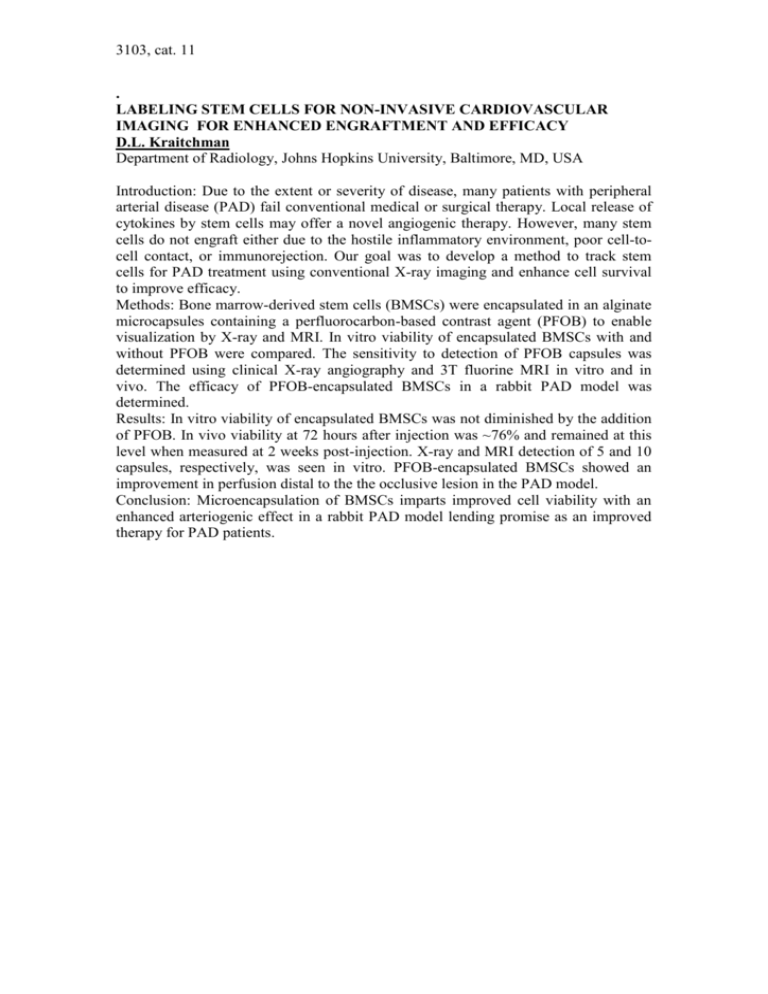
3103, cat. 11 . LABELING STEM CELLS FOR NON-INVASIVE CARDIOVASCULAR IMAGING FOR ENHANCED ENGRAFTMENT AND EFFICACY D.L. Kraitchman Department of Radiology, Johns Hopkins University, Baltimore, MD, USA Introduction: Due to the extent or severity of disease, many patients with peripheral arterial disease (PAD) fail conventional medical or surgical therapy. Local release of cytokines by stem cells may offer a novel angiogenic therapy. However, many stem cells do not engraft either due to the hostile inflammatory environment, poor cell-tocell contact, or immunorejection. Our goal was to develop a method to track stem cells for PAD treatment using conventional X-ray imaging and enhance cell survival to improve efficacy. Methods: Bone marrow-derived stem cells (BMSCs) were encapsulated in an alginate microcapsules containing a perfluorocarbon-based contrast agent (PFOB) to enable visualization by X-ray and MRI. In vitro viability of encapsulated BMSCs with and without PFOB were compared. The sensitivity to detection of PFOB capsules was determined using clinical X-ray angiography and 3T fluorine MRI in vitro and in vivo. The efficacy of PFOB-encapsulated BMSCs in a rabbit PAD model was determined. Results: In vitro viability of encapsulated BMSCs was not diminished by the addition of PFOB. In vivo viability at 72 hours after injection was ~76% and remained at this level when measured at 2 weeks post-injection. X-ray and MRI detection of 5 and 10 capsules, respectively, was seen in vitro. PFOB-encapsulated BMSCs showed an improvement in perfusion distal to the the occlusive lesion in the PAD model. Conclusion: Microencapsulation of BMSCs imparts improved cell viability with an enhanced arteriogenic effect in a rabbit PAD model lending promise as an improved therapy for PAD patients.
