MICROSCOPIC TEMPLATE FOR REPORTING RESECTED LUNG
advertisement
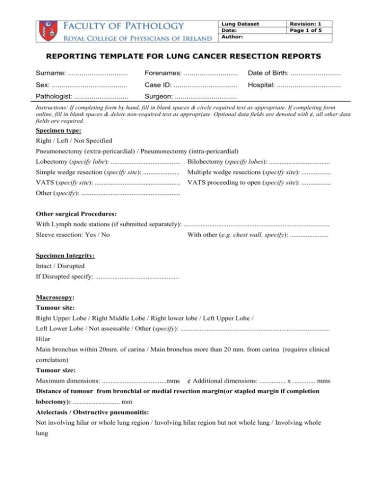
Lung Dataset Date: Author: Revision: 1 Page 1 of 5 REPORTING TEMPLATE FOR LUNG CANCER RESECTION REPORTS Surname: ............................... Forenames: ............................ Date of Birth: .......................... Sex: ....................................... Case ID: ................................. Hospital: ................................. Pathologist: ............................ Surgeon: ................................ Instructions: If completing form by hand, fill in blank spaces & circle required text as appropriate. If completing form online, fill in blank spaces & delete non-required text as appropriate. Optional data fields are denoted with ¢, all other data fields are required. Specimen type: Right / Left / Not Specified Pneumonectomy (extra-pericardial) / Pneumonectomy (intra-pericardial) Lobectomy (specify lobe): ........................................ Bilobectomy (specify lobes): ................................... Simple wedge resection (specify site): ..................... Multiple wedge resections (specify site): ................. VATS (specify site): ................................................. VATS proceeding to open (specify site): ................. Other (specify): ......................................................... Other surgical Procedures: With Lymph node stations (if submitted separately): .................................................................................... Sleeve resection: Yes / No With other (e.g. chest wall, specify): ...................... Specimen Integrity: Intact / Disrupted If Disrupted specify: ................................................ Macroscopy: Tumour site: Right Upper Lobe / Right Middle Lobe / Right lower lobe / Left Upper Lobe / Left Lower Lobe / Not assessable / Other (specify): ...................................................................................... Hilar Main bronchus within 20mm. of carina / Main bronchus more than 20 mm. from carina (requires clinical correlation) Tumour size: Maximum dimensions: .....................................mms ¢ Additional dimensions: ............... x ............. mms Distance of tumour from bronchial or medial resection margin(or stapled margin if completion lobectomy): ........................... mm Atelectasis / Obstructive pneumonitis: Not involving hilar or whole lung region / Involving hilar region but not whole lung / Involving whole lung Lung Dataset Date: Author: Revision: 1 Page 2 of 5 Tumour focality: Unifocal: Yes / No Separate tumour nodules in same lobe (specify number): ..................................................... (specify size range): .......................................... mm Separate tumour nodules in different lobes (specify sites): .......................................................... (specify number): ..................................................... (specify size range): .......................................... mm Microscopy (WHO Classification 2004): Histological Type (circle appropriate type from the following): Squamous Cell Carcinoma Adenocarcinoma: Pure subtype (specify type): ........................................................................................ Pure BAC: mucinous / non-mucinous / mixed (>10% of each) Mixed (specify components in %): ............................................................................. ¢ Adenoacarcinoma: Whole tumour size: ................................................... mm Invasive tumour size: ................................................ mm Large Cell Carcinoma Pleomorphic Carcinoma Other tumour: Specify (e.g. metastasis): ....................................... Typical Carcinoid / Atypical Carcinoid / Small Cell Carcinoma / Large Cell Neuroendocrine Carcinoma Combined Small Cell Carcinoma (specify): ................................................................................................... Tumour Grade: Cannot be assessed (GX) / Well (G1) / Moderate(G2) / Poor(G3) / Undifferentiated (G4) Separate Nodule(s): Present / Absent Site…………………………………………. Specify Histology: .......................................................................................................................................... Local Invasion: Visceral Pleura: Present (¢ indicate either PL1 or PL2 ) / Absent (PL0) Parietal pleura: Present (¢ PL3) / Absent / Not Applicable Chest wall: Submitted / Not submitted / Present (Endothoracic fascia / rib /muscle) / Absent Mention the following only if submitted and comment if invasion present or absent (Consult UICC manual for accurate staging) Mediastinal pleura, Pericardium (Parietal, Visceral), Diaphragm, Great Vessels (Aorta, Pulmonary trunk, SVC, IVC, Intrapericardial Pul. Arteries & veins), Atrium, Other ………………………………………………………………………………………………………………. Lymphovascular invasion: Present / Absent / Suspicious Lymph node spread: Lung Dataset Date: Author: Data from previous lymph node sampling procedures Revision: 1 Page 3 of 5 If Yes specify Specimen No: ………………... included: Yes / No Ipsilateral intrapulmonary/hilar (node stations Submitted: Yes / No Involved: Yes /No 10-14): Spread: Direct / Metastatic / Both Specify station status if sub. separately: .................................................................................................... Ipsilateral mediastinal (node stations 1-9): Submitted: Yes / No Involved: Yes /No Specify station status if sub. separately : …………………..................................................................... Extranodal extension by tumour: Present / Absent Contralateral mediastinal, hilar nodes, Ipsi or contralateral scalene or supraclavicular Submitted: Yes / No Involved: Yes /No nodes: Specify station status if sub. separately: …………………………………...................................... Margins (select all that apply): Bronchial: Uninvolved / Involved by Carcinoma in situ / Involved by Invasive Carcinoma Vascular: Uninvolved / Involved Chest wall/Parietal pleura: Not submitted / Uninvolved / Involved by Invasive carcinoma Parenchymal margin: Not submitted / Uninvolved / Involved by Invasive carcinoma Other Tissue margin: Specify: ................................ Uninvolved / Involved by Invasive CA ¢If all margins uninvolved by invasive carcinoma: Distance of invasive carcinoma from closest margin ……………..mm Margin specification…………………………………………………… Other Pathology: Emphysema: Yes / No Specify degree (mild/moderate/severe): ............................. Interstitial Fibrosis: Yes / No State cause (if known): ....................................................... Other (given details): ................................................................................................................................ ..................................................................................................................................................................... Distant Metastases: Unknown/M1a/M1b M1a (Circle as appropriate): separate tumour nodule in contralateral lung / tumour with pleural nodules / malignant pleural effusion (Lab Ref # ……..) M1b (Circle as appropriate): Distant mets outside the lung / pleura - Specify Site (Lab Ref # ………): .................................................................................................................... Involved Lung Dataset Date: Author: Revision: 1 Page 4 of 5 Ancillary Studies: Performed: Yes / No Comment: ……………………………….... Epidermal Growth Factor Receptor (EGFR) analysis performed: Yes / No Specify results (if known): ................................................................................... K-ras mutational analysis performed: Yes / No Specify results (if known): ................................................................................... Other: Yes / No If Yes, specify: ……………………………………………………....... ................................................................................. ¢ Additional comments: ............................................................................................................................... ......................................................................................................................................................................... Previous treatment (neoadjuvant chemotherapy / radiotherapy): Yes / No / Unknown Treatment Effect (circle appropriate response): N/A, >10% residual viable tumour, < 10% viable tumour, Cannot be determined Summary of Pathological Staging (Select highest stage from the above data) (y prefix : post treatment, m prefix: multiple primary tumours, r prefix: recurrent) Histopathological type: ………………………………………………………………………………….. UICC, TNM Classification 7th Edition: pT …….. N …….. M …….. (Delete pM if unknown) Complete resection at all margins No Yes Comment (if any): …………………………………………………………………………………………. Signature: ……………... Date: ............................... SNOMED codes: T ............... M ................. Lung Dataset Date: Author: Revision: 1 Page 5 of 5 Note on Biopsy Samples: It was decided that there would not be a template for biopsy samples. It is generally agreed however that sub-typing of NSCLC on biopsy material is important, particularly in this era of targeted therapy. In many cases the subtype is obvious but in others IHC and or histochemical stains are required. A suggested panel to help in these latter cases is as follows: CK5/6, p63, TTF-1, Mucin stain e.g. dPAS, mucicarmine If Small Cell Lung Cancer is being considered in this differential, CD56 should be included. Handling and Cut-up Guidelines: For guidelines on specimen handling, dissection and staging please refer to the Protocol for the examination of specimens from patients with primary Non-Small Cell Carcinoma , Small Cell Carcinoma or Carcinoid tumour of the lung, Butnor KJ et al1 and the Manual of Surgical Pathology, 2nd edition, Lester S 2. References: 1. Protocol for the examination of specimens from patients with primary Non-Small Cell Carcinoma , Small Cell Carcinoma or Carcinoid tumour of the lung Butnor KJ et al. Arch Pathol Lab Med Vol 133 Oct 2009 2. Lester S. Manual of Surgical Pathology, 2nd edition. Elsevier Churchill Livingstone, 2006;400. 3. Dataset for lung cancer histopathology reports (2nd Edition)(Sept 2007) Royal College of Pathologists. 4. TNM Classification of Malignant Tumours. 7th Edition. 5. Staging manual in Thoracic Oncology, Peter Goldstraw, Executive Editor International Association for the study of lung cancer. 2009. (Incorporates the TNM Classification of malignant tumours, 7th Edition 2009). 6. Tumours of the Lung, Pleura, Thymus & Heart. World Health Organisation Classification of Tumours. IARC press 2004.
