Note from MultiCare Health System This document contains
advertisement
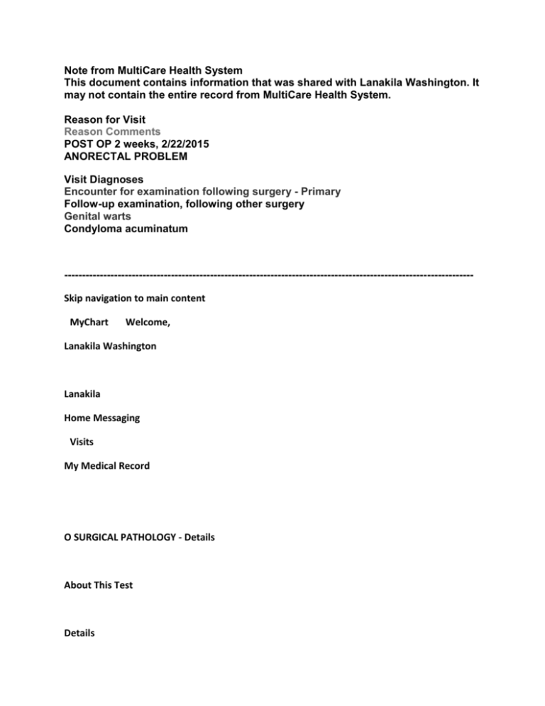
Note from MultiCare Health System This document contains information that was shared with Lanakila Washington. It may not contain the entire record from MultiCare Health System. Reason for Visit Reason Comments POST OP 2 weeks, 2/22/2015 ANORECTAL PROBLEM Visit Diagnoses Encounter for examination following surgery - Primary Follow-up examination, following other surgery Genital warts Condyloma acuminatum ------------------------------------------------------------------------------------------------------------------Skip navigation to main content MyChart Welcome, Lanakila Washington Lanakila Home Messaging Visits My Medical Record O SURGICAL PATHOLOGY - Details About This Test Details Past Results Graph of Past Results Component Results Component Standard Range Surgical Pathology Your Value Flag Accession Number S15-2556 FINAL DIAGNOSIS: 1) DISTAL RECTAL MASS, TRANSANAL DISC EXCISION: HIGH GRADE SQUAMOUS INTRAEPITHELIAL LESION (AIN 3). ANTERIOR AND POSTERIOR DISTAL MARGINS INVOLVED BY HIGH SQUAMOUS INTRAEPITHELIAL LESION. NO INVASIVE CARCINOMA IDENTIFIED. POLYPOID RECTAL MUCOSA WITH PROLAPSE CHANGES AND EROSIONS. 2) PERIANAL SKIN BIOPSY: HIGH GRADE SQUAMOUS INTRAEPITHELIAL LESION (AIN 3). MARGIN FOCALLY INVOLVED BY HIGH GRADE SQUAMOUS INTRAEPITHELIAL LESION. 3) PERIANAL CONDYLOMA BIOPSY: HIGH GRADE SQUAMOUS INTRAEPITHELIAL LESION (AIN 3). MARGIN FOCALLY INVOLVED BY HIGH GRADE SQUAMOUS INTRAEPITHELIAL LESION. QC:A RRR:cr COMMENT: This case was reviewed by Dr. Peterson, who agrees with the diagnosis. Electronically Signed By Rob R. Roth, MD HISTORY: Distal rectal mass. GROSS DESCRIPTION: PART 1: MASS, DISTAL RECTUM: (GF/ll) Received fresh from the operating room labeled Lanakila Washington, designated "distal rectal mass," is a previously pinned out portion of pale tissue measuring 4 x 2 cm. The specimen is anatomically oriented with color coated hypodermic needles, designated as follows: gray needle-proximal, orange needle-distal, green needle-posterior, and pink needle-anterior as noted on the requisition form. The mucosal surface is remarkable for a polypoid red-tan lesion which dominates the entire specimen. The margins are inked by quadrant as follows: anterior proximal-blue, anterior distal-black, posterior proximal-purple and posterior distal-green. The specimen is now perpendicularly sectioned along the anterior and posterior aspects. Entirely submitted sequentially progressing from anterior to posterior in cassettes 1a-1f. PART 2: PERIANAL SKIN LESION: (GF/ll) Received in formalin labeled Lanakila Washington, designated "perianal skin lesion," is an elliptoid portion of hair bearing gray-tan unoriented skin measuring 2 x 0.9 cm. The skin is smooth with a centrally located raised tan-brown soft to rubbery nodular mass measuring 1.1 cm in greatest dimension. The skin tips are inked blue and black respectively. Perpendicularly sectioned along its long axis. Entirely submitted in cassette 2a. PART 3: PERIANAL CONDYLOMA: (GF/ll) Received in formalin labeled Lanakila Washington, designated "perianal condyloma," is a purple-gray hair bearing fine granular unoriented skin portion measuring 0.7 x 0.5 cm. Marked black and trisected. Entirely submitted in cassette 3a. MICROSCOPIC DESCRIPTION: PART 1: MASS, DISTAL RECTUM: Sections examined. PART 2: PERIANAL SKIN LESION: Levels examined. PART 3: PERIANAL CONDYLOMA: Levels examined. Diagnosed at: Tacoma General Hospital 315 Martin Luther King Jr. Way Tacoma, WA 98405 General Information Collected: 02/19/2015 12:00 AM Resulted: 02/20/2015 2:33 PM Ordered By: Joshua E Levin, MD

