Methods: A retrospective study of 400 in
advertisement
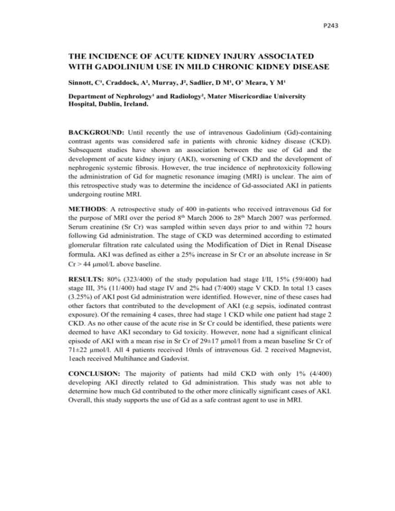
P243 THE INCIDENCE OF ACUTE KIDNEY INJURY ASSOCIATED WITH GADOLINIUM USE IN MILD CHRONIC KIDNEY DISEASE Sinnott, C¹, Craddock, A², Murray, J², Sadlier, D M¹, O’ Meara, Y M¹ Department of Nephrology¹ and Radiology², Mater Misericordiae University Hospital, Dublin, Ireland. BACKGROUND: Until recently the use of intravenous Gadolinium (Gd)-containing contrast agents was considered safe in patients with chronic kidney disease (CKD). Subsequent studies have shown an association between the use of Gd and the development of acute kidney injury (AKI), worsening of CKD and the development of nephrogenic systemic fibrosis. However, the true incidence of nephrotoxicity following the administration of Gd for magnetic resonance imaging (MRI) is unclear. The aim of this retrospective study was to determine the incidence of Gd-associated AKI in patients undergoing routine MRI. METHODS: A retrospective study of 400 in-patients who received intravenous Gd for the purpose of MRI over the period 8th March 2006 to 28th March 2007 was performed. Serum creatinine (Sr Cr) was sampled within seven days prior to and within 72 hours following Gd administration. The stage of CKD was determined according to estimated glomerular filtration rate calculated using the Modification of Diet in Renal Disease formula. AKI was defined as either a 25% increase in Sr Cr or an absolute increase in Sr Cr > 44 mol/L above baseline. RESULTS: 80% (323/400) of the study population had stage I/II, 15% (59/400) had stage III, 3% (11/400) had stage IV and 2% had (7/400) stage V CKD. In total 13 cases (3.25%) of AKI post Gd administration were identified. However, nine of these cases had other factors that contributed to the development of AKI (e.g sepsis, iodinated contrast exposure). Of the remaining 4 cases, three had stage 1 CKD while one patient had stage 2 CKD. As no other cause of the acute rise in Sr Cr could be identified, these patients were deemed to have AKI secondary to Gd toxicity. However, none had a significant clinical episode of AKI with a mean rise in Sr Cr of 29±17 µmol/l from a mean baseline Sr Cr of 71±22 µmol/l. All 4 patients received 10mls of intravenous Gd. 2 received Magnevist, 1each received Multihance and Gadovist. CONCLUSION: The majority of patients had mild CKD with only 1% (4/400) developing AKI directly related to Gd administration. This study was not able to determine how much Gd contributed to the other more clinically significant cases of AKI. Overall, this study supports the use of Gd as a safe contrast agent to use in MRI.
