Sample 1
advertisement
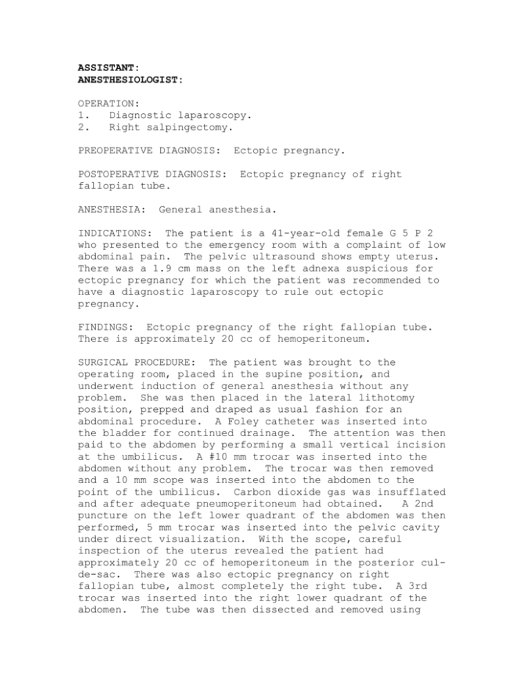
ASSISTANT: ANESTHESIOLOGIST: OPERATION: 1. Diagnostic laparoscopy. 2. Right salpingectomy. PREOPERATIVE DIAGNOSIS: POSTOPERATIVE DIAGNOSIS: fallopian tube. ANESTHESIA: Ectopic pregnancy. Ectopic pregnancy of right General anesthesia. INDICATIONS: The patient is a 41-year-old female G 5 P 2 who presented to the emergency room with a complaint of low abdominal pain. The pelvic ultrasound shows empty uterus. There was a 1.9 cm mass on the left adnexa suspicious for ectopic pregnancy for which the patient was recommended to have a diagnostic laparoscopy to rule out ectopic pregnancy. FINDINGS: Ectopic pregnancy of the right fallopian tube. There is approximately 20 cc of hemoperitoneum. SURGICAL PROCEDURE: The patient was brought to the operating room, placed in the supine position, and underwent induction of general anesthesia without any problem. She was then placed in the lateral lithotomy position, prepped and draped as usual fashion for an abdominal procedure. A Foley catheter was inserted into the bladder for continued drainage. The attention was then paid to the abdomen by performing a small vertical incision at the umbilicus. A #10 mm trocar was inserted into the abdomen without any problem. The trocar was then removed and a 10 mm scope was inserted into the abdomen to the point of the umbilicus. Carbon dioxide gas was insufflated and after adequate pneumoperitoneum had obtained. A 2nd puncture on the left lower quadrant of the abdomen was then performed, 5 mm trocar was inserted into the pelvic cavity under direct visualization. With the scope, careful inspection of the uterus revealed the patient had approximately 20 cc of hemoperitoneum in the posterior culde-sac. There was also ectopic pregnancy on right fallopian tube, almost completely the right tube. A 3rd trocar was inserted into the right lower quadrant of the abdomen. The tube was then dissected and removed using Harmonic instrument. The specimen was then removed out of the abdominal cavity. Irrigation was performed and hemostasis was found to be good. The procedure was completed by deflating the carbon dioxide gas. Local anesthesia was installed along the incision to prevent postop pain. The incision was then reapproximated using 40 Vicryl suture. The patient was then placed back in supine position, reversed from anesthesia, and transferred back to the recovery room in good general condition.
