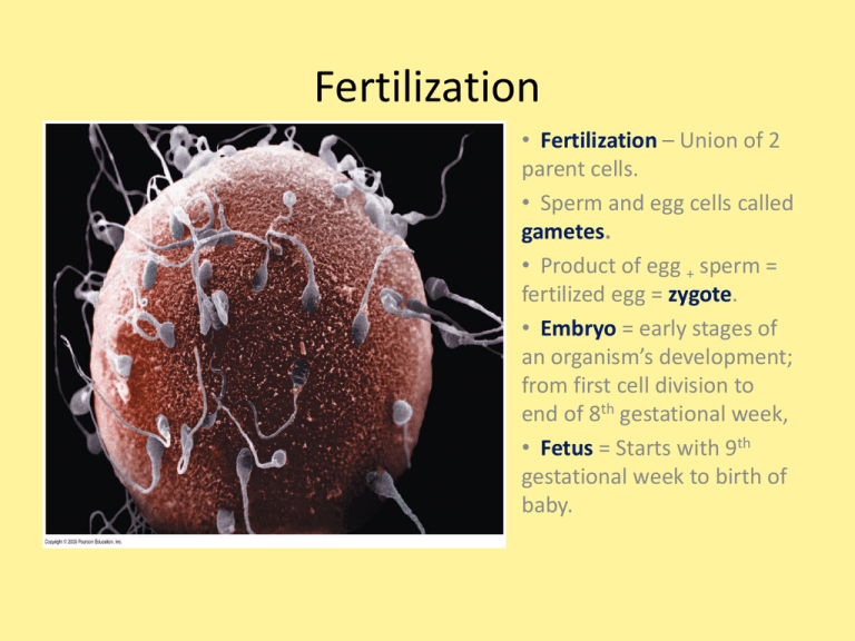
Fertilization
• Fertilization – Union of 2
parent cells.
• Sperm and egg cells called
gametes.
• Product of egg + sperm =
fertilized egg = zygote.
• Embryo = early stages of
an organism’s development;
from first cell division to
end of 8th gestational week,
• Fetus = Starts with 9th
gestational week to birth of
baby.
Gametes
• Sperm – small cell with
flagellum; nucleus &
mitochondria; ½
chromosomes.
• Ovum – larger cell;
cytoplasm contains yolk
(lipids &proteins); many
ribosomes, mitochondria; ½
chromosomes.
Fertilization-Activation
• Fertilization – Union of
two cells.
1. Sperm passes through
follicular cells.
2. Sperm passes through
jelly coat of ova by
releasing acrosomal
enzymes.
3. Proteins on sperm bind
to ova receptor
proteins; ensures single
species fertilization.
4. Sperm/egg plasma
membrane fuse;
membrane now
impenetrable by other
sperm.
5. Sperm nucleus enters
cytoplasm.
Fertilization/Activation
6. Fertilization envelope
forms; space between
outer membrane and
veteline layer fills with
water; further ensures
no fertilization by
other sperm.
7. Nuclei fusion activates
metabolic activity; cell
respiration, new
proteins etc.
8. Sperm and egg nuclei
fuse into zygotes.
9. http://bcs.whfreeman.
com/thelifewire/conte
nt/chp43/4301s.swf
Cleavage – Cell Growth
• Rapid succession of cell divisions.
• Produces multicellular ball of
cells from zygote.
• Mitosis produces may small cells;
many nuclei; cytoplasm
partitioned; size of embryo does
not change much.
• Multicellular ball = blastula.
• Cleavage partitions cells into
developmental regions.
Gastrulation - Differentiation
• Cells take up new locations that
later allow for formation of organs&
tissues.
•Cells migrate and form 3 layers.
1. Ectoderm
2. Endoderm
3. Mesoderm
•Gastrulation beginning of
differentiation ( cells become
different from each other); ends
when 3 tissue layers are formed.
•http://bcs.whfreeman.com/thelifew
ire/content/chp20/2002001.html
Embryonic tissue layers
Organs form after gastrulationMorphogenesis
• Morphogenesis- formation
tissues/organs.
•Two structures appear soon after
gastrulation.
1. Notochord - Arises from mesoderm;
establishes anterior/posterior axis (body
plan); later becomes backbone.
Frog Notochord
2. Neural Tube- Arises from ectoderm;
becomes brain, spinal cord and nerves.
Review- form teams of 4
1. What is fertilization
2. Describe how a sperm is specialized for its function.
3. What occurs to assure an egg can be fertilized by one sperm.
4. What is a gamete
5. Describe how an ovum is specialized for its function.
6. Describe why fertilization occurs within a single species.
7. What is a fertilized egg called.
8. Rapid succession of cell divisions in an embryo is called what.
9. What stage of embryo development is the beginning of differentiation.
10. What is the multicellular ball that is formed during cleavage called?
11. What type of cell reproduction forms the blastula?
12. What is an embryo
13. What are the 3 tissue layers formed during gastrulation.
14. Define differentiation.
15. Which tissue layer forms the skin – ectoderm
16. Which tissue layer forms the circulatory system – mesoderm
17. What is a fetus
18. Define morphogenesis
19. Into what structure does a notochord develop?
20. Into what structues do the neural tube develop?
21. Which tissue layer forms the reproductive system - endoderm
Checkpoint
• Discuss the definitions of fertilization, cell
growth (cleavage), differentiation
(gastrulation), and morphogenesis.
• How are these activities related and can they
occur independently?
Basic developmental pattern
• Two cell stage in snail; large cell produces mesoderm
and endoderm; small cell produces ectoderm. Neither
cell alone can produce a complete individual.
• Two cell stage in sea
star; cells identical and
both will produce all
tissue types & develop
into complete individual.
• Embryonic similarities reflect relatedness; more similarities in embryo
development = closer evolutionary relationship
Question
• Why do all animals have their head in the
anterior position?
• http://www.youtube.com/watch?v=LFGaLidT8s
Homeotic genes
• Despite developmental differences, morphogenesis in
animals follows similar genetic program; suggests common
ancestor.
• Emergence of body form with specialized organs and tissues
in the right places is controlled by master control genes.
• A class of similar control genes that help direct embryonic
body plan are called homeotic genes.
Homeotic genes
• Each homeotic gene acts on a different
body segment.
• Each gene contains one or more copies
of a 180 nucleotide sequence called a
homeobox.
• Each 180 base sequence codes for a 60
amino acid protein called a
homeodomain.
• This protein acts as a transcription
factor to regulate transcription of other
developmental genes.
• Homeotic genes are located in the
same order and correspond to analogous
body regions for a number of different
animals.
• Similarity in body plan formation shows
descent from common ancestor.
http://www.dnalc.org/view/167
60-Animation-37-Master-genescontrol-basic-body-plans-.html
Frog
sea star
human
Fetal development
1. Fertilization
occurs in
oviduct.
2. Cleavage starts
24 hours after
fertilization.
3. By 6th or 7th day,
embryo at 100
cell stage
reaches uterus.
4. 100 cell stage=
blastocyst=
hollow ball of
cells = blastula of
other animals.
Fetal development
1. Blastocyst has fluid filled cavity, a cell mass that
forms baby, and outer layer called trophoblast.
2. Trophoblast secretes enzymes to allow blastocyst
to implant in uterus 7 days after conception.
3. Trophoblast cells spread into
endometrium of uterus; becomes
placenta; provides nourishment and
O2 for embryo.
4. Extra embryonic membranes protect
embryo:
a. Purple cells in cell mass amnion
b. Yellow cells from cell mass yolk
sac.
c. From other trophoblast cells
chorion.
Fetal development
1. Embryo at 9 days after conception.
2. Gastrulation occurring in embryo.
3. Extra-embryonic membranes developing.
4. At one month, embryo life support from
extra-embryonic membranes.
5. Amnion (amniotic cavity) encloses embryo.
6. Yolk sac-helps produce first blood cells &
gametes.
7. Allantois- forms umbilical cord.
8. Chorion - contains other membranes;
merges with placenta; functions in gas
exchange.
Fetal development
1. Chorionic villi- projections
from chorion and blood
vessels from the lining of
mother’s uterus form the
placenta.
2. Placenta used to exchange
nutrients, gases, waste.
3. Baby is attached to
placenta by the umbilical
cord.
ra
First trimester
1. From 5 – 9 weeks grows
from 7mm to 5.5 cm in
size.
2. Embryo grows into
fetus; moves arms, legs,
turns head.
3. Dramatic changes occur.
Second trimester
1. Developmental changes
during 2nd trimester
involve increase in size
& refinement of human
features.
2. 20 wks – fetus is 19 cm
long and weighs ½ kb
Third trimester
1. Fetus continues to grow
rapidly; about 50 cm
long at birth and weighs
3-4 kg.
Birth defects
• Structural or functional abnormalities present at birth; cause physical or
mental disability; 1000’s of birth defects.
• Cause –
1. Defective genes
2. Defective chromosomes
3. Environmental causes; rubella, drugs, etc.
• Structural defect – abnormality in physical structure.
• cleft palate
spina bifida
Birth defects
• Functional defect – abnormality in how a body system works; can lead to
developmental disability
• Down’s syndrome
blindness
muscular dystrophy
• Treatments vary with defect.
Checkpoint- jeopardy
•
•
•
•
•
•
•
•
•
•
•
•
•
•
•
•
•
•
•
The similarities in animal morphogenesis suggests : a common ancestor
What are the genes that control body plan: homeotic
What is a homebox : 180 nucleotide sequence
What is a homeodomain: 60 amino acid protein
At what developmental stage does implantation occur?
The embryo at the 100 cell stage is called the ___________
What are the 3 parts of the blastocyst?
Trophoblast cells turn into the ______.
At 9 days, the embryo reaches which stage of development?
What is the function of amnion? – enclose baby
What is the function of yolk sac - blood cells gametes
What membrane forms the umbilical cord? allantois
What is the function of the chorion? Merge with placenta gas exchange
What is the function of the yolk sac : make blood cells and gametes
How is the baby attached to the placenta?
Which trimester has the most dramatic developmental change?
What are 3 causes of birth defects?
What are 2 major types of birth defects?
What are 3 forms of spina bifida
Objectives
• Topic 3: Mechanisms of Cell Differentiation
• Explain molecular hybridization.
• Compare the selective loss hypothesis and the genetic
equivalence hypothesis and describe which hypothesis
was supported by experimental evidence.
• Define determination and compare it to differentiation.
• Explain how differences in cytoplasm explain
determination.
• Summarize cell to cell interaction.
Differentiation- what happens to
genes that are not used.
• Selective gene loss hypothesis: differentiating
cells loose some genes.
• Genetic equivalence hypothesis: cells contain
same gene, but some become inactive during
differentiation.
• Research supports genetic equivalence
hypothesis.
Differentiation vs. Determination
•The developmental potential,
(potency) of a cell = range of
different cell types it CAN become.
•Zygote and its very early
descendents are totipotent - these
cells have the potential to develop
into a complete organism.
•As development proceeds,
developmental potential of
individual cells decreases until their
fate is determined.
.
Determination vs. Differentiation
• The determination of
different cell types (cell
fates) involves progressive
restrictions in their
developmental potentials.
• When a cell “chooses” a
particular fate, it is said to
be determined, although
it still "looks" just like its
undetermined neighbors.
• Determination implies a
stable change - the fate of
determined cells does not
change.
Determination vs. Differentiation
• Differentiation follows
determination, as the cell
elaborates a cell-specific
developmental program.
• Differentiation results in the
presence of cell types that
have clear-cut identities,
such as muscle cells, nerve
cells, and skin cells
Determination
• Snail at 2 cell stage – Large cell produces
mesoderm and endoderm; small cell produces
ectoderm.
•Determination has occurred ; commitment to a
path of development.
•Sea star at 2 cell stage - Either cell is
totipotent and can produce an entire organism.
•Determination has not occurred.
Cytoplasmic Determination
• Researchers determined the cytoplasm
looked different in the small lobe (cell) from
the large cell.
• Regulatory molecules (m RNA) are
distributed differently in cytoplasm of large
and small cells.
• The regulatory molecules control gene
expression and differentiation.
•http://bcs.whfreeman.com/thelifewire/cont
ent/chp20/2002002.html
Embryonic Induction
•
•
•
•
•
Action of one group of cells (inducing cells)
on another leads to a developmental
pathway in responding tissue; important in
embryo development of complex
organisms.
Many scientists believe molecules on
surface of responding tissues recognize
signal molecules on the surface of inducing
tissues.
Sometimes cell to cell contact is necessary;
other times a signal protein is sent from
inducing to responding cell.
Development of many tissues & organs
influenced by embryonic induction; eye and
ear structures, vertebral cartilage, kidneys.
Primary embryonic induction: During
gastrulation, proteins in the mesoderm
influence the ectoderm to develop into
neural tissue.
Embryonic Induction
• http://bcs.whfreeman.com/thelifewire/conten
t/chp20/2002s.swf
Checkpoint
• Explain the difference between selective gene
loss and genetic equivalence hypotheses.
• Explain how determination and differentiation
are related?
• What is embryonic induction?
Vocabulary
• Define the following:
• Asexual Reproduction:
1. Binary Fission:
2. Budding:
3. Fragmentation:
• Sexual Reproduction:
• Haploid
• Diploid
• What are the benefits to asexual reproduction?
• What are the benefits to sexual reproduction?
Objectives
• Topic 4: Asexual Reproduction
• Describe the types of asexual reproduction and
compare that to sexual reproduction
• Compare haploid (n) to diploid cells (2n).
• Differentiate between homologous
chromosomes, autosomes, and sex chromosomes
• Compare and contrast two chromatids of a
chromosome with two homologous
chromosomes.
• Differentiate between somatic cells and gametes.
Asexual Reproduction
• Requires one parent; offspring identical to parent
(clone).
•Budding – Organisms for buds which develop into
adult and breaks away from parent.
• Binary Fission – Single celled organisms divide into
two.
• Fragmentation - Organism breaks into pieces which
form new organism.
Sexual Reproduction
• Requires 2 parents;
offspring is not genetically
identical to parent.
• Each parent contributes a
haploid (n) gamete.
• Haploid = ½
chromosomes.
• Offspring are diploid (2n).
• Diploid = full set of
chromosomes.
Asexual vs. Sexual Reproduction
• Asexual reproduction beneficial in stable
environments; organisms unable to locate
mates; costs less energy metabolically.
• Sexual reproduction beneficial in changing
environments; adds genetic variety; meiosis
reduces inheritance of harmful genes.
Movie
• http://www.youtube.com/watch?v=Wns5OQR
74OQ
Vocabulary
•
•
•
•
•
Homologous chromosome
Autosome
Sex chromosome
Gamete
Somatic Cell
Chromosomes
• Homologous chromosomesMatched pair in diploid cell;
Possess the same genes
(alleles) at the same gene loci.
• Autosomes – Pairs 1-22 in
humans; non-gender related
chromosomes.
• Sex chromosome – Pair 23 in
humans; X and Y
chromosomes mediate gender.
Chromatids vs. Chromosomes
Somatic cell vs. Gamete
Gamete: Sex cell; eggs and sperm;
haploid
Somatic cell: Any cell not a sex
cell; diploid
Vocabulary review-Tic Tac Toe
Haploid
Somatic
Diploid
chromatids
reproduction
homologous chromosomes
autosome
gamete
sexchromosomes
Write a sentence for each of the 3 columns.
Write a sentence for each of the 3 rows.
Write a sentence for each diagonal.
Topic 5 Objectives
• Draw and describe each stage of meiosis.
• Explain how meiosis produces daughter cells that differ
from each other and the parent cell.
• Explain how variation in offspring is created through
independent assortment, crossing over, and random
fertilization.
• Explain how the purpose of meiosis differs from that of
mitosis.
• Draw and describe how meiosis creates gametes (4 sperm
cells, 1 egg cell).
• Differentiate between sexual and asexual reproduction and
compare the evolutionary advantages and disadvantages of
each.
Meiosis
Animation Meiosis
• http://highered.mcgrawhill.com/sites/0072495855/student_view0/ch
apter28/animation__how_meiosis_works.htm
l
• http://www.cellsalive.com/meiosis.htm
Meiosis I
• Meiosis is preceded by interphase.
• Chromosomes replicate.
• Centrioles duplicate
• Prophase I most complex and longest phase.
• Chromosomes condense, mitotic spindle forms,
nuclear envelope breaks down.
• Homologous chromosomes each with two sister
chromatids undergo synapsis to create a tetrad.
• Crossing over occurs.
Prophase I
• Synapsis – Process in which 2 replicated homologous
chromosomes pair together to form a structure called
a tetrad.
• Crossing over is the exchange of genetic material
between corresponding segments of homologous
chromosomes.
• It can occur because homologous chromosomes
are in close proximity.
• Chromosomes are recombinants; they carry
different genes than the ones originally donated by
parents.
Meiosis I
• Metaphase I – Homologous chromosomes line up in
center of cell and attach to spindle.
• Note one homologous chromosome (with 2 sister
chromatids) attaches to a spindle fiber and the other
homologus chromosomes attaches to a spindle fiber from
the opposite pole.
• Anaphase I - Chromosomes migrate to poles of
the cell.
• Only tetrads split up. Each spindle fiber holds one
replicated homologous chromosome from the pair.
Meiosis
• Telophase I - Chromosomes arrive at poles of the cell.
• Cytokinesis occurs.
• Each cell has a chromosome consisting of two sister
chromatids.
• Each cell has a haploid number of chromosomes.
• In some species, an interphase occurs; in others the
process proceeds directly to meiosis II.
• No replication of chromosomes occurs before meiosis
II.
Meiosis
• Meiosis II –
Phases are identical
to meiosis I.
• Difference in
outcome.
• Separation occurs
between sister
chromatids.
• After cytokinesis,
4 haploid cell with
one chromatid from
each of the
homologous pairs
results.
Pop Bead Meiosis
•
•
•
•
•
•
•
•
•
•
•
•
•
Create two homologous chromosomes in two different colors.
During interphase, DNA replication occurs. Replicate each of the chromosomes.
Question: How many chromatids are present? How many chromosomes?
During prophase I, tetrad formation and crossing over occurs. Cross over non-identical sister
chromatids.
Line up the homologous chromosomes on the metaphase I plate.
During anaphase I, homologous chromosomes begin to migrate toward opposite poles.
Homologous chromosomes arrive at opposite poles during telophase I.
Cytokinesis occurs.
Question: Are the two daughter cells identical? Why or why not? Are the daughter cells diploid or
haplioid
Prophase II and metaphase II: Line up the sister chromatids at the metaphase plate.
Anaphase II: Individual sister chromatids migrate toward the opposite poles.
Telophase II and cytokinesis: Sister chromatids complete their migration and cytokinesis occurs.
Question: Are the daughter cells diploid or haploid? Are the daughter cells genetically identical?


