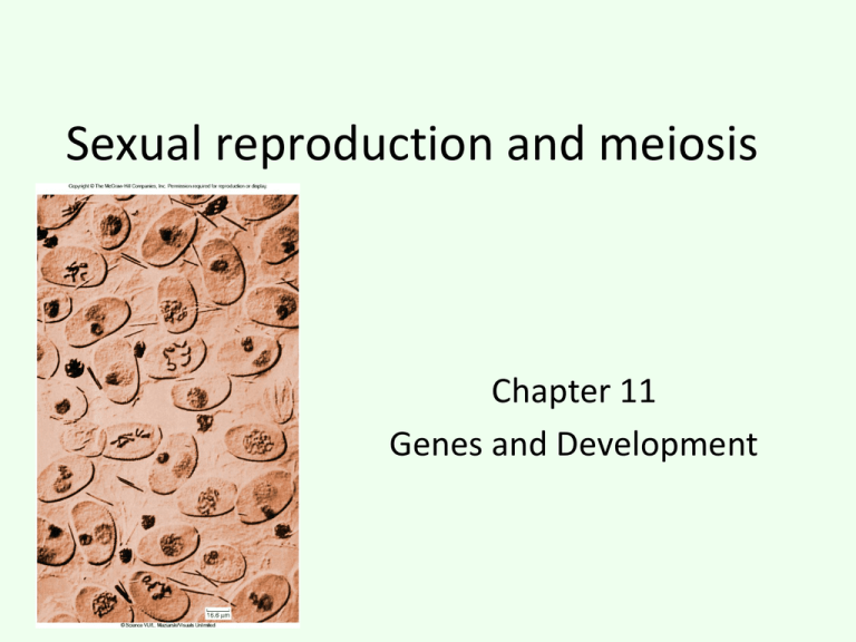
Sexual reproduction and meiosis
Chapter 11
Genes and Development
Fig. 11.1
Copyright © The McGraw-Hill Companies, Inc. Permission required for reproduction or display.
Haploid sperm
Paternal
homologue
Fertilization
Maternal
homologue
Diploid zygote
Haploid egg
Fig. 11.2
Copyright © The McGraw-Hill Companies, Inc. Permission required for reproduction or display.
Sperm
(haploid) n
Egg
(haploid) n
n
2n
Zygote
(diploid) 2n
MITOSIS
Somatic
cells
Germ-line
cells
Germ-line
cells
MITOSIS
Adult male
(diploid) 2n
Adult female
(diploid) 2n
Fig. 11.3b
Copyright © The McGraw-Hill Companies, Inc. Permission required for reproduction or display.
Diploid cell
Chromosome duplication
Meiosis I
Meiosis II
Haploid cells
c.
Fig. 11.3a-1
Copyright © The McGraw-Hill Companies, Inc. Permission required for reproduction or display.
Kinetochore
Sister chromatids
Synaptonemal
complex
Homologues
Centromere
a.
Fig. 11.3a
Copyright © The McGraw-Hill Companies, Inc. Permission required for reproduction or display.
Kinetochore
Sister chromatids
Synaptonemal
complex
Homologues
Centromere
a.
Synaptonemal Homologous
complex
chromosomes
b.
138 nm
b: Reprinted, with permission, from the Annual Review of Genetics, Volume 6 © 1972 by Annual Reviews, www.annualreviews.org
Fig. 11.4
Copyright © The McGraw-Hill Companies, Inc. Permission required for reproduction or display.
Site of crossover = Chiasmata
Fig. 11.7left-a
Copyright © The McGraw-Hill Companies, Inc. Permission required for reproduction or display.
MEIOSIS I
Prophase I
40 µm
Chromosome (replicated)
Spindle
Sister
chromatids
Paired homologous Chiasmata
chromosomes
In prophase I of meiosis I, the
chromosomes begin to
condense, and the spindle of
microtubules begins to form.
The DN A has been replicated,
and each chromosome consists
of two sister chromatids
attached at the centromere. In
the cell illustrated here, there are
four chromosomes, or two pairs
of homologues. Homologous
chromosomes pair up and
become closely associated
during synapsis. Crossing over
occurs, forming chiasmata,
which hold homologous
chromosomes together.
© Clare A. Hasenkampf/Biological Photo Service
Fig. 11.7left-b
Copyright © The McGraw-Hill Companies, Inc. Permission required for reproduction or display.
MEIOSIS I
Metaphase I
40 µm
Kinetochore microtubule
Homologue pair
on metaphase plate
In metaphase I, the pairs of
homologous chromosomes
align along the metaphase plate.
Chiasmata help keep the pairs
together and produce tension
when microtubules from
opposite poles attach to sister
kinetochores of each
homologue. A kinetochore
microtubule from one pole of the
cell attaches to one homologue
of a chromosome, while a
kinetochore microtubule from
the other cell pole attaches to
the other homologue of a pair.
© Clare A. Hasenkampf/Biological Photo Service
Fig. 11.7left-c
Copyright © The McGraw-Hill Companies, Inc. Permission required for reproduction or display.
MEIOSIS I
Anaphase I
40 µm
Sister chromatids
Homologous chromosomes
In anaphase I, kinetochore
microtubules shorten, and
homologous pairs are pulled
apart. One duplicated homologue
goes to one pole of the cell, while
the other duplicated homologue
goes to the other pole. Sister
chromatids do not separate.This
is in contrast to mitosis, where
duplicated homologues line up
individually on the metaphase
plate, kinetochore microtubules
from opposite poles of the cell
attach to opposite sides of one
homologue's centromere, and
sister chromatids are pulled apart
in anaphase.
© Clare A. Hasenkampf/Biological Photo Service
Fig. 11.7left-d
Copyright © The McGraw-Hill Companies, Inc. Permission required for reproduction or display.
MEIOSIS I
Telophase I
40 µm
Nonidentical sister chromatids
Chromosome
Homologous
chromosomes
In telophase I, the separated
homologues form a cluster at
each pole of the cell, and the
nuclear envelope re-forms
around each daughter cell
nucleus. Cytokinesis may occur .
The resulting two cells have half
the number of chromosomes as
the original cell: In this example,
each nucleus contains two
chromosomes (versus four in
the original cell). Each
chromosome is still in the
duplicated state and consists of
two sister chromatids, but sister
chromatids are not identical
because crossing over has
occurred.
© Clare A. Hasenkampf/Biological Photo Service
Fig. 11.7left
Copyright © The McGraw-Hill Companies, Inc. Permission required for reproduction or display.
MEIOSIS I
Prophase I
Metaphase I
40 µm
Chromosome (replicated)
Spindle
Telophase I
Anaphase I
40 µm
Kinetochore microtubule
40 µm
Sister chromatids
Sister
chromatids
Paired homologous Chiasmata
chromosomes
Nonidentical sister chromatids
Chromosome
Homologue pair
on metaphase plate
Homologous chromosomes
In prophase I of meiosis I, the
chromosomes begin to
condense, and the spindle of
microtubules begins to form.
The DN A has been replicated,
and each chromosome consists
of two sister chromatids
attached at the centromere. In
the cell illustrated here, there are
four chromosomes, or two pairs
of homologues. Homologous
chromosomes pair up and
become closely associated
during synapsis. Crossing over
occurs, forming chiasmata,
which hold homologous
chromosomes together.
40 µm
In metaphase I, the pairs of
homologous chromosomes
align along the metaphase plate.
Chiasmata help keep the pairs
together and produce tension
when microtubules from
opposite poles attach to sister
kinetochores of each
homologue. A kinetochore
microtubule from one pole of the
cell attaches to one homologue
of a chromosome, while a
kinetochore microtubule from
the other cell pole attaches to
the other homologue of a pair.
In anaphase I, kinetochore
microtubules shorten, and
homologous pairs are pulled
apart. One duplicated homologue
goes to one pole of the cell, while
the other duplicated homologue
goes to the other pole. Sister
chromatids do not separate.This
is in contrast to mitosis, where
duplicated homologues line up
individually on the metaphase
plate, kinetochore microtubules
from opposite poles of the cell
attach to opposite sides of one
homologue's centromere, and
sister chromatids are pulled apart
in anaphase.
© Clare A. Hasenkampf/Biological Photo Service
Homologous
chromosomes
In telophase I, the separated
homologues form a cluster at
each pole of the cell, and the
nuclear envelope re-forms
around each daughter cell
nucleus. Cytokinesis may occur .
The resulting two cells have half
the number of chromosomes as
the original cell: In this example,
each nucleus contains two
chromosomes (versus four in
the original cell). Each
chromosome is still in the
duplicated state and consists of
two sister chromatids, but sister
chromatids are not identical
because crossing over has
occurred.
Fig. 11.7right-e
Copyright © The McGraw-Hill Companies, Inc. Permission required for reproduction or display.
MEIOSIS II
Prophase II
40 µm
Spindle
Nuclear membrane breaking down
Following a typically brief
interphase, with no S phase,
meiosis II begins. During
prophase II, a new spindle
apparatus forms in each cell,
and the nuclear envelope
breaks down. In some species
the nuclear envelope does not
re-form in telophase I removing
the need for nuclear envelope
breakdown.
© Clare A. Hasenkampf/Biological Photo Service
Fig. 11.7right-f
Copyright © The McGraw-Hill Companies, Inc. Permission required for reproduction or display.
MEIOSIS II
Metaphase II
40 µm
Sister chromatids
Chromosome
In metaphase II, a completed
spindle apparatus is in place
in each cell. Chromosomes
consisting of sister chromatids
joined at the centromere align
along the metaphase plate in
each cell. No w , kinetochore
microtubules from opposite
poles attach to kinetochores of
sister chromatids.
© Clare A. Hasenkampf/Biological Photo Service
Fig. 11.7right-g
Copyright © The McGraw-Hill Companies, Inc. Permission required for reproduction or display.
MEIOSIS II
Anaphase II
40 µm
Kinetochore
microtubule
Sister chromatids
When microtubules shorten in
anaphase II, the centromeres
split, and sister chromatids
are pulled to opposite poles
of the cells.
© Clare A. Hasenkampf/Biological Photo Service
Fig. 11.7right-h
Copyright © The McGraw-Hill Companies, Inc. Permission required for reproduction or display.
MEIOSIS II
Telophase II
40 µm
Nuclear
membrane
re-forming
In telophase II, the nuclear
membranes re-form around
four di f ferent clusters of
chromosomes. After
cytokinesis, four haploid cells
result. No two cells are alike
due to the random alignment of
homologous pairs at
metaphase I and crossing over
during prophase I.
© Clare A. Hasenkampf/Biological Photo Service
Fig. 11.7right
Copyright © The McGraw-Hill Companies, Inc. Permission required for reproduction or display.
MEIOSIS II
Prophase II
Metaphase II
40 µm
Spindle
Nuclear membrane breaking down
Following a typically brief
interphase, with no S phase,
meiosis II begins. During
prophase II, a new spindle
apparatus forms in each cell,
and the nuclear envelope
breaks down. In some species
the nuclear envelope does not
re-form in telophase I removing
the need for nuclear envelope
breakdown.
Telophase II
Anaphase II
40 µm
Sister chromatids
Chromosome
In metaphase II, a completed
spindle apparatus is in place
in each cell. Chromosomes
consisting of sister chromatids
joined at the centromere align
along the metaphase plate in
each cell. No w , kinetochore
microtubules from opposite
poles attach to kinetochores of
sister chromatids.
40 µm
40 µm
Nuclear
membrane
re-forming
Kinetochore
microtubule
Sister chromatids
When microtubules shorten in
anaphase II, the centromeres
split, and sister chromatids
are pulled to opposite poles
of the cells.
© Clare A. Hasenkampf/Biological Photo Service
In telophase II, the nuclear
membranes re-form around
four di f ferent clusters of
chromosomes. After
cytokinesis, four haploid cells
result. No two cells are alike
due to the random alignment of
homologous pairs at
metaphase I and crossing over
during prophase I.
Fig. 11.8left-a
Copyright © The McGraw-Hill Companies, Inc. Permission required for reproduction or display.
MEIOSIS I
Prophase I
Metaphase I
Anaphase I
Telophase I
Parent cell (2n)
Chromosome
Paternal
replication
homologue
Homologous chromosomes pair;
synapsis and crossing over occur.
Paired homologous chromosomes
align on metaphase plate.
Homologous chromosomes separate;
sister chromatids remain together.
MITOSIS
Prophase
Metaphase
Anaphase
Telophase
Chromosome
Homologous
replication
chromosomes
Maternal
homologue
Two
daughter
cells
(each 2n)
Homologous chromosomes
do not pair.
Individual homologues align
on metaphase plate.
Sister chromatids separate, cytokinesis occurs, and two
cellsresult, each containing theoriginal number of homologues.
Fig. 11.8right-b
Copyright © The McGraw-Hill Companies, Inc. Permission required for reproduction or display.
MEIOSIS II
Prophase II
Metaphase II
Anaphase II
Telophase II
Four
daughter
cells
(each n)
Chromosomes align, sister chromatids separate, and four haploid cells result,
each containing half the original number of homologues.
Fig. 11.8right
Copyright © The McGraw-Hill Companies, Inc. Permission required for reproduction or display.
MEIOSIS II
Prophase II
Metaphase II
Anaphase II
Telophase II
Four
daughter
cells
(each n)
Chromosomes align, sister chromatids separate, and four haploid cells result,
each containing half the original number of homologues.
Fig. 11.5-1
Copyright © The McGraw-Hill Companies, Inc. Permission required for reproduction or display.
Mitosis
Meiosis I
Metaphase I
Chiasmata hold
homologues
together. The
kinetochores of
sister chromatids
fuse and function as
one. Microtubules
can attach to only
one side of each
centromere.
Metaphase
Homologues do
not pair;
kinetochores of
sister chromatids
remain separate;
microtubules
attach to both
kinetochores on
opposite sides of
the centromere.
Fig. 11.5
Copyright © The McGraw-Hill Companies, Inc. Permission required for reproduction or display.
Mitosis
Meiosis I
Metaphase I
Chiasmata hold
homologues
together. The
kinetochores of
sister chromatids
fuse and function as
one. Microtubules
can attach to only
one side of each
centromere.
Anaphase I
Metaphase
Homologues do
not pair;
kinetochores of
sister chromatids
remain separate;
microtubules
attach to both
kinetochores on
opposite sides of
the centromere.
Anaphase
Microtubules pull
the homologous
chromosomes
apart, but sister
chromatids are
held together.
Microtubules
pull sister
chromatids
apart.
Fig. 11.6
Copyright © The McGraw-Hill Companies, Inc. Permission required for reproduction or display.
Fig. 11.9-1
Copyright © The McGraw-Hill Companies, Inc. Permission required for reproduction or display.
SCIENTIFIC THINKING
Question:
Why are cohesin proteins at the centromeres of sister chromatids not destroyed at anaphase I of meiosis?
Fig. 11.9-2
Copyright © The McGraw-Hill Companies, Inc. Permission required for reproduction or display.
SCIENTIFIC THINKING
Question:
Why are cohesin proteins at the centromeres of sister chromatids not destroyed at anaphase I of meiosis?
Hypothesis: Meiosis-specific cohesin component Rec8 is protected by another protein at centromeres.
Fig. 11.9-3
Copyright © The McGraw-Hill Companies, Inc. Permission required for reproduction or display.
SCIENTIFIC THINKING
Question:
Why are cohesin proteins at the centromeres of sister chromatids not destroyed at anaphase I of meiosis?
Hypothesis: Meiosis-specific cohesin component Rec8 is protected by another protein at centromeres.
Prediction: If Rec8 and the centromere protecting protein are both expressed in mitotic cells, chromosome separation will be
prevented. This is lethal to a dividing cell.
Fig. 11.9-4
Copyright © The McGraw-Hill Companies, Inc. Permission required for reproduction or display.
SCIENTIFIC THINKING
Question: Why are cohesin proteins at the centromeres of sister chromatids not destroyed at anaphase I of meiosis?
Hypothesis: Meiosis-specific cohesin component Rec8 is protected by another protein at centromeres.
Prediction: If Rec8 and the centromere protecting protein are both expressed in mitotic cells, chromosome separation will be prevented.
This is lethal to a dividing cell.
Test: Fission yeast strain is designed to produce Rec8 instead of normal mitotic cohesin. These cells are transformed with a cDNA library
that expresses all cellular proteins. Transformed cells are duplicated onto media containing dye for dead cells (allows expression of Rec8
and cDNA), and media that will result in loss of plasmid cDNA (expresses only Rec8). Cells containing cDNA for protecting protein will be
dead in presence of Rec8.
Strain that expresses
Expresses cDNA + Rec8
Rec8 in mitosis
Red colony = dead cells
cDNA library that
expresses all proteins
Extract plasmid
containing cDNA
Expresses Rec8 alone
Fig. 11.9-5
Copyright © The McGraw-Hill Companies, Inc. Permission required for reproduction or display.
SCIENTIFIC THINKING
Question: Why are cohesin proteins at the centromeres of sister chromatids not destroyed at anaphase I of meiosis?
Hypothesis: Meiosis-specific cohesin component Rec8 is protected by another protein at centromeres.
Prediction: If Rec8 and the centromere protecting protein are both expressed in mitotic cells, chromosome separation will be prevented.
This is lethal to a dividing cell.
Test: Fission yeast strain is designed to produce Rec8 instead of normal mitotic cohesin. These cells are transformed with a cDNA library
that expresses all cellular proteins. Transformed cells are duplicated onto media containing dye for dead cells (allows expression of Rec8
and cDNA), and media that will result in loss of plasmid cDNA (expresses only Rec8). Cells containing cDNA for protecting protein will be
dead in presence of Rec8.
Strain that expresses
Expresses cDNA + Rec8
Rec8 in mitosis
Red colony = dead cells
cDNA library that
expresses all proteins
Extract plasmid
containing cDNA
Expresses Rec8 alone
Result: Transformed cells that die on the plates where Rec8 is coexpressed with cDNA identify the protecting protein. When the cDNA is
extracted and analyzed, the encoded protein localizes to the centromeres of meiotic cells.
Fig. 11.9-6
Copyright © The McGraw-Hill Companies, Inc. Permission required for reproduction or display.
SCIENTIFIC THINKING
Question: Why are cohesin proteins at the centromeres of sister chromatids not destroyed at anaphase I of meiosis?
Hypothesis: Meiosis-specific cohesin component Rec8 is protected by another protein at centromeres.
Prediction: If Rec8 and the centromere protecting protein are both expressed in mitotic cells, chromosome separation will be prevented.
This is lethal to a dividing cell.
Test: Fission yeast strain is designed to produce Rec8 instead of normal mitotic cohesin. These cells are transformed with a cDNA library
that expresses all cellular proteins. Transformed cells are duplicated onto media containing dye for dead cells (allows expression of Rec8
and cDNA), and media that will result in loss of plasmid cDNA (expresses only Rec8). Cells containing cDNA for protecting protein will be
dead in presence of Rec8.
Strain that expresses
Expresses cDNA + Rec8
Rec8 in mitosis
Red colony = dead cells
cDNA library that
expresses all proteins
Extract plasmid
containing cDNA
Expresses Rec8 alone
Result: Transformed cells that die on the plates where Rec8 is coexpressed with cDNA identify the protecting protein. When the cDNA is
extracted and analyzed, the encoded protein localizes to the centromeres of meiotic cells.
Conclusion: This screen identifies a protein with Rec8 protecting activity.
Fig. 11.9
Copyright © The McGraw-Hill Companies, Inc. Permission required for reproduction or display.
SCIENTIFIC THINKING
Question: Why are cohesin proteins at the centromeres of sister chromatids not destroyed at anaphase I of meiosis?
Hypothesis: Meiosis-specific cohesin component Rec8 is protected by another protein at centromeres.
Prediction: If Rec8 and the centromere protecting protein are both expressed in mitotic cells, chromosome separation will be prevented.
This is lethal to a dividing cell.
Test: Fission yeast strain is designed to produce Rec8 instead of normal mitotic cohesin. These cells are transformed with a cDNA library
that expresses all cellular proteins. Transformed cells are duplicated onto media containing dye for dead cells (allows expression of Rec8
and cDNA), and media that will result in loss of plasmid cDNA (expresses only Rec8). Cells containing cDNA for protecting protein will be
dead in presence of Rec8.
Strain that expresses
Expresses cDNA + Rec8
Rec8 in mitosis
Red colony = dead cells
cDNA library that
expresses all proteins
Extract plasmid
containing cDNA
Expresses Rec8 alone
Result: Transformed cells that die on the plates where Rec8 is coexpressed with cDNA identify the protecting protein. When the cDNA is
extracted and analyzed, the encoded protein localizes to the centromeres of meiotic cells.
Conclusion: This screen identifies a protein with Rec8 protecting activity.
Further Experiments: If the gene encoding the protecting protein is deleted from cells, what would be the expected phenotype? In mitotic
cells? In meiotic cells?


