John Eiler, California Institute of Technology: Reconstructing the
advertisement
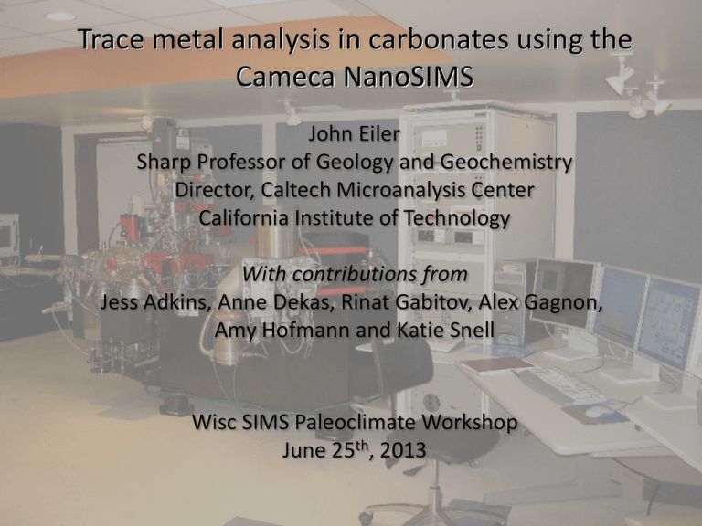
Trace metal analysis in carbonates using the Cameca NanoSIMS John Eiler Sharp Professor of Geology and Geochemistry Director, Caltech Microanalysis Center California Institute of Technology With contributions from Jess Adkins, Anne Dekas, Rinat Gabitov, Alex Gagnon, Amy Hofmann and Katie Snell Wisc SIMS Paleoclimate Workshop June 25th, 2013 Cation-exchange paleothermometry CaCO3 + Mgaq = MgCO3 + Caaq Keq ∞ Ca x[ ] [Mg ] Ca Mg Mitsuguchi et al., 1996 min fluid Global budgets Weathering Sediments Stanley and Hardie, 1998; model of Hardie, 1996 Hydrothermal Alteration Hitch #1: Vital effects Foraminifera Eggins et al., 2004; Deep-sea coral Gagnon et al., 2007 Hitch #2: Diagenesis PA: Primary Aragonite; SA: Secondary Aragonite; SC: Secondary Calcite Cements vs. ‘Primary’ Allison et al., 2007 The Cameca NanoSIMS What puts the ‘nano’ in nanoSIMS Geometry of focusing and extraction lenses • Short working distance promotes small, dense probe • Extraction optics easily contaminated or damaged Minimum spot size Si metal in Al matrix <<1 pA Cs+; ca. 30 nm resolution Illustration of the ‘84/16 %’ definition 1 µm • Nominally 50 nm for Cs+, 150 nm for O- (‘84/16 %’ definition) • Actual minimum ca. 20 nm for Cs+, ca. 100 nm for O• Brighter beams needed for trace element mapping typically in 100-300 nm range • Actually tricky to measure in many samples; assume it is ~500 nm unless proven otherwise Connection between beam current and resolution TiCN; Cs+ primary beam 2 pA; 93 nm resolution 1 µm TiCN; Cs+ primary beam <<1 pA; 22 nm resolution 1 µm • Images sharpen by minimizing beam current and carefully tuning primary beam • Count rates decrease and errors increase in proportion to beam current Even carefully tuned images ‘broaden’ features you can see clearly by SEM Sub-micron rutile inclusion in zircon Work of Amy Hoffman The analyzer and detector array of the nanoSIMS are also distinctive High-dispersion multi-collection Fixed collector Minimum mass spacing—1:58 Maximum mass spacing —22:1 • Conceived of as a tool for elemental mapping with exact spatial correlation of measured species • Also enables true multi-collection of almost any element/element ratio Transmission and mass-resolving power Relative sensitivity (%) 100 10 1 0 5000 10000 15000 20000 25000 30000 Mass resolving power Absolute sensitivity comparison Data from CIT nanoSIMS 50L; image from Frank Stadermann Design purpose: Composition mapping at µm scale Organic matter in 0.85 Ga Bitter Springs fm. Chert; Oehler et al., 2006 • Field of view: 200x200 µm in principle; 20x20 in practice. 10x10 or 5x5 is ideal • Discretization: 64x64 to 1024x1024; typically set so 1 pixel ~ beam radius Design purpose: Composition mapping at µm scale Science, 2006 • Fundamental data of interest is presence/absence of signal and spatial associations. Quantification is a secondary issue • Most imaging artifacts are of secondary importance and cannot be seen in scaled images Three dimensional ion imaging 3 % 15N 5 Interior Edge Work of Anne Dekas Common tuning conditions for ‘spot’ analyses result in horrendous imaging artifacts Zr/Si ratio of zircon 0.2 94Zr/28Si 0.1 10 20 Distance (µm) • Clearly unacceptable for even semi-quantitative ion imaging • Must be corrected by tuning the primary beam octapole (‘stigmator’) and various immersion lens electrodes Work of Amy Hofmann Even after careful tuning for ‘flatness’, edge effects are generally still present After tuning for ‘flatness’ 0.2 94Zr/28Si Before tuning for ‘flatness’ 0.1 10 20 Distance (µm) • Improves by pre-sputtering large area and keeping image ≤ 10 µm • No recognized solution, other than ‘gating’ or culling data • May be obscured by saturation of images • Community should insist on demonstrations that stoichiometric ratios yield roughly ‘flat’ images in domains of interest Work of Amy Hofmann The accuracy of ‘good’ images 40Ca ion intensity image of Oka carbonatite 42Ca/40Ca Cps 88Sr/40Ca Image ‘Spots’ 6.44E-3 6.52E-3 2.42E-4 2.78E-4 ~ several % artifacts in average element and isotope abundance ratios are common Work of Alex Gagnon The accuracy of ‘good’ images Integrals of 20x20 µm ion images of carbonate standards and samples Oka carbonatite 1.2 88Sr+ 42Ca+ 0.8 0.4 BCC carbonatite standard Coral 5 10 15 Sr/Ca (mmol/mol) • Images can provide quantitative data at ~% level accuracy with effort, but the community should insist that this accuracy is tested and demonstrated on a caseby-case basis Work of Alex Gagnon Where do spot measurements by nanoSIMS fit into our stable of tools for element/element ratio analysis? 1 ppm 100 ppm 1% Precision (1 s.e.) 10 % 1% 0.1 % 0.01 % 1nm 1µm Spatial resolution (m) 1mm 10-7 10-5 10-3 Concentration E-probe ATEM LA-ICPMS, conv. SIMS Bulk (e.g., solution ICP) M. Baker, pers. com; Cavosie et al., 2006; Klemme et al., 2008; Sobolev and Hofmann 2007; Hart and Cohen, 1997 10-1 ‘Spot’ analyses of element/element ratios in carbonates Internal errors Various nominally homogeneous calcite standards; O- beam; 1-2 µm spots Nano-SIMS 50L 0 -0.5 24_42 of ppm) 88 88_42 Sr/42Ca (1000’s of ppm) 138_42 138 Ba/42Ca (~1-10 ppm) Count. Stat. 24Mg/42Ca (100’s (1 error) Log log(2sigma/mean) 10 % -1 -1.5 1% -2 -2.5 1 22 Log Ni.Nj 33 44 55 log[NX*N42/(NX+N42)] X ~ Ni for trace species 6 7 1 m = Ni + N j (X0.5/X = external error for ratio [I]/[j]) Follows counting statistics down to ~3 ‰ 1.s.e. error, across a wide range in concentration Work of Rinat Gabitov There are limits imposed by drift in ratios during long sputtering Nano-SIMS (five single acquisitions) Oka Carbonatite; analyzed50L on the NanoSIMS with a 2 µm rastered spot of O- log(2sigma/mean) Internal error (1 0 -0.5 10 % -1 88Sr/ 42Ca 88/42 -1.5 Count.Stat. 1% -2 -2.5 1 22 33 Log Ni.Nj 44 55 66 7 7 ~ Log (Ni) for trace species Ni +log[NX*N42/(NX+N42)] Nj (X0.5/X = external error for ratio [I]/[j]) • Reflects gradual ‘drift’ in intensity and ratios after reaching nominal steady-state sputtering • Not sufficiently reproducible to correct completely by matching drift with standards • Appears to limit precision to no better than ~0.15 % 1 s.e., relative Work of Rinat Gabitov Point-to-point reproducibility log (rel. external stdev multiple spots) Point-to-point reproducibility (1) 0 -0.5 10 %-1 24Mg/42Ca 24/42 88Sr/42Ca 88/42 138/4242 138 -1.5 Ba/ Ca 1 %-2 -2.5 -2.5 -2 -1.5 -1 -0.5 1 % 10 % log (rel. mean internal er multiple spots) 0 Average internal error (1 s.e.) • Tracks counting statistics errors down to ~0.3-1.0 %, 1 s.e. • Likely only applies to central half of 1” rounds and central 3/4 of 1 cm rounds • Illustrated data didn’t require heroic efforts at polishing, but normal caution regarding topography effects is appropriate Slopes of calibration curves vary session-to-session much more than for other SIMS instruments • This could reflect fractionations associated with changing the acceptance angle when the immersion lens stack is tuned • Session-to-session reproducibility for secondary standards (i.e., assuming given value for a primary standard) follows internal errors down to ~0.8 % 1 s.e.. Many of the carbonate standards available for trace element measurements by SIMS are crap ID-ICPMS data for sub-samples AG-1, NBS-19, 135-CC, HUJ-AR & UCI-CC also examined • Oka is poor overall, but its calcite matrix has the best long-term reproducibility (±0.8 %) • BlueCC is a close second, and lacks ‘nuggets’ of exotic carbonates • All of the 7 other commonly used standards we explored are much worse • 1 %-level accuracy requires independent analysis of the same crystal Where do spot measurements by nanoSIMS fit into our stable of tools for element/element ratio analysis? 1nm 1µm 1mm 1 ppm 100 ppm 1% Precision (1 s.e.) 10 % 1% NanoSIMS 0.1 % 0.01 % 10-9 10-8 10-7 10-6 10-5 10-4 10-3 10-7 10-5 10-3 Concentration Spatial resolution (m) E-probe ATEM LA-ICPMS, conv. SIMS nanoSIMS Bulk (e.g., solution ICP) 10-1 Semi-quantitative imaging of growth banding in biogenic carbonates Foraminifera Kunioka, 2006 Surface coral Mg/Ca Meibom et al., 2004 Intergrown CaCO3 and Ca0.55Mg0.45CO3 in a sea urchin’s tooth! 5x5 µm ion image Ma et al., 2009 Extended example of applied use as a quantitative tool Experimental studies of vital effects 1. 2. 43Ca • Calcein marks start of growth during experiment; later layers should carry ‘spikes’ • The sub-micron resolution of the NanoSIMS reduces culture time from several months to a few days Gagnon et al. 2013 Ion images of overgrowths Calcein stain 43Ca/42Ca Demonstrations of image ‘flatness’ Gagnon et al., 2012 A conceptual model of coral biomineralization (after McConnaughey) (extracellular calcifying fluid) Well-resolved gradients should measure residence time in ECF ‘mother liquor’ Budget for a metal in ECF [Ca]SW Flow out [Ca] Flow in P Precipitation External solution Spike Precipitated carbonate Instantaneous 2 hrs Natural Time Gagnon et al., 2012 Growth Axis Measured growth rates are generally faster than can be resolved by beam width Model of a sharp boundary, given beam ‘broadening’ Calcium turnover time (1/2) is less than 2 hrs, possibly much faster Fastest growing measured foram 1/2 = 1.2 hrs Comparison of profiles should provide evidence for mechanisms of uptake Mg2+ Ca2+ Growth Axis Growth Axis Delivery of metals to site of growth appears to be dominated by transport of seawater to growing crystal surface Uptake of 25Mg and 43Ca spikes specific pumping Growth Axis seawater transport Growth Axis This conclusion is corroborated by uptake of spikes that are unlikely to be biologically ‘pumped’ across membranes Gagnon et al., 2012 43Ca spike 159Tb spike Extracting quantitative partition coefficients from ion images of ~µm overgrowths Corals grown over a range of carbonate-ion concentrations Gagnon et al., 2013 Related work has demonstrated that growth zonation is controlled by variable extents of Rayleigh distillation on carbonate growth from ECF Flow in ECF Flow out Keqarag-fluid Precipitation Septa of deep-sea corals; Gagnon et al., 2007 Surface corals; Gaetani et al., 2011 This advance creates the possibility that models of vital effects will be quantitative tools of paleoclimate reconstructions Flow in Flow out ECF Keqarag-fluid Precipitation Fit to Rayleigh distillation model Gaetani et al., 2011 Thermometry using implied Kd controlling the distillation fractionation Using the NanoSIMS to unravel the thorny problem of diagenetic modifications Fossil 34-91; a Paleocene mollusk from the Bighorn Basin Modified aragonite growth plates Mix of secondary calcite and entombed growth plates Work of Katie Snell Using the NanoSIMS to unravel the thorny problem of diagenetic modifications Fossil 34-91; a Paleocene mollusk from the Bighorn Basin 55Mn16O/40Ca16O Work of Katie Snell Summary and parting thoughts • NanoSIMS may be uniquely suited to quantitative trace element analysis of carbonates with µm to sub-µm zonation (synchrotron µ-XRF may be comparable) • Imaging can yield several-per cent errors at scales down to 300 nm; much better is likely unrealistic • 0.8-1 % (1 s.e.) long-term external precision for ~1 µm domains are demonstrated and half that seems possible • Real limitation at present is poor quality of interlaboratory standards • Relatively casual standardization could easily result in ~10 % errors • There is no established community-wide practice for achieving and documenting precision and accuracy; for the time being, this needs to be approached as an experimental tool, and data in the literature should be approached with a ‘show me!’ attitude.
