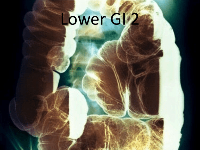
Lower GI 2
Barium Enema
• The radiographic study of the large intestine is commonly
termed a barium enema. It requires the use of contrast media
to demonstrate the large intestine and its components.
Alternative names include BE (BaE) and lower GI series.
• The purpose of the barium enema is to radiographically study
the form and function of the large intestine to detect any
abnormal conditions. Both the single-contrast and the
double-contrast barium enema involve study of the entire
large intestine
Contraindications
• The two strict contraindications for the barium enema are
similar to those described for the small bowel series. These
have been described as a possible perforated hollow viscus
and a possible large bowel obstruction. These patients
should not be given barium as a contrast media agent.
Although not as radiopaque as barium sulfate, watersoluble contrast media can be used for these conditions.
• Careful review of the patient's chart and clinical history
may help to prevent problems during the procedure. The
radiologist should be informed of any conditions or disease
processes noted in the patient's chart. This information
may dictate the type of study that will be performed
Also important is to review the patient's chart to determine
whether the patient had a sigmoidoscopy or a colonoscopy
before undergoing the barium enema. If a biopsy of the colon
was performed during these procedures, the involved section of
the colon wall may be weakened, which may lead to perforation
during the barium enema. The radiologist must be informed of
this situation before beginning the procedure.
PATHOLOGIC INDICATIONS (BARIUM ENEMA)
• Colitis is an inflammatory condition of the large intestine that
may be caused by many factors, including bacterial infection,
diet, stress, and other environmental conditions. The intestinal
mucosa can appear rigid and thick, and haustral markings may
be missing along the involved segment. Because of chronic
inflammation and spasm, the intestinal wall has a “saw-tooth” or
jagged appearance.
• Ulcerative colitis describes a severe form of colitis that is most
common among young adults. It is a chronic condition that often
leads to development of coinlike ulcers within the mucosal wall.
Along with Crohn's disease, it is one of the most common forms
of inflammatory bowel disease. These ulcers may be seen during
the barium enema as multiple ring-shaped filling defects that
create a “cobblestone” appearance along the mucosa. Patients
with long-term bouts of ulcerative colitis may develop
“stovepipe” colon, in which haustral markings and flexures are
most often absent.
Diverticulum
• A diverticulum (di′-ver-tik′-u-lum) is an outpouching of
the mucosal wall that may result from herniation of
the inner wall of the colon. Although this is a relatively
benign condition, it may become widespread
throughout the colon but is most prevalent in the
sigmoid colon. It is most common among adults over
40 years of age. The condition of having numerous
diverticula is termed diverticulosis. If these diverticula
become infected, the condition then is referred to as
diverticulitis. Inflamed diverticula may become a
source of bleeding, in which case surgical removal may
be necessary. A patient may develop peritonitis if a
diverticulum perforates the mucosal wall.
Intussusception
• Intussusception is a telescoping or invagination of one
part of the intestine into another. It is most common in
infants younger than 2 years of age but can occur in
adults. A barium enema or an air/gas enema may play
a therapeutic role in reexpanding the involved bowel.
Radiographically, the barium column terminates into a
“mushroom-shaped” dilation with very little
barium/gas passing beyond it. This dilation marks the
point of obstruction. Intussusception must be resolved
quickly so it does not lead to obstruction and necrosis
of the bowel. If the condition recurs, surgery may be
needed.
Neoplasm
• Neoplasms are common in the large intestine.
Although benign tumors do occur, carcinoma of the
large intestine is a leading cause of death among both
men and women. Most carcinomas of the large
intestine occur in the rectum and sigmoid colon. These
cancerous tumors often encircle the lumen of the
colon, producing an irregular channel through it. The
radiographic appearance of these tumors as
demonstrated during a barium enema has led to the
use of descriptive terms such as “apple-core” or
“napkin-ring” lesions. Both benign and malignant
tumors may begin as polyps.
Polyps
• Polyps are saclike projections similar to
diverticula except that they project inward into
the lumen rather than outward, as do diverticula.
Similar to diverticula, polyps can become
inflamed and may be a source of bleeding, in
which case they may have to be surgically
removed. Barium enema, endoscopy, and
computed tomography colonography (CTC) are
the most effective modalities used to
demonstrate neoplasms in the large intestine.
Volvulus
• Volvulus is a twisting of a portion of the intestine on
its own mesentery, leading to a mechanical type of
obstruction. Blood supply to the twisted portion is
compromised, leading to obstruction and necrosis, or
localized death of tissue. A volvulus may be found in
portions of the jejunum or ileum or in the cecum and
sigmoid colon. Volvulus is more likely to occur in men
than in women and is most common between the ages
of 20 and 50 years. The classic sign is called a “beak”
sign, a tapered narrowing at the volvulus site as
demonstrated during a barium enema. A volvulus will
produce an air-fluid level, as is well demonstrated on
an erect abdomen projection.
CONDITION OR
DISEASE
MOST COMMON RADIOGRAPHIC
EXAM
POSSIBLE RADIOGRAPHIC APPEARANCE
ADJ
Colitis
Single- and double-contrast
(preferred) BE
Thickening of mucosal wall with loss of haustral
markings
None
Ulcerative colitis
Single- and double-contrast
(preferred) BE
“Cobblestone” and possible “stovepipe” appearance
with severe forms
None
Diverticula
(diverticulosis/diverti Double-contrast BE recommended
culitis)
Barium-filled circular defects projecting outward from
colon wall; jagged or “sawtooth” appearance of the
None
mucosa
Intussusception
Single or air/gas contrast enema
recommended
“Mushroom-shaped” dilation at the distal aspect of
the intussusception, with very little barium or gas
passing beyond it
None
Neoplasm
Double-contrast BE recommended
to detect small polyps; computed
tomography; colonography
Filling defects; narrowness or tapering of lumen;
“apple-core” or “napkin-ring” lesions
None
Polyps
Double-contrast BE recommended;
Barium-filled, saclike projections projecting inward
computed tomography;
into the lumen of the bowel
colonography
Volvulus
Single-contrast BE
Tapered or “corkscrew” appearance, with air-filled
distended region of the intestine
None
PATIENT PREPARATION
Preparation of the patient for a barium enema is more
involved than is preparation for the stomach and small bowel.
The final objective, however, is the same. The section of
alimentary canal to be examined must be empty. Thorough
cleansing of the entire large bowel is of paramount
importance for a satisfactory contrast media study of the large
intestine.
CONTRAINDICATIONS TO LAXATIVES (CATHARTICS)
Certain conditions contraindicate the use of very effective
cathartics or purgatives needed to thoroughly cleanse the
large bowel. These exceptions include (1) gross bleeding, (2)
severe diarrhea, (3) obstruction, and (4) inflammatory
conditions such as appendicitis.
A laxative is a substance that produces frequent soft or liquid
bowel movements. These substances increase peristalsis in the
large bowel and occasionally in the small bowel as well by
irritating sensory nerve endings in the intestinal mucosa. This
increased peristalsis dramatically accelerates the passage of
intestinal contents through the digestive system
Barium Enema Containers
A closed-system enema container is used to administer barium
sulfate or an air and barium sulfate combination during the
barium enema. This closed-type disposable barium enema bag
system has replaced the older open-type system for
convenience and for reducing the risk of cross-infection.
This system, which is shown in the photograph, includes the
disposable enema bag with a pre-measured amount of barium
sulfate. Once mixed, the suspension travels down its own
connective tubing, and flow is controlled by a plastic stopcock.
An enema tip is placed on the end of the tubing and is inserted
into the patient's rectum.
After the examination has been completed, much of the barium
can be drained back into the bag by lowering the system to
below tabletop level. The entire bag and tubing are disposed of
after a single use.
LATEX ALLERGIES
Today, most products are primarily latex-free, but
determination of whether the patient is sensitive to natural
latex products is still important. Patients with sensitivity
toward latex experience anaphylactoid-type reactions that
include sneezing, redness, rash, difficulty in breathing, and
even death.
If the patient has a history of latex sensitivity, the
technologist must ensure that the enema tip, tubing, and
gloves are latex-free. Even the dust produced by removal of
gloves may introduce into the air latex protein, which may
be inhaled by the patient.
CONTRAST MEDIA
• Barium sulfate is the most common type of positivecontrast medium used for the barium enema. The
concentration of the barium sulfate suspension
varies according to the study performed. A standard
mixture used for single–contrast media barium
enemas ranges between 15% and 25% weight-tovolume (w/v). The thicker barium used for doublecontrast barium enemas has a weight-to-volume
concentration between 75% and 95% or higher.
Negative-Contrast Agent
• The double-contrast media uses a number of negativecontrast agents, in addition to barium sulfate. Room air,
nitrogen, and carbon dioxide are the most common forms of
negative-contrast media used. Carbon dioxide is gaining wide
use because it is well tolerated by the large intestine and is
absorbed rapidly after the procedure. Carbon dioxide and
nitrogen gas are stored in a small tank and can be introduced
into the rectum through an air-contrast retention enema tip.
• An iodinated, water-soluble contrast media may be used in
the case of a perforated or lacerated intestinal wall, or when
the patient is scheduled for surgery after the barium enema.
Remember that a medium-range kV (80 to 90) should be used
with a water-soluble, negative-contrast agent.
Contrast Media Preparation
• The mixing instructions as supplied by the manufacturer
should be followed precisely.
• A debate has evolved over the temperature of the water
used to prepare the barium sulfate suspension. Some
experts recommend the use of cold water (40°F to 45°F) in
the preparation of contrast media. Cold water is reported
to have an anesthetic effect on the colon and to increase
the retention of contrast media. Critics have stated that the
use of cold water may lead to colonic spasm.
• Room temperature water (85°F to 90°F) is recommended
by most experts for completion of a more successful
examination with maximal patient comfort.
• The technologist should NEVER use hot water to prepare
contrast media. Hot water may scald the mucosal lining of
the colon.
• Because barium sulfate produces a colloidal suspension,
shaking the enema bag before tip insertion is important for
preventing separation of barium sulfate from water.
Spasm during the barium enema is a common side
effect. Patient anxiety, overexpansion of the intestinal
wall, discomfort, and related disease processes all may
lead to colonic spasm. To minimize the possibility of
spasm, a topical anesthetic such as lidocaine may be
added to the contrast media. If spasm does occur during
the study, glucagon can be given intravenously and
should be kept in the department for these situations.
• After the fluoroscopic room and the contrast media
have been completely prepared, the patient is escorted
to the examination room. Before insertion of the
enema tip, a pertinent history should be taken and the
examination carefully explained. Because complete
cooperation is essential and this examination can be
somewhat embarrassing, every effort should be made
to reassure the patient at every stage of the exam.
• Previous radiographs should be made available to the
radiologist. The patient is placed in Sims' position
before the enema tip is inserted.
ENEMA TIP INSERTION
• Before the enema tip is inserted, the opening in the back of
the patient's gown should be adjusted to expose only the
anal region. The rest of the patient should be well covered
when the rectal tube is inserted. The patient's modesty
should be protected in any way possible during the barium
enema examination. The right buttock should be raised to
open the gluteal fold and expose the anus. The patient
should take in a few deep breaths before actual insertion of
the enema tip. If the tip will not enter with gentle pressure,
the patient should be asked to relax and assist if possible.
The tip should NEVER be forced in a manner that could
cause injury to the patient. Because the abdominal muscles
relax on expiration, the tip should be inserted during the
exhalation phase of respiration.
• The rectum and anal canal present a double curvature;
therefore, the tube is inserted first in a forward direction
approximately 1 to 1½ inches (2.5 to 4 cm). This initial
insertion should be aimed toward the umbilicus. After the
initial insertion, the rectal tube is directed superiorly and
slightly anteriorly to follow the normal curvature of the
rectum. The total insertion of the tip should not exceed 3 to 4
cm to prevent possible injury to the wall of the rectum. The
rectal tube may be taped in place or held to prevent it from
slipping out while the patient turns back into a supine
position for the start of fluoroscopy. This position is usually
supine but may be prone, depending on the preference of the
radiologist.
• If the retention-type tip is necessary, most departments allow
the technologist to instill one or two puffs of air into the
balloon end to help hold it in place. However, the bulb should
be filled to its maximum only under fluoroscopic control as
the fluoroscopy procedure begins. As the procedure begins,
the IV pole supporting the enema bag should be no higher
than 24 inches (60 cm) above the radiographic table.
SUMMARY OF ENEMA TIP INSERTION PROCEDURE
• Step 1 Describe the tip insertion procedure to the patient. Answer
any questions.
• Step 2 Place the patient in Sims' position. Patient should lie on the
left side, with the right leg flexed at the knee and hip.
• Step 3 Shake enema bag once more to ensure proper mixing of
barium-sulfate suspension. Allow barium to flow through the tubing
and from the tip to remove any air in the system.
• Step 4 Wearing gloves, coat enema tip well with water-soluble
lubricant. Wrap proximal aspect of enema tip in paper towel.
• Step 5 On expiration, direct enema tip toward the umbilicus
approximately 1 to 1½ inches (2.5 to 4 cm).
• Step 6 After initial insertion, advance up superiorly and slightly
anteriorly. The total insertion should not exceed 3 to 4 cm. Do NOT
force enema tip.
• Step 7 Tape tubing in place to prevent slippage. Do NOT inflate
retention tip unless directed by radiologist.
• Step 8 Ensure that IV pole/enema bag is no more than 24 inches (60
cm) above the table. Ensure that tubing stopcock is in the closed
position, and that no barium flows into the patient.
SINGLE-CONTRAST BARIUM ENEMA PROCEDURE
• The single-contrast barium enema is a procedure in which
only a positive-contrast media is used. In most cases, the
contrast material is barium sulfate in a thin mixture.
Occasionally, the contrast media must be a water-soluble
contrast material. For example, if the patient is to be taken to
surgery after undergoing the single-contrast enema
procedure, then a water-soluble contrast media must be
used.
DOUBLE-CONTRAST BARIUM ENEMA
PROCEDURE
• A second common type of barium enema procedure is the
double-contrast type. Double-contrast studies are more
effective in demonstrating polyps and diverticula than is the
single-contrast barium enema procedure. Radiographic and
fluoroscopic procedures for a double-contrast barium enema
are somewhat different in that both air and barium must be
introduced into the large bowel. An absolutely clean large
bowel is essential to the double-contrast study, and a much
thicker barium mixture is required. Although exact ratios
depend on the commercial preparations used, the ratio
approaches a one-to-one mix, so that the final product is like
heavy cream.
SUMMARY OF SAFETY CONCERNS
•
•
•
•
•
Review Patient's Chart: Note any pertinent clinical history on the exam requisition,
and inform the radiologist about whether the patient underwent a sigmoidoscopy
or colonoscopy before the barium enema was given, especially if a biopsy was
performed. Determine whether the patient has any known allergies to the contrast
media or the natural latex products. Diabetic patients shall not be given glucagon
prior to or during procedure unless ordered by physician.
Never Force Enema Tip Into Rectum: This action may lead to a perforated rectum.
The radiologist inserts the enema tip under fluoroscopic guidance, if needed.
Height of Enema Bag Does Not Exceed 24 Inches (60 cm) Above Table: This
distance should be maintained before the procedure is begun. The radiologist may
wish to raise bag height during the procedure based on rate of flow of the contrast
media.
Verify Water Temperature of Contrast Media: Water that is too hot or too cold
may injure the patient or compromise the procedure.
Escort Patient to the Restroom After Completion of the Study: A barium enema
can be stressful for some patients. Patients have been known to faint during or
after evacuation.
PA PROJECTION: SMALL BOWEL SERIES
Pathology Demonstrated
• Inflammatory processes, neoplasms, and obstructions of the
small intestine are shown
PA, 15 or 30 minutes—centered
approximately 2 inches (5 cm) above
iliac crest.
PA, hourly—centered to iliac crest
Central Ray
• CR is perpendicular to IR. (1) 15 or 30 min: Center to about 2 inches (5 cm)
above iliac crest. (2) Hourly: Center CR and midpoint of IR to iliac crest.
• Center IR to CR.
• Minimum SID is 40 inches (100 cm).
Respiration
Suspend respiration and expose on expiration.
Note: Timing begins with ingestion of barium. Timed intervals of radiographs
depend on transit time of the specific barium preparation used and on
department protocol. For the first 30-minute radiograph, center high to
include the entire stomach.
Subsequent 30-minute interval radiographs are taken until barium reaches
the large bowel (usually 2 hours). The study generally is completed once the
contrast media reaches the cecum and/or the ascending colon.
Fluoroscopy and spot imaging of the ileocecal valve and terminal ileum
after barium reaches this area are commonly included in the small bowel
series routine. This procedure, however, is determined by the radiologist's
preference and by department routines.
PA SBS—30 minutes (most barium located in stomach and jejunum). Note: Large
(12″) ascariasis (parasitic roundworm) in jejunum
PA SBS—1 hour (most barium located in jejunum).
PA, 2 hour (most barium located in ileum and proximal colon).
Structures Shown:
Entire small intestine is demonstrated on each radiograph, with the stomach
included on the first 15- and/or 30-minute radiograph.
Position:
No rotation is present. The ala of the ilium and the lumbar vertebrae are
symmetric.
Collimation and CR:
Only minimal collimation margins are seen on all four sides for adults. • CR
is centered approximately 2 inches (5 cm) above the iliac crest for the initial
radiographs. • CR is centered at iliac crest for remaining radiographs.
Exposure Criteria and Markers:
Appropriate technique is employed to visualize the contrast-filled small
intestine without overexposing those parts that are filled only partially with
barium. • Sharp structural margins indicate no motion. • Patient ID
information, time interval markers, and R or L marker are visible without
superimposition of essential anatomy.
Barium Enema
• PA and/or AP
• RAO
• LAO
• LPO and RPO
• Lateral rectum and ventral
decubitus
• R lateral decubitus
• L lateral decubitus
• PA (AP) postevacuation
• AP or LPO axial (butterfly)
• PA or RAO axial (butterfly)
PA AND/OR AP PROJECTION: BARIUM ENEMA
Pathology Demonstrated
Obstructions, including ileus, volvulus, and intussusception, are demonstrated. Doublecontrast media barium enema is ideal for demonstrating diverticulosis, polyps, and
mucosal changes.
Radiographic Criteria
Structures Shown: • The transverse colon should be primarily barium-filled on the PA and
air-filled on the AP with a double-contrast study. • Entire large intestine, including the left
colic flexure, should be visible.
RAO POSITION: BARIUM ENEMA
Radiographic Criteria
Structures Shown: • The right colic flexure and the ascending and sigmoid colon are seen
“open” without significant superimposition. • The entire large intestine is included, with the
possible exception of the left colic flexure, which is best demonstrated in LAO position (or may
require a second image centered higher). • The rectal ampulla should be included on the lower
margin of the radiograph.
LAO POSITION: BARIUM ENEMA
Radiographic Criteria
Structures Shown: • The left colic flexure should be seen as open without significant
superimposition. • The descending colon should be well demonstrated. • The entire large
intestine should be included
LPO AND RPO POSITIONS: BARIUM ENEMA
Radiographic Criteria
Structures Shown: • LPO—The right colic (hepatic) flexure and the ascending and
rectosigmoid portions should appear “open” without significant superimposition. • RPO—The
left colic (splenic) flexure and the descending portions should appear “open” without
significant superimposition. (A second IR centered lower to include the rectal area is required
on most adult patients if this area is to be included on these postfluoroscopy overheads.) • The
rectal ampulla should be included on the lower margins of the radiograph. • Entire contrastfilled large intestine, including the rectal ampulla, should be included
LEFT LATERAL: BARIUM ENEMA
Radiographic Criteria
Structures Shown: • Contrast-filled rectosigmoid
region is demonstrated.
RIGHT LATERAL DECUBITUS POSITION (AP OR PA): BARIUM ENEMA–DOUBLE
CONTRAST
R LATERAL DECUB : BARIUM ENEMA
Radiographic Criteria
Structures Shown: • Entire large intestine is demonstrated to include air-filled left
colic flexure and descending colon.
L LATERAL DECUB : BARIUM ENEMA
Structures Shown:
• Entire large intestine is demonstrated, with air-filled right colic flexure, ascending
colon, and cecum.
PA (AP) PROJECTION—POSTEVACUATION: BARIUM ENEMA
Radiographic Criteria
Structures Shown: • Entire large intestine should be visualized with only a residual
amount of contrast media
AP AXIAL OR AP AXIAL OBLIQUE (LPO) PROJECTIONS: BARIUM ENEMA
“Butterfly” Positions
Central Ray
• Angle CR 30° to 40° cephalad.
Direct CR 2 inches (5 cm) inferior to level
of ASIS and to MSP.
Radiographic Criteria
Structures Shown: • Elongated views of the rectosigmoid segments should be
visible with less overlapping of sigmoid loops than with a 90° AP projection.
PA AXIAL OR PA AXIAL OBLIQUE (RAO) PROJECTIONS: BARIUM ENEMA
“Butterfly” Positions
Central Ray
Angle CR 30° to 40° caudad.
•PA: • Align CR to exit at level of ASIS and MSP.

