British Heart Valve Society Carcinoid Heart Disease
advertisement
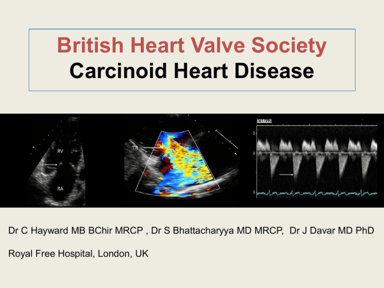
British Heart Valve Society Carcinoid Heart Disease Dr C Hayward MB BChir MRCP , Dr S Bhattacharyya MD MRCP, Dr J Davar MD PhD Royal Free Hospital, London, UK Case Presentation Clinical History • 60 year old female. • 6 month history of flushing, diarrhoea, fatigue and dyspnoea on exertion. NYHA Class III at presentation. Investigations • CT abdomen: multiple liver metastases and a small bowel mesenteric mass. Liver Biopsy: consistent with low grade carcinoid tumour. • 24 hour Urinary 5-HIAA: 800µmol/24 hours. Cardiac Investigations • ECG – sinus tachycardia. Normal axis. • CXR – Cardiothoracic ratio > 50%. • Echocardiogram: – Right Ventricle: dilated and mildly impaired (TAPSE 13cm). – Tricuspid Valve: severe “free flowing” tricuspid regurgitation. – Pulmonary Valve: severe pulmonary regurgitation, moderate pulmonary stenosis. – NT-proBNP: 700 pg/ml. Management Medical •Reduction of peripheral oedema with diuretics. Valve Surgery •Replacement of tricuspid and pulmonary valve: Pulmonary homograft. Pericardial tissue valve – tricuspid valve. Length of hospital stay 5 days. Required permanent pacemaker for complete heart block. Outcome 6 months post surgery •Diuretics weaned off. •Functional NYHA Class I. Climb > 5 flights of stairs. Clinical Manifestations • Carcinoid syndrome consists of a triad: flushing, diarrhoea and bronchospasm. • Between 20-50% of all patients with carcinoid syndrome will develop carcinoid heart disease. • Vasoactive substances such as 5-hydroxytryptamine produced by neoplastic cells are able to travel to the right heart via the hepatic vein/IVC and are thought to be responsible for deposition of endocardial plaques of fibrous tissue. • Classically patients develop signs and symptoms of right heart failure: fatigue, oedema and ascites. Pathology – “Carcinoid Plaque” • Right-sided lesions more common than left. • Preferential right-sided involvement due to inactivation of vasoactive substances by lungs. • 5–10% have left-sided valvular pathology due to either high tumour load, bronchial carcinoid or patent foramen ovale. • Plaque - composed of smooth muscle cells + myofibroblasts forming white fibrous layer (arrow) lining endocardial surface of cardiac valves superficial to normal valve Echocardiographic Features – Tricuspid Valve • Typically thickened, retracted, valve leaflets. Leaflets do not co-apt (arrow). • Anatomical features leads to predominantly tricuspid regurgitation (TR). • Classical “Dagger” shaped Doppler profile of severe TR (arrow). Echocardiographic Features – Pulmonary Valve • Fixed, thickened cusps (arrow). • Non-coaptation of cusps (*). • Predominantly pulmonary stenosis with varying degrees of regurgitation (arrow). Biochemical Markers • Elevated urinary 5-hydroxyindolacetic acid is a highly sensitive but poorly specific maker of carcinoid heart disease. • NT-proBNP > 260pg/ml has greater than 90% sensitivity and negative predictive value for significant carcinoid heart disease. This may allow its use as a screening test. • NT-proBNP also correlated with disease severity and NYHA Class. Management Medical Management •Poor outcome when managed medically. •3 year survival 68% without cardiac involvement compared to 31% with cardiac involvement. •Diuretics mainstay of therapy. Valve Surgery •High peri-operative risk (10% 20% depending on institution). •Valve replacement improves symptom status (functional NYHA Class). •Emerging data suggest may improve prognosis. Conclusions • Carcinoid heart disease = common complication of carcinoid syndrome but is a rare cause of all acquired valvular heart disease • 5-HT is produced by metastatic tumour cells in the liver → deposition of endocardial plaques. • Right sided valvular dysfunction is common and presents with characteristic echocardiographic appearances. Left sided valve lesions in 5-10% of cases of carcinoid heart disease. • Medical management alone is associated with poor survival. • Valve surgery improves symptoms and may improve prognosis. Further Reading 1. 2. 3. 4. 5. 6. Bhattacharyya S, Davar J, Dreyfus G, Caplin ME. Carcinoid Heart Disease. Circulation 2007; 116:2860-2865. Lundin L, Norheim I, Landelius J, Oberg K, Theodorsson-Norheim E. Relationship of circulating vasoactive substances to ultrasound detectable cardiac abnormalities. Circulation 1988;77:264-269. Bhattacharyya S, Toumpanakis D, Burke M, Taylor AM, Caplin ME, Davar J. Features of carcinoid heart disease identified by 2- and 3-dimensional echocardiography and cardiac MRI. Circ Cardiovasc Imaging 2010:3:103111. Korse CM, Taal BG, de Groot CA, Bakker RH, Bonfrer JM. ChromograninA and N-terminal pro-brain natriuretic peptide: an excellent pair of biomarkers for diagnostics in patients with neuroendocrine tumor. J Clin Oncol. 2009;27:4293-4299. Bhattacharyya S, Toumpanakis C, Caplin M, Davar J. Usefulness of NTerminal Brain Natriuretic Peptide As A Biomarker Of The Presence Of Carcinoid Heart Disease. American Journal of Cardiology 2008;102:938942. Moller JE, Pellikka PA, Bernheim AM, Schaff HV, Rubin J, Connolly HM. Prognosis of carcinoid heart disease: An analysis of 200 cases over two decades. Circulation 2005;112:3320-3327.


