James L. Angtuaco, MD, DPPS, DPSPC, FPCC
advertisement
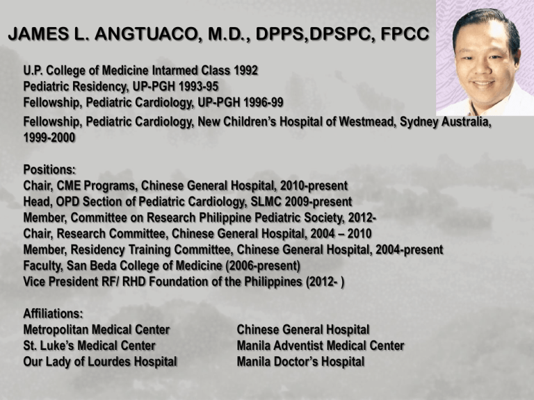
JAMES L. ANGTUACO, M.D., DPPS,DPSPC, FPCC U.P. College of Medicine Intarmed Class 1992 Pediatric Residency, UP-PGH 1993-95 Fellowship, Pediatric Cardiology, UP-PGH 1996-99 Fellowship, Pediatric Cardiology, New Children’s Hospital of Westmead, Sydney Australia, 1999-2000 Positions: Chair, CME Programs, Chinese General Hospital, 2010-present Head, OPD Section of Pediatric Cardiology, SLMC 2009-present Member, Committee on Research Philippine Pediatric Society, 2012Chair, Research Committee, Chinese General Hospital, 2004 – 2010 Member, Residency Training Committee, Chinese General Hospital, 2004-present Faculty, San Beda College of Medicine (2006-present) Vice President RF/ RHD Foundation of the Philippines (2012- ) Affiliations: Metropolitan Medical Center St. Luke’s Medical Center Our Lady of Lourdes Hospital Chinese General Hospital Manila Adventist Medical Center Manila Doctor’s Hospital Pulmonary Hypertension: Guidelines in the Diagnosis and Treatment James L. Angtuaco, M.D., DPPS, DPSPC, FPCC Pediatric Cardiologist DISCLAIMER OBJECTIVES: 1. To discuss the definition of Pulmonary arterial hypertension 2. To discuss the different pathology / pathobiology of Pulmonary arterial hypertension 3. To describe the classifications of Pulmonary arterial hypertension 4. To discuss the clinical presentations of Pulmonary arterial hypertension 5. To discuss the different diagnostic modalities for Pulmonary arterial hypertension 6. To discuss the different treatment modalities for Pulmonary arterial hypertension Pulmonary Hypertension • Definition: – increase in mean pulmonary arterial pressure > 25 mmHg at rest as assessed by right heart catheterization – normal mean pulmonary arterial pressure is 14+3 mmHg. with an upper limit of ~20 mmHg. – gray zone : 21-24 mmHg. CLINICAL CLASSIFICATION OF PULMONARY ARTERIAL HYPERTENSION (The Task Force for the Diagnosis and Treatment of Pulmonary Hypertension of the European Society of Cardiology and the European Respiratory Society, endorsed by the International Society of Heart & Lung Transplantation 2009) European Heart Journal 2009 • GROUP 1: PULMONARY ARTERIAL HYPERTENSION – idiopathic – Heritable • BMPR2 (bone morphogenesis protein receptor 2 gene) • ALK1 (activin receptor like kinase type 1 gene), endoglin (with or without hereditary hemorrhagic telangiectasia) • Unknown – Drugs and toxins induced (weight loss drugs) CLINICAL CLASSIFICATION OF PULMONARY ARTERIAL HYPERTENSION (The Task Force for the Diagnosis and Treatment of Pulmonary Hypertension of the European Society of Cardiology and the European Respiratory Society, endorsed by the International Society of Heart & Lung Transplantation 2009) European Heart Journal 2009 • GROUP 1: PULMONARY ARTERIAL HYPERTENSION – associated with: • • • • • • connective tissue disease HIV infection portal hypertension Congenital Heart Disease Schistosomiasis Chronic hemolytic anemia – Persistent pulmonary hypertension of the newborn CLINICAL CLASSIFICATION OF PULMONARY ARTERIAL HYPERTENSION (The Task Force for the Diagnosis and Treatment of Pulmonary Hypertension of the European Society of Cardiology and the European Respiratory Society, endorsed by the International Society of Heart & Lung Transplantation 2009) European Heart Journal 2009 • GROUP 1’ : PULMONARY VENO-OCCLUSIVE DISEASE WITH PULMONARY CAPILLARY HEMANGIOMATOSIS CLINICAL CLASSIFICATION OF PULMONARY ARTERIAL HYPERTENSION (The Task Force for the Diagnosis and Treatment of Pulmonary Hypertension of the European Society of Cardiology and the European Respiratory Society, endorsed by the International Society of Heart & Lung Transplantation 2009) European Heart Journal 2009 • GROUP 2: PULMONARY HYPERTENSION DUE TO LEFT HEART DISEASE – systolic dysfunction – diastolic dysfunction – valvular disease CLINICAL CLASSIFICATION OF PULMONARY ARTERIAL HYPERTENSION (The Task Force for the Diagnosis and Treatment of Pulmonary Hypertension of the European Society of Cardiology and the European Respiratory Society, endorsed by the International Society of Heart & Lung Transplantation 2009) European Heart Journal 2009 • GROUP 3: PULMONARY HYPERTENSION DUE TO LUNG DISEASE AND/ OR HYPOXIA – chronic obstructive pulmonary disease – interstitial lung disease – other pulmonary diseases with mixed restrictive and obstructive pattern – sleep-disordered breathing – alveolar hypoventilation disorders – chronic exposure to high altitude – developmental abnormalities CLINICAL CLASSIFICATION OF PULMONARY ARTERIAL HYPERTENSION (The Task Force for the Diagnosis and Treatment of Pulmonary Hypertension of the European Society of Cardiology and the European Respiratory Society, endorsed by the International Society of Heart & Lung Transplantation 2009) European Heart Journal 2009 • GROUP 4: CHRONIC THROMBOEMBOLIC PULMONARY HYPERTENSION (CTEPH) CLINICAL CLASSIFICATION OF PULMONARY ARTERIAL HYPERTENSION (The Task Force for the Diagnosis and Treatment of Pulmonary Hypertension of the European Society of Cardiology and the European Respiratory Society, endorsed by the International Society of Heart & Lung Transplantation 2009) European Heart Journal 2009 • GROUP 5 : PULMONARY HYPERTENSION WITH UNCLEAR AND/OR MULTIFACTORIAL MECHANISMS – hematologic disorders : myeloproliferative disorders, splenectomy – systemic disorders : sarcoidosis, pulmonary Langerhans cell histiocytosis, lymphangioleiomyomatosis, neurofibromatosis, vasculitis – metabolic disorders: glycogen storage disease: Gaucher disease, thyroid disorders – others: tumoural obstruction, fibrosing mediastinitis, chronic renal failure on dialysis PATHOLOGY OF PULMONARY HYPERTENSION • Group 1: PAH – affects the distal pulmonary arteries (<500 um in diameter) – medial hypertrophy – intimal proliferation and fibrotic changes (concentric and eccentric) – adventitial thickening with moderate perivascular inflammatory infiltrates – plexiform dilated lesions – thrombotic lesions – PULMONARY VEINS ARE CLASSICALLY UNAFFECTED Pietra GG et. al. JACC 2004 Tuder RM et. al. JACC 2009 PATHOLOGY OF PULMONARY HYPERTENSION • Group 1’: Pulmonary Veno-Occlusive Disease – – – – – – – – involves septal veins and pre-septal venules occlusive fibrotic lesions venous muscularization frequent capillary proliferation (patchy) pulmonary edema occult alveolar hemorrhage lymphatic dilatation / lymph node enlargement distal pulmonary arteries have medial hypertrophy, intimal fibrosis Pietra GG et. al. JACC 2004 Tuder RM et. al. JACC 2009 PATHOLOGY OF PULMONARY HYPERTENSION • Group 2: Left Heart Disease – enlarged, thickened pulmonary veins – pulmonary capillary dilatation – interstitial edema – alveolar hemorrhage – lymphatic vessel and lymph node enlargement – distal pulmonary artery may have medial hypertrophy and intimal fibrosis Pietra GG et. al. JACC 2004 Tuder RM et. al. JACC 2009 PATHOLOGY OF PULMONARY HYPERTENSION • Group 3: PAH due to lung disease – medial hypertrophy and intimal obstructive proliferation of the distal pulmonary arteries – variable degree of destruction of the vascular bed in emphysematous or fibrotic areas Pietra GG et. al. JACC 2004 Tuder RM et. al. JACC 2009 PATHOLOGY OF PULMONARY HYPERTENSION • Group 4: CTEPH – organized thrombi attached to the medial layer of the elastic pulmonary arteries – may cause complete occlusion, stenosis, formation of webs or bands – collateral vessels from bronchial, costal, diaphragmatic or coronary arteries may develop to reperfuse the distal segments Pietra GG et. al. JACC 2004 Tuder RM et. al. JACC 2009 PATHOLOGY OF PULMONARY HYPERTENSION • Group 5: Idiopathic – unclear pathology Pietra GG et. al. JACC 2004 Tuder RM et. al. JACC 2009 PATHOBIOLOGY OF PULMONARY HYPERTENSION • Group 1: PAH – vasoconstriction, proliferative, and obstructive remodelling of pulmonary vessel wall – inflammation and thrombosis – abnormal function or expression of potassium channels in smooth muscle cells – endothelial dysfunction – impaired production of vasodilator and anti-proliferative agents (e.g. NO and prostacyclin) – overexpression of vasoconstrictor & proliferative substances (e.g.. thromboxane A2 and endothelin-1) Humbert M. et. al. JACC 2004 Hassoun PM et. al. JACC 2009 Morrell N. et. al JACC 2009 PATHOBIOLOGY OF PULMONARY HYPERTENSION • Group 1: PAH – elevated vascular tone – promote vascular remodelling by proliferative changes (includes endothelial, smooth muscle cells, and fibroblasts) – increased production of extracellular matrix including: collagen, elastin, fibronectin, and tenascin – prothrombotic activity Humbert M. et. al. JACC 2004 Hassoun PM et. al. JACC 2009 Morrell N. et. al JACC 2009 PATHOBIOLOGY OF PULMONARY HYPERTENSION • Group 2: PAH due to left heart disease – vasoconstrictive reflexes from stretch receptors in the left atrium and pulmonary veins – endothelial dysfunction of pulmonary arteries favour vasoconstriction and proliferation of vessel wall cells Humbert M. et. al. JACC 2004 Hassoun PM et. al. JACC 2009 Morrell N. et. al JACC 2009 PATHOBIOLOGY OF PULMONARY HYPERTENSION • Group 3: PAH due to lung disease – hypoxic vasoconstriction – mechanical stress of hyperinflated lungs – loss of capillaries – inflammation and toxic effects of cigarette smoke – endothelin derived vasoconstrictor – vasodilator imbalance Humbert M. et. al. JACC 2004 Hassoun PM et. al. JACC 2009 Morrell N. et. al JACC 2009 PATHOBIOLOGY OF PULMONARY HYPERTENSION • Group 4: CTEPH – abnormalities in the clotting cascade, endothelial cells, and platelets – shear, stress, pressure, inflammation, and release of cytokines and vasculotropic substances Humbert M. et. al. JACC 2004 Hassoun PM et. al. JACC 2009 Morrell N. et. al JACC 2009 PATHOBIOLOGY OF PULMONARY HYPERTENSION • Group 5: – unknown Humbert M. et. al. JACC 2004 Hassoun PM et. al. JACC 2009 Morrell N. et. al JACC 2009 DIAGNOSIS • CLINICAL PRESENTATION – NON-SPECIFIC SYMPTOMS: • • • • • • • breathlessness fatigue weakness angina syncope abdominal distension symptoms at rest only in very advanced cases Rich S et. al Ann Intern Med 1987 DIAGNOSIS • CLINICAL PRESENTATION – SIGNS: • • • • left parasternal lift accentuated P2 holosystolic murmur of tricuspid regurgitation diastolic murmur of pulmonary regurgitation Rich S et. al Ann Intern Med 1987 DIAGNOSIS • CLINICAL PRESENTATION – SIGNS in more advanced cases: • • • • • • S3 gallop jugular vein distension hepatomegaly peripheral edema ascites cool extremities Gaine SP et al. Lancet 1998 DIAGNOSIS • CLINICAL PRESENTATION – ASSOCIATED SIGNS: • telangiectasia, digital ulceration, sclerodactyly (SCLERODERMA) • inspiratory crackles (INTERSTITIAL LUNG DISEASE) • spider nevi, testicular atrophy, palmar erythema (CHRONIC LIVER DISEASE) • clubbing (IPAH, CHD, PVOD) Rich S et. al Ann Intern Med 1987 DIAGNOSIS • ELECTROCARDIOGRAM: – suggestive or supportive evidence • • • • RV hypertrophy (87%) and strain Right axis deviation (79%) RA dilatation atrial flutter and atrial fibrillation (ADVANCED STAGES) – absence does NOT rule out disease – insufficient sensitivity (55%) and specificity (70%) Rich S et. al Ann Intern Med 1987 DIAGNOSIS • CHEST RADIOGRAPH: – central pulmonary arterial dilatation – pruning (loss of) distal pulmonary vessels – RA and RV enlargement (ADVANCED STAGES) – can be used to exclude moderate to severe lung diseases or pulmonary venous hypertension due to left heart disease – cannot assess the degree of PAH Rich S et. al Ann Intern Med 1987 DIAGNOSIS • PULMONARY FUNCTION TESTS and ABG – decreased lung diffusion capacity of carbon monoxide (40-80% of predicted) – mild to moderate reduction of lung volume • may indicate interstitial lung disease if coupled with above – – – – – arterial oxygen tension is normal or slightly lower at rest arterial carbon dioxide tension is decreased irreversible airflow obstruction increased residual volumes overnight oximetry or polysomnography is with OSA DIAGNOSIS • ECHOCARDIOGRAPHY – transthoracic echocardiography – estimation of PAP: • peak pressure gradient of TR = 4x(TR velocity)2 • PAP = PG TR jet + estimated RA Pressure – RA Pressure is estimated at 5-10 mmHg. • mPAP = 0.61 x PA systolic pressure + 2 mmHg – may use contrast echocardiography if difficult to assess TR – few studies have been done to find an accurate correlation between these and RHC Fisher MR et. al. Am J. Resp Crit Care 2009 DIAGNOSIS • ECHOCARDIOGRAPHY – other findings: increased velocity of pulmonary valve regurgitation short acceleration time of RV ejection into the PA increased dimension of right heart chambers abnormal shape and function of the interventricular septum • increased RV wall thickness • dilated main PA • • • • Murkejee D et al. Rheumatology 2004 VENTILATION-PERFUSION SCAN • useful for potentially treatable CTEPH • higher sensitivity than CT • normal or low-probability VQ scan – excludes CTEPH (sensitivity 90-100% / specificity 94-100%) Tunariu N. et. al. J Nucl Med 2007 HIGH-RESOLUTION CT, CONTRASTENHANCED CT, AND PULMONARY ANGIOGRAPHY • detailed view of lung parenchyma interstitial lung disease and emphysema • suspected PVOD: interstitial edema with diffuse central central ground-glass opacification and thickening of interlobular septa; lymphadenopathy and pleural effusion Resten A et. al. Am J Roentgenol 2004 HIGH-RESOLUTION CT, CONTRASTENHANCED CT, AND PULMONARY ANGIOGRAPHY • contrast CT angiography of the PA – to check for evidence of surgically accessible CTEPH – complete obstruction, bands and webs, and intimal irregularities (similar to digital subtraction angiography) • traditional pulmonary angiography – required to identify patients who may benefit from pulmonary endarterectomy – evaluation for vasculitis or pulmonary AVMs Dartevelle P et. al. Eur Respir J 2004 CARDIAC MRI • image RV size, morphology and function • non-invasive assessment of blood flow : including – – – – stroke volume cardiac output distensibility of PA RV mass • decreased stroke volume, increased RV enddiastolic volume* and decreased LV end-diastolic volume (predictors of poor prognosis) * most appropriate marker Torbicki A et. al. Eur Heart J 2007 BLOOD TESTS AND IMMUNOLOGY • serologic test to identify CTD, HIV and hepatitis • limited scleroderma – anti-centromere antibodies, dsDNA, anti-Ro, U3-RNP, B23, Th/To, U1-RNP • diffuse scleroderma – U3-RNP • SLE – anti-cardiolipin antibodies • Thrombophilia in CTEPH – anti-phospholipid antibodies, lupus anticoagulant, anticardiolipin antibodies Rich S et. al. JACC 1986 Chu JW et. al. Chest 2002 ABDOMINAL ULTRASOUND • liver cirrhosis and / or portal hypertension Albrecht T. et. al. Lancet 1999 RIGHT HEART CATHETERIZATION • REQUIRED to diagnose PAH – assess severity of hemodynamic impairment – test for vasoreactivity of the pulmonary circulation – PAP (systolic, diastolic, mean), right atrial pressure, PWP, and RV Pressure – Cardiac output RIGHT HEART CATHETERIZATION • REQUIRED to diagnose PAH – to identify who may benefit from long-term therapy with CCBs. – acute vasodilator challenge with short-acting, safe, and easy to administer drugs with no or limited systemic effects • nitric oxide • intravenous epoprostenol • intravenous adenosine (risky for systemic vasodilator effect) Galie N et. al Am J. Cardiol 1995 RIGHT HEART CATHETERIZATION • REQUIRED to diagnose PAH – positive acute response: • reduction mean PAP > 10 mmHg. to reach an absolute value of mean PAP < 40 mmHg. ; with • an increased or unchanged Cardiac Output • long-term responders to CCBs Sitbon O et. al Circ 2005 exertional dyspnea syncope angina progressive limitation of exercise capacity Bone Morrphogenetic Protein Receptor 2, Activin receptor –Like Kinase type 1, Endoglin Family History TREATMENT • GENERAL MEASURES: – degree of social isolation – encourage patients and family members to join patient support groups positive effect on coping, confidence, outlook TREATMENT • GENERAL MEASURES: – PHYSICAL ACTIVITY and SUPERVISED REHABILITATION: • active within symptom limits (mild breathlessness is acceptable; avoid severe breathlessness, exertional dizziness, or chest pain) • training program to improve exercise performance • avoid excessive physical activities • when physically deconditioned supervised exercise rehabilitation Mereles D. et. al. Circulation 2006 TREATMENT • GENERAL MEASURES: – PREGNANCY, BIRTH CONTROL, AND POSTMENOPAUSAL HORMONAL THERAPY • pregnancy : 30-50% mortality in patients with PAH • barrier method: unpredictable but safe • progesterone only preparations e.g. medroxyprogesterone acetate and etonogestrel are effective • NOTE: endothelin receptor antagonist bosentan reduces efficacy of contraceptives • Mirena coil is effective but can cause Vasovagal reaction The Task Force on the Management of Cardiovascular Disease During Pregnancy Eur Heart J 2003 Beclard E. et. al Eur Heart J 2009 TREATMENT • GENERAL MEASURES: – TRAVEL • may need in –flight O2 if WHO-FC III/IV and arterial blood O2 <60 mmHg. • 2L / min. sufficient • avoid going to altitudes above 1500-2000 m without supplemental O2 • travel with a written information about the PAH TREATMENT • GENERAL MEASURES: – PSYCHOSOCIAL SUPPORT • REFER for Psychiatric evaluation if with severe anxiety and depression • Patient Support Groups – INFECTION PREVENTION • influenza and pneumococcal vaccine – ELECTIVE SURGERY • suggest epidural vs. general anesthesia • shift to i.v. or nebulized meds then shift back to oral Rich S. eta. al Ann Intern Med 1998 TREATMENT • SUPPORTIVE THERAPY – Oral Anticoagulation • its use is based on the non-specific risk for venous thromboembolism (e.g. heart failure and immobility) versus the risk of bleeding (e.g. portopulmonary hypertension) • useful in patients with IPAH, heritable PAH, and PAH due to anorexigens Fuster V et. al Circ 1984 TREATMENT • SUPPORTIVE THERAPY – Diuretics • no RCTs on its use • benefit in fluid overloaded patients with raised CVP, hepatic congestion, ascites, and peripheral edema • monitor renal function and blood biochemistry • avoid hypokalemia and decreased intravascular volume TREATMENT • SUPPORTIVE THERAPY – Oxygen • no RCTs on its use on a long-term basis • advise to maintain an O2 arterial blood pressure of > 60 mmHg. at least 15h/day – Digoxin • improves cardiac output acutely in IPAH • efficacy is unknown when taken chronically • slow ventricular rate in atrial tachyarrhythmia Weitzenblum E. et. al. Am Rev Respir Dis 1985 Rich S. Chest 1998 TREATMENT • SPECIFIC DRUG THERAPY – CALCIUM CHANNEL BLOCKERS (CCBs) • choice depends on baseline heart rate • relative bradycardia favouring NIFEDIPINE and AMLODIPINE; relative tachycardia favouring DILTIAZEM • daily doses in IPAH : 120-240 mg. for NIFEDIPINE; or 240720 mg. in DILTIAZEM, or 20 mg. in AMLODIPINE • low initial doses: 30 mg. Nifedipine slow release BID, or 60 mg. Diltiazem TID, or 2.5 mg. Amlodipine OD – cautiously increase to maximum tolerated dose – care for systemic hypotension and peripheral edema Sitbon O et. al. Circ 2005 TREATMENT • SPECIFIC DRUG THERAPY – PROSTANOIDS • Prostacyclin produced in endothelial cells inducing potent vasodilation of all vascular beds • most potent endogenous inhibitor of platelet aggregation • potent cytoprotective and antiproliferative activities TREATMENT • SPECIFIC DRUG THERAPY – PROSTANOIDS • EPOPROSTENOL – freeze dried preparation dissolved in alkaline buffer for IV infusion – short half-life (3-5 mins.) – stable at room temperature for only 8 hours – administered continuously via infusion pump and a permanent tunnelled catheter – improves symptoms, exercise capacity and hemodynamics – only treatment showing improvement in survival in IPAH Barst J et. al. NEJM 1996 TREATMENT • SPECIFIC DRUG THERAPY – PROSTANOIDS • EPOPROSTENOL – initial dose: 2-4 ng/kg/min. – doses increasing at a rate limited by the side effects (flushing, headache, diarrhea, leg pain) – optimal dose : 20-40 ng/kg/min. – serious adverse events: pump malfunction, local site infection, catheter obstruction, sepsis – avoid abrupt interruption of infusion rebound PH with symptomatic deterioration and even death McLaughlin V et al. Circ 2002 TREATMENT • SPECIFIC DRUG THERAPY – PROSTANOIDS • ILOPROST – STABLE prostacyclin analogue (i.v., oral, and aerosol) – inhaled route advantageous selective for pulmonary circulation – daily repetitive inhalation 2.5 – 5 ug/inhalation 6-9x/day ; median dose 30 ug/day – increase in exercise capacity and improvement of symptoms, PVR, and clinical events – iv as effective as Epoprostenol, but oral form has not been assessed AIR Olschewski H et al NEJM 2002 TREATMENT • SPECIFIC DRUG THERAPY – PROSTANOIDS • TREPROSTINIL – tricyclic benzidine analogue of epoprostenol – stable at ambient temperature; iv. and subQ routes – initial dose: 1-2 ng/kg/min. SQ increasing dose limited by side effects (local site pain, flushing, headache); optimal dose 20-80 ng/kg/min – more convenient as reservoir is changed every 48 hours – inhaled treprostinil improved exercise capacity with bosentan or sildenafil – improves exercise capacity and hemodynamics Simmoneau G et al. Am J Respir Crit Care Med 2002 Barst RJ et. al. Eur Respir J 2006 TREATMENT • SPECIFIC DRUG THERAPY – PROSTANOIDS • BERAPROST – – – – improvement in exercise capacity persists only for 3-6 months no hemodynamic benefits headache, flushing, jaw pain, diarrhea Galie N et al. JACC 2002 TREATMENT • SPECIFIC DRUG THERAPY – ENDOTHELIN RECEPTOR ANTAGONIST • Endothelin-1 exerts vasoconstrictor and mitogenic effects by binding to distinct receptors (endothelin-A and B) in the pulmonary vascular smooth muscle cells • activation of Endothelin B receptors in endothelial cells result in release of vasodilators and antiproliferative substances (e.g. NO and prostacyclin) TREATMENT • SPECIFIC DRUG THERAPY – ENDOTHELIN RECEPTOR ANTAGONIST • BOSENTAN – oral active dual (ETa and ETb) receptor antagonist for PAH (IPAH, CTD associated PAH, Eisenmenger’s syndrome) – start at 62.5 mg. twice daily and uptitrated to 125 mg. bid after 4 weeks – improvement in exercise capacity, functional class, hemodynamics, echocardiographic and doppler variables, and time to clinical worsening – increases in hepatic transaminases, reduction in hemoglobin, and impaired spermatogenesis Galie N et. al. Circ 2006 TREATMENT • SPECIFIC DRUG THERAPY – ENDOTHELIN RECEPTOR ANTAGONIST • SITAXENTAN – selective orally active ETa receptor antagonist for PAD (IPAH, PAH associated with CTD or CHD) – 100 mg once daily – improvement in exercise capacity and hemodynamics – 3-5% abnormal liver function – interacts with warfarin need to reduce warfarin dose Benza RL et. al. Chest 2008 TREATMENT • SPECIFIC DRUG THERAPY – ENDOTHELIN RECEPTOR ANTAGONIST • AMBRISENTAN – non-sulfonamide, propanoic acid-class selective ETa – improves symptoms, exercise capacity, hemodynamics, and time to clinical worsening – 5 mg. once daily to 10 mg. once daily if tolerated at the initial dose – 0.8 – 3% with abnormal liver function test – increased episode of peripheral edema McGoon M et. al. Chest 2009 TREATMENT • SPECIFIC DRUG THERAPY – PHOSPHODIESTERASE TYPE 5 INHIBITORS • inhibition of cGMP-degrading enzyme phosphodiesterase type 5 vasodilation thru NO/cGMP pathway • exerts antiproliferative effects • significant pulmonary vasodilation occurs after 60 (Sildenafil), 75-90 (Tadalafil), and 40-45 (Vardenafil) minutes TREATMENT • SPECIFIC DRUG THERAPY – PHOSPHODIESTERASE TYPE 5 INHIBITORS • SILDENAFIL – orally active potent selective inhibitor – 20 mg. tid with up titration to 40-80 mg. tid (where durability of effects up to a year is seen) (Pediatric : 0.35 mg/kg/dose q 4H)/ (0.25 -1 mg/kg QID) – improvement in exercise capacity, symptoms, and hemodynamics – headache, flushing, epistaxis – 5-year survival rate 81%; more than 75% did not reach the composite end-point of hospitalization for RV Failure and death for a 5-year period Palma G. et. al. Tex Heart Inst J 2011 Tilman H. et. al. Circ 2005 SUPER-2 Rubin L et. al. Chest 2012 Yanagisawa R et. al Circulation J 2012 TREATMENT • SPECIFIC DRUG THERAPY – PHOSPHODIESTERASE TYPE 5 INHIBITORS • TADALAFIL – once daily dispensed selective phosphodiesterase type 5 inhibitor – 5, 10, 20, or 40 mg. OD (in Pediatrics: 1 mg/kg/day) – favourable result in exercise capacity, symptoms, hemodynamics, and time to clinical worsening – side effects: headache, nausea, myalgia, nasal congestion, flushing Galie N. et. al. Circulation 2009 Takatsuki S. et. al. Pediatr Cardiol 2012 TREATMENT • EXPERIMENTAL COMPOUNDS – PHASE II and III studies: • • • • • • • NO-independent stimulators cGMP activators inhaled vasoactive intestinal peptide non-prostanoid prostacyclin receptor agonists tissular dual ERA tyrosine kinase inhibitors serotonin antagonists TREATMENT • EXPERIMENTAL COMPOUNDS – earlier stage of development • • • • • • rho-kinase inhibitors vascular endothelial growth factor receptor inhibitors angiopoietin-1 inhibitors elastase inhibitors Gene therapy Stem cell therapy TREATMENT • COMBINATION THERAPY: – standard of care but long-term safety and efficacy have not been amply explored – BREATHE-2 study: epoprostenol-bosentan – STEP-1 study: iloprost – bosentan with marginal improvement in 6MWT vs. iloporost after 12 weeks of treatment – TRIUMPH study: inhaled treprostinil + bosentan/sildenafil: improvement of 6MWT only – PACES study: epoprostenol + sildenafil : improvement in 6 MWT and time to clinical worsening – PHIRST study: tadalafil + bosentan: improved 6MWT Humbert M et. al. Eur Respr J 2004 MacLaughlin V. et. al. Am J Respir Crit Care Med 2006 McLaughlin V et. al. Am J Respir Crit Care Med 2009 Simmoneau G et. al Ann Intern Med 2008 Galie N. et. al Circ 2009 TREATMENT • TREATMENT OF ARRYTHMIAS – atrial flutter and atrial fibrillation may occur – restore stable sinus rhythm is associated with a favourable long term outcome – atrial fibrillation associated with a 2-year mortality of >80% Tongers J et. al Am Heart J 2007 TREATMENT • BALLOON ATRIAL SEPTOSTOMY – inter-atrial communication (right to left shunting) decompresses the right heart chambers and increases the LV preload and cardiac output – improves systemic O2 transport, and 6MWT – decreases sympathetic hyperactivity – graded balloon dilation and septostomy – benefits WHO FC IV with right heart failure refractory to medical therapy or severe syncopal symptoms; those awaiting transplantation or when medical therapy is unavailable Sandoval J. et. al. JACC 1998 TREATMENT • BALLOON ATRIAL SEPTOSTOMY – avoid in end stage patients with: • baseline mean RA pressure of > 20 mmHg. • O2 saturation at rest of <80% in room air – should be on optimal medical therapy • pre-conditioning with iv inotropes – indications: severe IPAH, PAH associated with surgically corrected CHD, CTD, Distal CTEPH, PVOD, and pulmonary capillary hemangiomatosis Kurzyna M. et. al Chest 2007 TREATMENT • TRANSPLANTATION: – those who fail to improve on disease-specific therapy and who remain in WHO FC III or IV – heart-lung transplantation and double lung transplantation (due to shortage of donors, double lung is considered more) – RV and LV systolic functions do not improve immediately hemodynamic instability Orens JB et. al. J Heart Lung Transplant 2006 TREATMENT • TRANSPLANTATION: – single lung transplantation developing complications severe hypoxemia – vast majority undergo bilateral lung transplantation – overall 5-year survival 45-50% with evidence of good quality of life Trulock EP et. al J Heart Lung Transplant 2006 SAS AND PAH • prevalence of 27% of PAH in SAS (sleep apnea syndrome) • positive correlation with Body Weight, BMI, TSTSaO2 <90% (total sleep time spent at this O2sat), PaCO2 • severity and duration of nocturnal desaturation remodelling and restructuring of the pulmonary arteriolar walls permanent daytime pulmonary HPN Bady E. et. al. Thorax 2000 SAS AND PAH • increased levels of ET-1 in patients with OSAS compared to controls • decreased with CPAP therapy • vasoconstrictor and mitogenic effects of ET-1 contribute to the increased vascular risk in SAS Phillips BG et. al. J. Hypertens 1999 Lerman A. et. al. Circ 1991
