Induction
advertisement
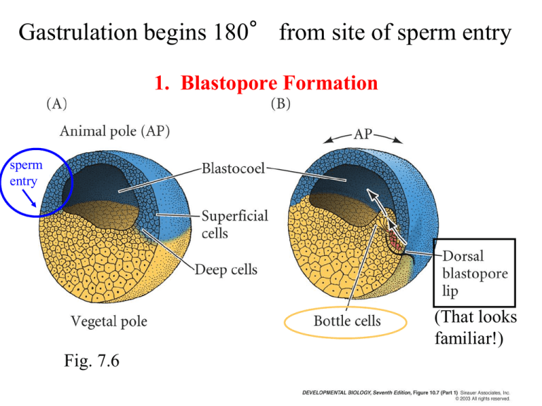
Gastrulation begins 180° from site of sperm entry 1. Blastopore Formation sperm entry (That looks familiar!) Fig. 7.6 How do cells choose fates before gastrulation? The dorsal-ventral axis in the frog Animal pole A frog’s mother sets up the initial axis of polarity Vegetal pole An outline of frog gastrulation Future Ventral Side (belly) Sperm always enters at the animal hemisphere Future Dorsal side (back) Sperm entry is the critical asymmetric cue in setting up the dorsal-ventral axis of the frog Xenopus See Figure 7.1 Sperm entry triggers a rotation of the outer cortex relative to the inner cytoplasm, fixing the site of gastrulation initiation Sperm entry point Animal cytoplasm Vegetal cortex Yolky cytoplasm Gray crescent = junction of vegetal cortex with animal cytoplasm Animal cortex 30 gastrulation initiates here. cortex rotation Rotation occurs during the first division, and requires the sperm centriole Experimental alteration of cortical rotation changes the blastopore location Sperm entry 1. Rotate egg so that sperm entry point is now on top. Sperm entry point point 2. Prevent normal cortical rotation Sperm entry point gray crescent 3. Gravity rearranges cytoplasm so that the junction of vegetal pole cortex with animal pole cytoplasm is next to sperm entry point instead of opposite it. 4. Gastrulation now occurs next to sperm entry point Information from neighbors: Mother cell Cell division Cell type A Cell type B Cell division places daughter cells in different environments, which can lead to different cell fate choices Induction: information from neighbors influences cell fate responder inducer Competence: ability to respond to a certain inductive signal responder Cell not competent to respond inducer Succesive inductions: can generate many cell types from just a few interactions inducer Cell not competent to respond responder Types of signals Inducer Responder Release into the Fallopian tube Signals can act globally throughout the body Signals can also act in a graded fashion Induction: An initial difference can be amplified into many cell types How is cell fate determined -- the dorsal-ventral axis in newts See p. 255 Spemann & Mangold: the organizer can influence neighboring cells to form a secondary body axis: Figure 7.17 Induction of the organizer can influence neighboring cells to form a secondary body axis: If one transplants a second inducer of the organizer the embryo forms two body axes Figure 7.19 Induction of mesoderm in the frog embryo Initial asymmetry of Xenopus egg leads to some cell fate differences Animal pole Vegetal pole Mesoderm arises at endoderm/ ectoderm junction Neither vegetal or animal pole alone can make mesoderm. Experiment: Figure 7.18 Vegetal pole cells can induce animal pole cells to make mesoderm Fig. 7.18 Conclusion: a signal in the vegetal part is needed to induce mesoderm in the animal cap. Induction of dorsal mesoderm by dorsalmost vegetal cells The 3-step model of mesoderm induction #3 #1 #2 Figure 7.18 The 3-step model of mesoderm induction #3 #2 #1 Model for mesoderm induction and organizer formation Figure 7.23 Neurulation separates the ectoderm into neural and epidermal cell fates Neurulation is driven by cell shape and adhesion changes similar to those that drive gastrulation Alberts Figure 20.26 Dorsal-Most Cells Neural tube formation Figure 9.4 Differential adhesion helps the neural tube and skin to separate from each other ectoderm E-cadherin N-cadherin See Fig 9.6 Hatta and Takeichi (1986) Nature Experiment: Differential adhesion helps the neural tube and surface ectoderm separate from each other ectoderm Inject N-cadherin mRNA so that surface ectoderm expresses N-cadherin Fig 9.6 Neural crest cells migrate from the CNS to form the peripheral nervous system Defects in neural tube closure are among the most common birth defects Figure 9.5 Numbers indicate regions of neural tube closure. It is estimated that 50% of human neural tube defects would be prevented if all women of childbearing age take supplemental folic acid (vitamin B12) 400 mg/day Expression of a folate receptor in mice neural tubes prior to fusion Figure 9.7
