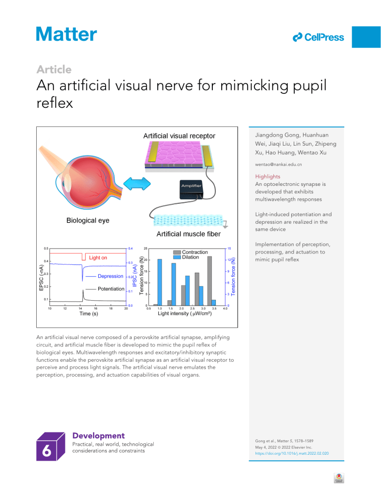
ll Article An artificial visual nerve for mimicking pupil reflex Jiangdong Gong, Huanhuan Wei, Jiaqi Liu, Lin Sun, Zhipeng Xu, Hao Huang, Wentao Xu wentao@nankai.edu.cn Highlights An optoelectronic synapse is developed that exhibits multiwavelength responses Light-induced potentiation and depression are realized in the same device Implementation of perception, processing, and actuation to mimic pupil reflex An artificial visual nerve composed of a perovskite artificial synapse, amplifying circuit, and artificial muscle fiber is developed to mimic the pupil reflex of biological eyes. Multiwavelength responses and excitatory/inhibitory synaptic functions enable the perovskite artificial synapse as an artificial visual receptor to perceive and process light signals. The artificial visual nerve emulates the perception, processing, and actuation capabilities of visual organs. Gong et al., Matter 5, 1578–1589 May 4, 2022 ª 2022 Elsevier Inc. https://doi.org/10.1016/j.matt.2022.02.020 ll Article An artificial visual nerve for mimicking pupil reflex Jiangdong Gong,1 Huanhuan Wei,1 Jiaqi Liu,1 Lin Sun,1 Zhipeng Xu,1 Hao Huang,1 and Wentao Xu1,2,* SUMMARY Progress and Potential Research on bionic eyes is of great importance for neuroprosthetics, biorobotics, and autonomous intelligent electronics. However, implementation of an artificial visual nerve by emulating the architecture and functions of biological eyes remains challenging. Here we demonstrate a p-i-n perovskite optoelectronic synapse capable of implementing excitatory and inhibitory light-mediated synaptic functions. The optoelectronic synapse exhibits a distinct response to visible light at multiple wavelengths (435, 545, and 700 nm). Additionally, an artificial visual reflex arc is established to integrate perception, processing, and actuation of light signals, successfully mimicking a pupil reflex that is adaptively controlled by different muscles (dilator iridis and sphincter pupillae). This artificial visual system simplifies the architecture of an artificial optical nerve, proving the feasibility of emulating complex physiological behaviors involving diverse nerves and effectors. Our work may offer a new strategy for constructing intelligent visual system and sensory neuromorphic electronics. Perovskite optoelectronic synapses are attractive for their potential in constructing bionic eyes. However, developing lightinhibitory perovskite synaptic devices remains challenging because light irradiation will excite charge carriers in materials to elevate the current. This study proposes a p-i-n perovskite optoelectronic synapse with lightmediated excitatory and inhibitory functions. An artificial visual nerve is implemented by combining the perovskite synaptic device with artificial muscle fiber to emulate the pupil reflex of biological eyes. Specifically, lightinduced excitatory and inhibitory synaptic functions, as realized in one device, enrich the function of an artificial visual system. This work may provide guidance to design optoelectronic synaptic devices and systems. INTRODUCTION Human eyes are the most sophisticated and elaborate optical imaging system, composed of diverse receptors, nerves, and effectors.1 They can recognize millions of colors from visual images and decode them with high resolution in a short time. More than 80% of information for our brain is acquired by the visual process.2,3 As a result, this complex and powerful system with robust sensing and processing capabilities, has attracted much attention regarding development of biomimetic eyes, which is of great significance for neuroprosthetics, bioinspired robots, and unmanned technology.4–7 To date, tremendous effort has been dedicated to components and systems of biomimetic eyes, such as photonic synapses, bioinspired sensory systems, and artificial afferent and efferent nerves, boosting rapid development of vision-related neuromorphic electronics.8–15 Because of their excellent optoelectronic characteristics, such as tunable band gap, high mobility, and robust light harvesting, organic-inorganic hybrid perovskites (OHPs) have yielded remarkable results in optoelectronic synapses.16–18 They show enormous potential for construction of artificial vision systems.19,20 Some studies have combined an artificial synapse with sensors or actuators that performed well in mimicking the afferent and efferent nerves.21,22 For example, Lee et al.23 constructed a neuromuscular system by combining a stretchable organic nanowire synaptic transistor with a photodetector and actuator to realize light-mediated muscle tension responses. Wan et al.12 developed a bimodal artificial sensory 1578 Matter 5, 1578–1589, May 4, 2022 ª 2022 Elsevier Inc. ll Article neuron to integrate optic and pressure information to actuate a robotic hand. Some higher-order physiological events, including unconscious behaviors, require a synergistic effect of excitatory and inhibitory synapses.24 Multiple responses to light stimuli are beneficial to realize versatile synaptic functions.25 Hence, research on excitatory and inhibitory optoelectronic synapses is crucial for implementation of artificial visual system in terms of the diverse functions of the biological counterparts. We developed an OHP-based optoelectronic synapse with a p-i-n structure that exhibited a broad wavelength response to visible light. By applying external voltage on the device to modulate photogenerated current, excitatory and inhibitory synaptic functions in response to light stimuli at the same wavelength are implemented. As a result, excitatory postsynaptic current (EPSC), inhibitory postsynaptic current (IPSC), paired-pulse facilitation (PPF), and dynamic filtering were achieved. This strategy of achieving EPSC and IPSC in response to light stimuli with the same wavelength is also applicable for other perovskite optoelectronic devices with a similar structure. In addition, an artificial visual nerve was implemented by combining the optoelectronic synapse with a Ni-Ti alloy artificial muscle fiber (a core component of the artificial pupil), successfully mimicking the pupil reflex to light irradiation. We believe that this work may provide a new strategy for development of neuromorphic sensing and artificial visual systems. RESULTS The pupil reflex is a complex physiological process that involves diverse nerves and muscles (Figure 1A). Generally, light signals with varied intensity can be collected by photoreceptors, cones and rods in the retina, and converted into electrical signals and transmitted to our brain through nerves (Figure 1B). Biological effectors, including the sphincter pupillae and dilator iridis, are triggered to control dilation of the pupil, regulating the incident light flux. In biology, the sphincter pupillae appears in the free margin of iris in a cricoid pattern, and the dilator iridis radiates around the iris. Strong light triggers the sphincter pupillae to shrink, leading to pupil contraction. Weak light triggers the dilator iridis to shrink, leading to pupil dilation (Figure 1C).26,27 An artificial visual system composed of an optoelectronic synapse, amplifying circuit, and artificial muscle fiber is developed to mimic light-mediated pupil reflex process (Figures 1D and 1E). Similarly, light signals can be captured and transformed into postsynaptic current by an optoelectronic device that serves as a retinal photoreceptor, then transmitted through the amplifying circuit to actuate the artificial muscle fiber, regulating the diameter of the artificial pupil. The direction and amplitude of the motion of the artificial pupil is determined by a Ni-Ti alloy fiber. Thus, the optoelectronic synapse that controls construction of the Ni-Ti alloy fiber enables it as a key component in the whole system. The optoelectronic synapse with a vertical laminated structure of the top electrode (TE)/poly(3-hexylthiophene) (P3HT)/MAPbI3/SnO2/indium tin oxide (ITO) was developed via solution processing to mimic a biological synapse (Figure 2A).28 The corresponding layers are illustrated in a cross-sectional scanning electron microscopy (SEM) image. The thickness of the OHP film was 625 nm, which is sufficient to absorb incident photons (Figure 2B). The P3HT and SnO2 layers served as hole transport layer (HTL) and electron transport layer (ETL), respectively. According to atomic force microscopy (AFM) images (Figures S1 and S2), their thickness was 16.3 and 26.7 nm, respectively. As seen in the energy band diagram (Figure 2C), the energy level of MAPbI3 matches with P3HT and SnO2 that photogenerated carriers in MAPbI3 can be separated through the HTL and ETL by the built-in potentials. The 1Institute of Photoelectronic Thin Film Devices and Technology of Nankai University, Key Laboratory of Photoelectronic Thin Film Devices and Technology of Tianjin, Engineering Research Center of Thin Film Photoelectronic Technology, Ministry of Education, #38 Tongyan Road, Jinnan District, Tianjin 300350, P.R. China 2Lead contact *Correspondence: wentao@nankai.edu.cn https://doi.org/10.1016/j.matt.2022.02.020 Matter 5, 1578–1589, May 4, 2022 1579 ll Figure 1. Schematics of the human visual system and an artificial visual nerve (A) Schematic of the human eye. (B) Schematic of the retina. (C) The sphincter and dilator muscle of the pupil. (D) Configuration of OHP and the OHP-based artificial synapse. (E) Schematic of the Ni-Ti alloy actuator. X-ray diffraction (XRD) pattern illustrated in Figure 2D shows distinct diffraction peaks at 14.1 and 28.5 , which correspond to the (002) and (004) planes of MAPbI3, respectively. These results indicate that the film is pure tetragonal b-phase perovskite. A homogeneous and pinhole-free surface can be seen in the top-view SEM image (Figure 2E), indicating a crystalline OHP film that is consistent with the XRD result. The roughness of the OHP film is defined by AFM, which shows a smooth surface with a root-mean-square (RMS) roughness of 3.74 nm (Figure 2F). Optical properties were investigated by ultraviolet-visible (UV-vis) spectroscopy and photoluminescence (PL) spectroscopy. MAPbI3 and SnO2/MAPbI3/P3HT exhibit an obvious light absorption edge at 780 nm. The optical band gap, therefore, can be calculated as 1.6 eV from the absorption spectrum.29 Comparison of UV-vis spectra for three types of films indicated that the presence of P3HT and SnO2 only reduces the absorption intensity but does not change the band gap of MAPbI3 (Figure 2G). The steady-state PL spectrum of MAPbI3 shows an obvious peak at 760 nm, which is a slight blueshift compared with absorption cutoff edge (Figure 2H). Because of the relative low exciton binding energy (Eb) for 3D bulk MAPbI3, more free carriers will be generated upon photoexcitation rather than excitons.30 The reduction in PL intensities in other MAPbI3-based heterojunction films indicates faster extraction of charge carriers. Time-resolved PL (TRPL) was conducted to verify the charge carrier dynamics (Figure 2I). From the fitting curves, the lifetime of photogenerated carriers obviously decreases in MAPbI3-based heterojunction films. The shorter lifetime is mainly relative to the fast separation of photogenerated carriers at the interface, especially in SnO2/MAPbI3/P3HT. 1580 Matter 5, 1578–1589, May 4, 2022 Article Article ll Figure 2. Detailed structure and characterization of the p-i-n perovskite synaptic device (A) Schematic of a biological synapse and OHP-based artificial synapse. (B) Cross-sectional SEM image of the OHP-based synaptic device. (C) Band energy diagrams for the OHP-based synaptic device. (D) XRD pattern of the MAPbI 3 film. (E) Top-view SEM image of the MAPbI3 film. (F) AFM image of the MAPbI 3 film. (G) UV-vis spectrum of three types of films. Inset: Tauc plot to estimate optical band gap. (H) PL spectra of the MAPbI 3 , SnO 2 /MAPbI 3 , MAPbI 3 /P3HT, and SnO 2 /MAPbI 3 /P3HT films. (I) TRPL spectra of the MAPbI 3 , SnO 2 /MAPbI 3 , MAPbI 3 /P3HT, and SnO 2 /MAPbI 3 /P3HT films. Current-voltage (I-V) characteristics were determined by constructing a metal probe on the bottom electrode (BE) for voltage sweeping and a metal probe on the TE for grounding. The current increases rapidly as the voltage sweeps from 0 to 1 V but changes slightly on the counterpart (Figure S3A). In contrast, a I-V sweep was conducted in a perovskite-only device (Figure S3B). The perovskite-only device exhibits a larger current, and the current value increases rapidly to approach 0.6 mA as the voltage sweeps from 0 to 1 V, which is obviously different with a p-i-n synaptic device. This phenomenon coincides with the unidirectional conductivity of the P-N junction.31 A series of voltage sweeps on the optoelectronic synaptic device was conducted to verify the repeatability of I-V curves. Hysteresis loops can be observed in the positive and negative voltage sweeps (Figures S4A and S4B). This may be Matter 5, 1578–1589, May 4, 2022 1581 ll related to the ionic migration in MAPbI3. Under successive voltage sweeping (from 0 to 1 V), halide ions with relatively lower activation energy (Ea) are constantly moving in MAPbI3, causing formation of highly conductive states. When the sweep voltage turns to the opposite (from 0 to 1 V), halide ions begin to go back to their initial positions. The sweep loops become small as the number of scans increases. The current value in the negative voltage sweep is almost three orders of magnitude larger than the positive one because of the effect of built-in potential. 1,000 consecutive voltage spikes ( 1 V, 51 ms) were applied to the optoelectronic synaptic device to investigate endurance. Sight fluctuations in the current plots can be observed, which suggests a reliable synaptic device (Figure S5). The direction of photogenerated current (Iph) is confined by the ETL and HTL. Under the effect of built-in potential, photo-generated electrons can only be transported from SnO2 to the BE, whereas photogenerated holes can only be transported from P3HT to the TE. Therefore, external voltages with different amplitudes and polarities were applied to the TE to modulate Iph. As a result, excitatory and inhibitory synaptic functions can be realized simultaneously; schematics are shown in Figure S6. With negative bias ( 0.1 V) applied to the TE, the external electric field is in the same direction of build-in potential (Figure S6A). The potential barrier will be elevated because of the reversed bias. As a result, photogenerated carriers will move rapidly under the synergetic effect of the external electric field and built-in potential, forming an EPSC that is defined as –IE Iph. When the bias turns positive (0.3 V), the external electric field is in the opposite direction of the built-in potential (Figure S6B). The potential barrier will downshift so that the band bending of perovskite becomes weak. The separation of photogenerated carriers will be suppressed temporarily because of the forward bias. Photogenerated carriers will first neutralize voltageinduced carriers, forming an IPSC that is defined as IE Iph. Because of the unique lattice structure and appropriate band-gap width, OHPs exhibit a robust harvesting ability of visible light.32 Consecutive light pulses, including a single pulse (wavelength, 435 nm; duration, 71 ms; light intensity, 3.38 mW/cm2) and a pair of pulses (interval, 143 ms) were applied to the device to explore fundamental synaptic functions. The EPSC triggered by a single light pulse increases from 0.08 nA to 0.17 nA and decays rapidly to its initial state as the reading voltage is set to 0.1 V. There is a distinct enhancement of the second light pulse when a pair of light pulses irradiates the device, indicating establishment of neural facilitation. This enhancement is related to the slow decay of Iph. When the first light pulse was applied to the device, photogenerated carriers are separated. Because part of the carriers remained, the collected Iph did not decay to the initial level. When the second light pulse was applied, more carriers were separated and collected sequentially, which further increased the postsynaptic current. Therefore, the optoelectronic device can operate as an artificial visual receptor to capture and transform the light signals into artificial EPSC signals. The same results also can be observed with the reading voltage set to positive (0.3 V) (Figure 3A). In particular, the net value for EPSC corresponding to the synaptic weight change is 0.09 nA, which shows a smaller reduction compared with the IPSC response (0.11 nA). The number of photocarriers is fixed such that slight change in the net value for the EPSC and IPSC response may be caused by the reading voltage with different polarity and amplitude to influence the collection efficiency of photogenerated carriers.33,34 Neural facilitation was verified by the PPF index: A2/A13100%, where A2 is the value of the second postsynaptic current (PSC), and A1 is the value of the first PSC. As seen in the fitting curves in Figure 3B, the neural facilitation effect becomes 1582 Matter 5, 1578–1589, May 4, 2022 Article Article ll Figure 3. Emulation of synaptic plasticity (A) EPSC/IPSC triggered by a single light pulse and a pair of light pulses. (B) PPF index versus time interval for potentiation and depression. (C) EPSC triggered by consecutive light pulses with different pulse widths. (D) EPSC triggered by consecutive light pulses with different pulse numbers. (E) EPSC triggered by consecutive light pulses with different pulse frequencies. (F) EPSC versus duration for 3 different wavelengths of light pulses. (G) PPF index versus time interval for 3 different wavelengths of light pulses. (H) EPSC gain versus frequency for 3 different wavelengths of light pulses. weak when prolonging the interval between two consecutive light pulses. The PPF index calculated in potentiation is larger than in depression whenever the interval time varies. Synaptic plasticity can also be tuned by changing the form of the input light pulse, such as light intensity, duration, or number of light pulses. As shown in Figure 3C, the EPSC gradually elevated to 0.4 nA as the duration increased from 71 ms to 643 ms because the Iph is determined by the number of photogenerated carriers. Long-term irradiation will cause more carriers to be excited and collected, leading to a large EPSC response. Similarly, the EPSC value and decay time are obviously prolonged by increasing the number of light pulses (Figure 3D). The same results also can be achieved in inhibitory mode (Figures S7 and S8). Matter 5, 1578–1589, May 4, 2022 1583 ll In biology, a synapse can serve as a filter to process dynamic real-time information, which is of great significance for memory and learning.35 In our OHP-based synaptic device, high-pass filtering and low-pass filtering characteristics can be realized by setting the excitatory and inhibitory modes. Figure 3E illustrates the EPSC triggered by consecutive light pulses with different frequencies. An obvious enhancement of the EPSC value can be observed as the frequency of light pulses increases. The filtering characteristic is verified by defining A10/A1 as EPSC gain, where A10 and A1 are the amplitude of the 10th and the first EPSC, respectively. With the frequency of light pulses increased from 1.27 Hz to 6.99 Hz, EPSC gain increased from 1.37 to 1.65, indicating a synapse-like behavior of high-pass filtering (Figure S9). The enhancement of the EPSC response triggered by light pulses with increasing frequency is caused by the motion of photoexcited carriers in MAPbI3. Under light irradiation, photogenerated electrons and holes drift to the interfaces in MAPbI3 and are separated by ETL and HTL. When the light is off, no extra charge carriers are excited, and the rest of the electrons and holes continue to drift and and are collected until exhausted or the light is turned on. Increasing the frequency of light pulses is equivalent to reducing the duration of light off while maintaining the duration of light on. As a result, more photogenerated carriers are formed in MAPbI3 to induce a large EPSC response. The same results can also be observed in the inhibitory counterpart that corresponds to low-pass filtering behavior (Figures S10 and S11). According to the absorption spectrum (Figure 2G), light pulses with various wavelengths are applied to the optoelectronic synapse to investigate the influence of light frequency on synaptic plasticity. Figure 3F shows that the EPSC responded to light pulses of 435, 545, 545, and 700 nm, which corresponds to the three primary colors. The EPSC triggered by the blue light is the largest, which is consistent with the absorption spectrum. There is an obvious enhancement in EPSC gain for blue light as the duration time increases. The PPF index exhibits similar results for blue and green light and an obvious decline for red light, indicating that the neural facilitation effect can be enhanced under short-wave light illumination (Figure 3G). The same result can also be observed in EPSC gain versus frequency of incident light pulses (Figure 3H). In summary, this p-i-n perovskite artificial synapse glitters in two perspectives (Table S1). First, excitatory and inhibitory synaptic functions under light stimuli at the same wavelength are realized by modulation of the external voltage on Iph, and this method is applicable to other perovskite synaptic devices with a similar structure.36 Second, this p-i-n perovskite artificial synapse is sensitive to light irradiation because of the synergetic effect of built-in potential and external voltage, which is desirable for energy-efficient applications. In biology, the pupil reflex is divided into two categories: contraction and dilation, which controlled by the sphincter pupillae and dilator iridis, respectively.37 For example, our pupil contracts to reduce light flux when exposed to an environment where the light intensity is increasing. On the contrary, dark conditions enable the pupil to dilate enough for more induced light flux. Figure 4A shows a complete diagram of the artificial visual system for mimicking the pupil reflex. Light signals are collected and transformed by the artificial visual receptor and are then transmitted through the amplifying circuit to activate the artificial muscle, regulating the diameter of the artificial pupil. There are two connection modes for Ni-Ti alloy artificial muscle fiber that resemble the sphincter pupillae and dilator iridis: radial and ring like. 1584 Matter 5, 1578–1589, May 4, 2022 Article Article ll Figure 4. Implementation of the light-mediated pupil reflex (A) Schematic of the OHP-based artificial visual system (OHP-based optoelectronic synapse, amplifying circuit, and Ni-Ti alloy artificial muscle fiber). (B) EPSC triggered by consecutive light pulses with increasing light intensity and the corresponding tension force of the Ni-Ti alloy actuator. (C) Normalized maximum force versus light intensity. Inset: artificial pupil connected to the Ni-Ti alloy fiber in a ring-like fashion. (D) EPSC triggered by consecutive light pulses with decreasing light intensity and the corresponding tension force of the Ni-Ti alloy actuator. (E) Normalized maximum force versus light intensity. Inset: artificial pupil connected to the Ni-Ti alloy fiber in a radial fashion. To mimic the construction of the pupil, an excitatory optoelectronic synapse is implemented by tuning the reading voltage to negative ( 0.1 V). The Ni-Ti artificial muscle fiber is wrapped around the artificial pupil in a circle. Consecutive light pulses with increased light intensity are applied to the synaptic device. As shown in Figure 4B, the EPSC obviously increased as the light intensity increased (from 0.91 mW/cm2 to 3.38 mW/cm2). Under the stimulus of a larger EPSC, the Ni-Ti alloy artificial muscle fiber begins to contract with the transformation from martensite to austenite. Tension force increases correspondingly. Therefore, the circumference of the Ni-Ti alloy artificial muscle fiber was reduced to shrink the diameter of artificial pupil. Normalized force maximum (Fmax) versus light intensity was used to analyze the change in tension force for the Ni-Ti alloy artificial muscle. With increased light intensity, the normalized Fmax increased from 0.6 N to 21.6 N, which corresponded to the motion of the sphincter pupillae. As a result, the artificial pupil began to contract as the input light intensity increased (Figure 4C). Matter 5, 1578–1589, May 4, 2022 1585 ll The inhibitory optoelectronic synapse is set up by turning the reading voltage to positive (0.3 V) to mimic the dilation process of the pupil. The Ni-Ti alloy artificial muscle fiber is connected to the artificial pupil in a radial pattern. Consecutive light pulses with decreased light intensities are applied to the synaptic device. The inhibitory effect became weak as the light intensity decreased (from 3.38 mW/cm2 to 0.91 mW/cm2). Correspondingly, the IPSC value increased gradually with the decrease in light intensity. Tension force in the Ni-Ti alloy artificial muscle increased similarly during light irradiation with different intensities (Figure 4D). As a result, the normalized Fmax increased from 1.5 N to 12.2 N as the light intensity decreased from 3.38 mW/cm2 to 0.91 mW/cm2. The artificial pupil began to dilate under contraction of the Ni-Ti alloy artificial muscle fiber, which corresponded to the motion of the dilator iridis (Figure 4E). These results indicate that higher-order physiological behaviors involved in excitatory and inhibitory functions can be realized in our artificial visual system with dual operation modes. DISCUSSION We demonstrated an OHP-based optoelectronic synapse capable of simulating excitatory and inhibitory functions. The optoelectronic synapse exhibited a broad wavelength response to visible light. For light stimuli of the same wavelength, potentiation and depression synaptic behavior can be realized simultaneously because of the modulation of the external electric field on Iph. The optoelectronic synapse can operate under light irradiation with extremely low intensity (0.91 mW/cm2) under the synergetic effect of the external electric field and built-in potential. Diverse synaptic behaviors, including EPSC, IPSC, PPF, and dynamic filtering, were achieved, which enriched the functionalities of neuromorphic electronics. More importantly, an artificial visual system was implemented by combining the optoelectronic synapse with a Ni-Ti alloy artificial muscle fiber through an amplifying circuit to mimic the pupil reflex. This artificial visual nerve integrates the lightsensitive synaptic device and artificial muscle, providing guidelines for emulating advanced functions of human sensorimotor nerves. Our work may pave the way for neuromorphic electronics, especially vision-related artificial systems with complex and intelligent functions. EXPERIMENTAL PROCEDURES Resource availability Lead contact Additional data and files are available from the lead contact, Wentao Xu (wentao@ nankai.edu.cn). Materials availability This study did not generate new unique reagents. Data and code availability This study did not generate any datasets/code. Materials synthesis and synaptic device fabrication P3HT was purchased from Sigma-Aldrich. Methylammonium iodide (MAI), lead iodide (PbI2), and ITO were purchased from Advanced Electronic Technology. The tin (IV) oxide colloid precursor was purchased from Alfa Aesar. 1.1 mol/L MAPbI3 solution was prepared by dissolving MAI and PbI2 (molar ratio, 1:1) in methylamine ethanol and acetonitrile (volume ratio, 3:2) and then stirred for 1 h at room 1586 Matter 5, 1578–1589, May 4, 2022 Article Article ll temperature. 5 mg/mL P3HT solution was prepared by dissolving P3HT in anhydrous chlorobenzene and stirred at 50 C for 2 h. All regents were used without further purification. Firstly, the ITO substrate was cleaned in deionized water, acetone, and isopropanol (IPA) for 15 min each by ultrasonic. After drying by nitrogen flow, the substrate was treated with UV-ozone for 20 min. Then 100 mL SnO2 solution (mixed with NH3$H2O at a volume ratio of 1:5) was spin coated on the as-prepared substrate at 4,000 rpm for 30 s and annealed at 150 C for 30 min in ambient. 100 mL MAPbI3 solution was spin coated on SnO2 at 4,000 rpm for 60 s. Then 70 mL P3HT solution was spin coated on MAPbI3 film at 2,500 rpm for 30 s and annealed at 60 C for 10 min. Finally, 100nm Au electrodes were deposited by thermal evacuation through a predesigned shadow mask. Fabrication of the Ni-Ti alloy actuator and artificial pupil First, Ni and Ti metal were mixed at a mass ratio of 6:4 and smelted 5 times to form an original ingot. The ingot was annealed in a vacuum at 1,000 C for 12 h and hot forged into a steel bar at 850 C, then cooled to room temperature. After that, the steel bar was drawn into an alloy filament (r = 0.3 mm) at 200 C. The alloy filament was then annealed to 950 C for 15-min solid-solution treatment. After cooling, the alloy filament was treated by cold-drawing four times. Finally, the filament was covered with a sliver thermal conductive layer to form a Ni-Ti alloy artificial muscle fiber with 3 mm diameter. The artificial pupil model was fabricated using fused deposition modeling (FDM) 3D printing technology. The thermoplastic polyurethane elastomer (TPU) was extruded through a nozzle and then deposited on the printer platform according to the predefined path, forming the target model in a layer-by-layer mode. Device characterization SEM images were obtained using a Thermo Scientific field emission microscope (Apero S). AFM images were obtained by using a Veeco NanoScope microscope (NanoNavi-SPA400). The XRD pattern was obtained using a Rigaku Ultima IV instrument (MAX-2500). The optical absorption spectrum was acquired using an UV-vis near-infrared (NIR) spectrophotometer (Cary 5000). PL and TRPL spectra were measured using a fluorescence spectrometer (Edinburgh FS5). Light pulses were generated using a xenon source (Zolix, GLORIA-Bright). Tension force was measured using an Aidebao HP-100 forcemeter and Keithley 2400 digital source meter. Electrical measurements were conducted using a Keithley 4200A semiconductor parameter analyzer. SUPPLEMENTAL INFORMATION Supplemental information can be found online at https://doi.org/10.1016/j.matt. 2022.02.020. ACKNOWLEDGMENTS The authors thank Dr. Chengpeng Jiang, Dr. Haiyang Yu, and Prof. Yi Ding from Nankai University for discussions and modifications. This research was supported by the National Science Fund for Distinguished Young Scholars of China (T2125005), Tianjin Science Foundation for Distinguished Young Scholars (19JCJQJC61000), and Shenzhen Science and Technology Project (JCYJ20210324121002008). Matter 5, 1578–1589, May 4, 2022 1587 ll Article AUTHOR CONTRIBUTIONS W.X. conceived the work. J.G. and W.X. designed the experiments. J.L. fabricated the devices and performed electrical measurements. H.W., J.L., Z.X., H.H., and L.S. contributed to analysis and discussion of the data. J.G., H.W., and W.X. wrote the manuscript with input from all other authors. W.X. supervised the research. All authors discussed the results and commented on the manuscript. DECLARATION OF INTERESTS The authors declare no competing interests. Received: November 8, 2021 Revised: February 7, 2022 Accepted: February 18, 2022 Published: March 17, 2022 REFERENCES 1. Georgiev, G.A., Eftimov, P., and Yokoi, N. (2017). Structure-function relationship of tear film lipid layer: a contemporary perspective. EEyeR 163, 17–28. 2. Pocock, D.C.D. (1981). Sight and knowledge. Trans. Inst. Br. Geogr. 6, 385–393. 3. Miyawaki, Y., Uchida, H., Yamashita, O., Sato, M.-a., Morito, Y., Tanabe, H.C., Sadato, N., and Kamitani, Y. (2008). Visual image reconstruction from human brain activity using a combination of multiscale local image decoders. Neuron 60, 915–929. 11. Kwon, S.M., Cho, S.W., Kim, M., Heo, J.S., Kim, Y.-H., and Park, S.K. (2019). Environmentadaptable Artificial visual perception behaviors using a light-adjustable optoelectronic neuromorphic device array. Adv. Mater. 31, 1906433. 12. Wan, C., Cai, P., Guo, X., Wang, M., Matsuhisa, N., Yang, L., Lv, Z., Luo, Y., Loh, X.J., and Chen, X. (2020). An artificial sensory neuron with visual-haptic fusion. Nat. Commun. 11, 4602. 4. Li, J., Wang, Y., Liu, L., Xu, S., Liu, Y., Leng, J., and Cai, S. (2019). A biomimetic soft lens controlled by electrooculographic signal. Adv. Funct. Mater. 29, 1903762. 13. Li, Y., Wang, Y., Yin, L., Huang, W., Peng, W., Zhu, Y., Wang, K., Yang, D., and Pi, X. (2021). Silicon-based inorganic-organic hybrid optoelectronic synaptic devices simulating cross-modal learning. Sci. China Inf. Sci. 64, 162401. 5. Wang, T., Yu, W., Li, C., Zhang, H., Xu, Z., Lu, Z., and Sun, Q. (2012). Biomimetic compound eye with a high numerical aperture and antireflective nanostructures on curved surfaces. Opt. Lett. 37, 2397–2399. 14. Wang, Y., Zhu, Y., Li, Y., Zhang, Y., Yang, D., and Pi, X. (2021). Dual-Modal optoelectronic synaptic devices with versatile synaptic plasticity. Adv. Funct. Mater. 32, 2107973. 6. Lee, G.J., Choi, C., Kim, D.-H., and Song, Y.M. (2018). Bioinspired artificial eyes: optic components, digital cameras, and visual prostheses. Adv. Funct. Mater. 28, 1705202. 7. Gu, L., Poddar, S., Lin, Y., Long, Z., Zhang, D., Zhang, Q., Shu, L., Qiu, X., Kam, M., Javey, A., et al. (2020). A biomimetic eye with a hemispherical perovskite nanowire array retina. Nature 581, 278–282. 8. Park, H.-L., Kim, H., Lim, D., Zhou, H., Kim, Y.-H., Lee, Y., Park, S., and Lee, T.-W. (2020). Retina-inspired carbon nitride-based photonic synapses for selective detection of UV light. Adv. Mater. 32, 1906899. 9. Wang, Y., Lv, Z., Chen, J., Wang, Z., Zhou, Y., Zhou, L., Chen, X., and Han, S.-T. (2018). Photonic synapses based on inorganic perovskite quantum dots for neuromorphic computing. Adv. Mater. 30, 1802883. 10. Hao, D., Zhang, J., Dai, S., Zhang, J., and Huang, J. (2020). Perovskite/organic semiconductor-based photonic synaptic transistor for artificial visual system. ACS Appl. Mater. Interf 12, 39487–39495. 1588 Matter 5, 1578–1589, May 4, 2022 15. Yang, F., Sun, L., Duan, Q., Dong, H., Jing, Z., Yang, Y., Li, R., Zhang, X., Hu, W., and Chua, L. (2021). Vertical-organic-nanocrystal-arrays for crossbar memristors with tuning switching dynamics toward neuromorphic computing. SmartMat 2, 99–108. 16. Yin, L., Huang, W., Xiao, R., Peng, W., Zhu, Y., Zhang, Y., Pi, X., and Yang, D. (2020). Optically stimulated synaptic devices based on the hybrid structure of silicon nanomembrane and perovskite. Nano Lett. 20, 3378–3387. 17. Wang, K., Dai, S., Zhao, Y., Wang, Y., Liu, C., and Huang, J. (2019). Light-stimulated synaptic transistors fabricated by a facile solution process based on inorganic perovskite quantum dots and organic semiconductors. Small 15, 1900010. accelerated learning at low power inspired by dopamine-facilitated synaptic activity. Adv. Funct. Mater. 29, 1806646. 20. Huang, W., Hang, P., Wang, Y., Wang, K., Han, S., Chen, Z., Peng, W., Zhu, Y., Xu, M., Zhang, Y., et al. (2020). Zero-power optoelectronic synaptic devices. Nano Energy 73, 104790. 21. Kim, S., Roe, D.G., Choi, Y.Y., Woo, H., Park, J., Lee, J.I., Choi, Y., Jo, S.B., Kang, M.S., Song, Y.J., et al. (2021). Artificial stimulus-response system capable of conscious response. Sci. Adv. 7, eabe3996. 22. Wei, H., Shi, R., Sun, L., Yu, H., Gong, J., Liu, C., Xu, Z., Ni, Y., Xu, J., and Xu, W. (2021). Mimicking efferent nerves using a graphdiyne-based artificial synapse with multiple ion diffusion dynamics. Nat. Commun. 12, 1068. 23. Lee, Y., Oh, J.Y., Xu, W., Kim, O., Kim, T.R., Kang, J., Kim, Y., Son, D., Tok, J.B.-H., Park, M.J., et al. (2018). Stretchable organic optoelectronic sensorimotor synapse. Sci. Adv. 4, eaat7387. 24. Yang, C.-M., Chen, T.-C., Verma, D., Li, L.-J., Liu, B., Chang, W.-H., and Lai, C.-S. (2020). Bidirectional all-optical synapses based on a 2D Bi2O2Se/graphene hybrid structure for multifunctional optoelectronics. Adv. Funct. Mater. 30, 2001598. 25. Zhang, Z., Wang, S., Liu, C., Xie, R., Hu, W., and Zhou, P. (2022). All-in-one two-dimensional retinomorphic hardware device for motion detection and recognition. Nat. Nanotechnol. 17, 27–32. 26. Ellis, C.J. (1981). The pupillary light reflex in normal subjects. Br. J. Ophthalmol. 65, 754. 18. Sun, Y., Qian, L., Xie, D., Lin, Y., Sun, M., Li, W., Ding, L., Ren, T., and Palacios, T. (2019). Photoelectric synaptic plasticity realized by 2D perovskite. Adv. Funct. Mater. 29, 1902538. 27. Chen, S.K., Badea, T.C., and Hattar, S. (2011). Photoentrainment and pupillary light reflex are mediated by distinct populations of ipRGCs. Nature 476, 92–95. 19. Ham, S., Choi, S., Cho, H., Na, S.-I., and Wang, G. (2019). Photonic organolead halide perovskite artificial synapse capable of 28. Wang, K., Wu, C., Hou, Y., Yang, D., Ye, T., Yoon, J., Sanghadasa, M., and Priya, S. (2020). Isothermally crystallized perovskites at ll Article room-temperature. Energy Environ. Sci. 13, 3412–3422. 29. Makuła, P., Pacia, M., and Macyk, W. (2018). How to correctly determine the band gap energy of modified semiconductor photocatalysts based on UV– vis spectra. J. Phys. Chem. Lett. 9, 6814– 6817. 30. Umari, P., Mosconi, E., and De Angelis, F. (2018). Infrared dielectric screening determines the low exciton binding energy of metal-halide perovskites. J. Phys. Chem. Lett. 9, 620–627. 31. Wang, H., Yi, G., Zu, X., Qin, P., Tan, M., and Luo, H. (2016). Photoelectric characteristics of the p–n junction between ZnO nanorods and polyaniline nanowires and their application as a UV photodetector. Mater. Lett. 162, 83–86. 32. Green, M.A., Ho-Baillie, A., and Snaith, H.J. (2014). The emergence of perovskite solar cells. Nat. Photon. 8, 506–514. 33. Eames, C., Frost, J.M., Barnes, P.R.F., O’Regan, B.C., Walsh, A., and Islam, M.S. (2015). Ionic transport in hybrid lead iodide perovskite solar cells. Nat. Commun. 6, 7497. 34. Xiao, Z., Yuan, Y., Shao, Y., Wang, Q., Dong, Q., Bi, C., Sharma, P., Gruverman, A., and Huang, J. (2015). Giant switchable photovoltaic effect in organometal trihalide perovskite devices. Nat. Commun. 14, 193–198. 35. Gupta, I., Serb, A., Khiat, A., Zeitler, R., Vassanelli, S., and Prodromakis, T. (2016). Realtime encoding and compression of neuronal spikes by metal-oxide memristors. Nat. Commun. 7, 12805. 36. Wang, Y., Yin, L., Huang, W., Li, Y., Huang, S., Zhu, Y., Yang, D., and Pi, X. (2021). Optoelectronic synaptic devices for neuromorphic computing. Adv. Intell. Syst. 3, 2000099. 37. Hakerem, G., and Sutton, S. (1966). Pupillary response at visual threshold. Nature 212, 485–486. Matter 5, 1578–1589, May 4, 2022 1589

![[doi 10.1109%2FEI2.2018.8582096] Liu, Linping; Chen, Siyu -- [IEEE 2018 2nd IEEE Conference on Energy Internet and Energy System Integration (EI2) - Beijing (2018.10.20-2018.10.22)] 2018 2](http://s3.studylib.net/store/data/025229574_1-ea860691491e3e454418b88e0739fe0c-300x300.png)
