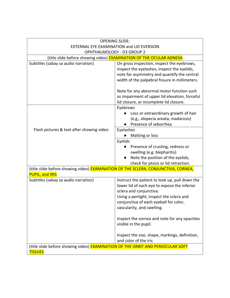
OPENING SLIDE: EXTERNAL EYE EXAMINATION and LID EVERSION OPHTHALMOLOGY - D3 GROUP 2 (title slide before showing video) EXAMINATION OF THE OCULAR ADNEXA Subtitles (sabay sa audio narration) On gross inspection, inspect the eyebrows, inspect the eyelashes, inspect the eyelids, note for asymmetry and quantify the central width of the palpebral fissure in millimeters. Note for any abnormal motor function such as impairment of upper lid elevation, forceful lid closure, or incomplete lid closure. Eyebrows ● Loss or extraordinary growth of hair (e.g., alopecia areata, madarosis) ● Presence of seborrhea Flash pictures & text after showing video Eyelashes ● Matting or loss Eyelids ● Presence of crusting, redness or swelling (e.g. blepharitis) ● Note the position of the eyelids, check for ptosis or lid retraction. (title slide before showing video) EXAMINATION OF THE SCLERA, CONJUNCTIVA, CORNEA, PUPIL, and IRIS Subtitles (sabay sa audio narration) Instruct the patient to look up, pull down the lower lid of each eye to expose the inferior sclera and conjunctiva. Using a penlight, inspect the sclera and conjunctiva of each eyeball for color, vascularity, and swelling. Inspect the cornea and note for any opacities visible in the pupil. Inspect the size, shape, markings, definition, and color of the iris. (title slide before showing video) EXAMINATION OF THE ORBIT AND PERIOCULAR SOFT TISSUES Subtitles (sabay sa audio narration) Prior to palpation, inspect both orbits and eyes for position, alignment, symmetry, size, and shape. In cases of suspected orbital trauma, infections, or neoplasms, palpate the bony orbital rim and periocular soft tissues. Inspect and palpate for inflammatory signs such as swelling, erythema, warmth, and tenderness. Auscultate for orbital bruit, as this may be a sign of an underlying neurovascular pathology. (title slide before showing video) EXAMINATION OF THE GLOBE Subtitles (sabay sa audio narration) Standing behind the patient, look for proptosis by instructing the patient to tilt back the head. Examine the height of the cornea in relation to the brow for asymmetry between two eyes. (title slide before showing video) OTHER DIAGNOSTICALLY RELEVANT EXAMINATIONS Subtitles (sabay sa audio narration) Lastly, depending on the circumstances, checking for enlarged pre and post-auricular (Sabay sabay pakita yung tatlong video in one lymph nodes, sinus tenderness, temporal screen) artery prominence, or skin and mucous membrane abnormalities may be diagnostically relevant. (title slide before showing video) LID EVERSION Subtitles (sabay sa audio narration) For lid eversion, the patient is positioned and instructed to look down. Using the thumb and index finger of one hand, gently grasp the upper lashes. Using the other hand, position the applicator handle just above the superior edge of the tarsus. The lid is everted by applying slight downward pressure with the applicator, as the lash margin is simultaneously lifted. The patient continues to look down, and the lashes are held pinned to the skin overlying ENDING SLIDE the superior orbital rim as the applicator is withdrawn. The tarsal conjunctiva is then examined. To undo eversion, the lid margin is gently stroked downward as the patient looks up. References: Riordan-Eva & Augsburger (2018). Vaughan & Ashbury’s General Ophthalmology. 19th ed. UST FMS Department of Medicine (2020). MED1 Eye Examination Checklist.
