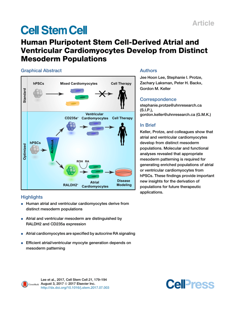
Article Human Pluripotent Stem Cell-Derived Atrial and Ventricular Cardiomyocytes Develop from Distinct Mesoderm Populations Graphical Abstract Authors Jee Hoon Lee, Stephanie I. Protze, Zachary Laksman, Peter H. Backx, Gordon M. Keller Correspondence stephanie.protze@uhnresearch.ca (S.I.P.), gordon.keller@uhnresearch.ca (G.M.K.) In Brief Keller, Protze, and colleagues show that atrial and ventricular cardiomyocytes develop from distinct mesoderm populations. Molecular and functional analyses revealed that appropriate mesoderm patterning is required for generating enriched populations of atrial or ventricular cardiomyocytes from hPSCs. These findings provide important new insights for the derivation of populations for future therapeutic applications. Highlights d Human atrial and ventricular cardiomyocytes derive from distinct mesoderm populations d Atrial and ventricular mesoderm are distinguished by RALDH2 and CD235a expression d Atrial cardiomyocytes are specified by autocrine RA signaling d Efficient atrial/ventricular myocyte generation depends on mesoderm patterning Lee et al., 2017, Cell Stem Cell 21, 179–194 August 3, 2017 ª 2017 Elsevier Inc. http://dx.doi.org/10.1016/j.stem.2017.07.003 Cell Stem Cell Article Human Pluripotent Stem Cell-Derived Atrial and Ventricular Cardiomyocytes Develop from Distinct Mesoderm Populations Jee Hoon Lee,1,2,6 Stephanie I. Protze,1,6,* Zachary Laksman,3 Peter H. Backx,4,5 and Gordon M. Keller1,2,7,* 1McEwen Centre for Regenerative Medicine and Princess Margaret Cancer Center, University Health Network, Toronto, ON M5G 1L7, Canada 2Department of Medical Biophysics, University of Toronto, Toronto, ON M5G 1L7 Canada 3Department of Medicine, University of British Columbia, Vancouver, BC V6E 1M7, Canada 4Department of Biology, York University, Toronto, ON M3J 1P3, Canada 5Division of Cardiology and the Peter Munk Cardiac Centre, University Health Network, Toronto, ON M5G 2N2, Canada 6These authors contributed equally 7Lead Contact *Correspondence: stephanie.protze@uhnresearch.ca (S.I.P.), gordon.keller@uhnresearch.ca (G.M.K.) http://dx.doi.org/10.1016/j.stem.2017.07.003 SUMMARY The ability to direct the differentiation of human pluripotent stem cells (hPSCs) to the different cardiomyocyte subtypes is a prerequisite for modeling specific forms of cardiovascular disease in vitro and for developing novel therapies to treat them. Here we have investigated the development of the human atrial and ventricular lineages from hPSCs, and we show that retinoic acid signaling at the mesoderm stage of development is required for atrial specification. Analyses of early developmental stages revealed that ventricular and atrial cardiomyocytes derive from different mesoderm populations that can be distinguished based on CD235a and RALDH2 expression, respectively. Molecular and electrophysiological characterization of the derivative cardiomyocytes revealed that optimal specification of ventricular and atrial cells is dependent on induction of the appropriate mesoderm. Together these findings provide new insights into the development of the human atrial and ventricular lineages that enable the generation of highly enriched, functional cardiomyocyte populations for therapeutic applications. INTRODUCTION Access to cardiovascular cells from human pluripotent stem cells (hPSCs) has transformed our approach to studying heart development and disease, as it enables, for the first time, the use of patient-derived human cells to model these processes in vitro (Burridge et al., 2012; Josowitz et al., 2011). Additionally, hPSC-derived cardiomyocytes offer a new avenue for developing novel cell-based therapies to replace heart tissue damaged by age or disease (Burridge et al., 2012; Laflamme and Murry, 2005). Modeling studies to date have demonstrated that it is possible to re-create aspects of cardiovascular diseases in vitro using patient-specific induced pluripotent stem cells (iPSCs), while transplantation experiments have shown that hPSC-derived cardiomyocytes can remuscularize infarcted regions of recipient hearts (Caspi et al., 2007; Chong et al., 2014; Laflamme et al., 2007; Shiba et al., 2016). Although successful, most of these studies have been carried out with mixed cardiovascular populations that contain ventricular-like cells together with small numbers of pacemaker- and atrial-like cells. The use of mixed populations for such studies is problematic, as contaminating cell types can easily influence disease outcomes in vitro and alter the behavior of cardiovascular grafts in vivo. To be able to model and treat diseases that affect specific regions of the heart, it is essential to develop differentiation strategies that promote the generation of each of these cardiomyocyte subtypes. The efficient and reproducible derivation of pure populations of atrial, ventricular, and pacemaker cardiomyocytes from hPSCs depends on our ability to successfully translate the principles of cardiovascular development in the early embryo to the hPSC differentiation cultures. The heart develops from mesodermal cells that migrate anterolaterally from the primitive streak to a position under the developing headfold where they form an epithelial structure known as the cardiac crescent (Buckingham et al., 2005; Christoffels et al., 2000). This crescent fuses at the midline to establish the primitive heart tube that is patterned along the anterior-posterior axis to form distinct anterior and posterior poles containing progenitors that give rise to different regions of the adult organ (Christoffels et al., 2000; Rosenthal and Xavier-Neto, 2000; Vincent and Buckingham, 2010). The anterior progenitors differentiate first and give rise to the ventricles, whereas those positioned in the posterior pole differentiate at a slightly later time and contribute to the atria and sinus venosa (Bruneau et al., 2000; Rosenthal and Xavier-Neto, 2000; Vincent and Buckingham, 2010). Studies using different model organisms have provided evidence that these different progenitors are specified early during gastrulation, likely at the stage of mesoderm induction in the primitive streak (Lescroart et al., Cell Stem Cell 21, 179–194, August 3, 2017 ª 2017 Elsevier Inc. 179 Cardiomyocytes hPSCs Primitive Streak Cardiac Mesoderm 0 1 Activin A BMP4 bFGF IWP2 VEGF 3 5 7 20 12 Relative Expression B Days Differentiation 4 2000 MYL2 1500 1000 80 60 40 20 ** * 0 5.7 30 CTNT(APC) ** H day3 RA MYL2 ** IRX4 C on tro da l y da 3 y da 5 da y7 y1 2 FV FA MYH7 NR2F2 500nM RA 0.3 92 TBX5 F NPPA 1.0 0.0 6.7 2.0 % MLC2V+ 100 80 MYL7 60 CACNA1D 40 20 0 KCNJ3 ** KCNA5 2.0 96 GJA5 -2 CTNT(APC) Control G Control C o da ntro y3 l R A 63 NKX2-5:GFP 7.0 D ## 0 MLC2V (PE) NKX2-5:GFP MLC2V (PE) 5.0 1.3 0 CTNT day3 RA 87 200 500nM RA KCNJ3 1 ## Control 1.0 400 2 500nM RA E CTNT 600 3 C on tr da ol y da 3 y da 5 da y7 y1 2 FV FA C Relative Expression 500nM RA 800 C on tro da l y da 3 y da 5 da y7 y1 2 FV FA A 0.82 3 day3 RA MLC2V CTNT Merged+DAPI MLC2V CTNT Merged+DAPI COUPTFII CTNT Merged+DAPI COUPTFII CTNT Merged+DAPI Figure 1. RA Signaling Promotes Atrial-like Cardiomyocyte Development (A) Schematic of the hPSC cardiomyocyte differentiation protocol indicating developmental stages and timing of RA addition. (B and C) qRT-PCR analysis of the expression levels of (B) a pan-cardiomyocyte gene and (C) ventricular-specific (MYL2), and atrial-specific (KCNJ3) genes in NKX2-5+SIRPa+CD90 cells isolated from day 20 EB populations induced with 10 ng/mL BMP4 and 6 ng/mL Activin A (10B/6A) and treated with RA at the indicated time points (n = 3) and in fetal tissue controls (n = 6) (t test, *p < 0.05 and **p < 0.01 versus DMSO control and ##p < 0.01 F-V versus F-A). (D) Heatmap comparing the gene expression profiles of NKX2-5+SIRPa+CD90 cells isolated from day 20 EBs (10B/6A induced) and treated with RA or DMSO (control) between days 3 and 5 (n = 5). Values represent log10 of expression levels relative to the housekeeping gene TBP. (E) Representative flow cytometric analyses of the proportion of NKX2-5+/CTNT+ and MLC2V+/CTNT+ cells in day 20 EB populations induced with 10B/6A and treated between days 3 and 5 with RA or DMSO (control). (legend continued on next page) 180 Cell Stem Cell 21, 179–194, August 3, 2017 2014; Wei and Mikawa, 2000). Lineage-tracing experiments, based on ISL1, FGF10, and HCN4 expression, have identified two distinct progenitor populations known as the first heart field (FHF) and second heart field (SHF) that show differential contribution to the atria, the ventricles, and the outflow tract (Cai €ter et al., 2003; Kelly et al., 2001; Meilhac et al., 2004; Spa et al., 2013). A more recent study, using an inducible labeling strategy based on Mesp1 expression, demonstrated that these progenitors are generated at different times during gastrulation (Lescroart et al., 2014). The earliest progenitors to develop (embryonic day [E]6.25–6.75) appear to contribute exclusively to the left ventricle, whereas those specified at slightly later times (E7.25) give rise to right ventricular, atrial, and outflow tract cardiomyocytes. Collectively, these observations provide strong evidence that the different cardiomyocyte subtypes in the heart derive from distinct mesodermal populations that are induced in a defined temporal pattern. While the pathways that regulate the development of these different progenitors are not fully understood, studies in the early mouse and chick embryo have identified retinoic acid (RA) as a key regulator of cardiovascular cell fate (Moss et al., 1998; Xavier-Neto et al., 2000). These studies showed that embryos exposed to excessive levels of RA displayed severe cardiac malformations characterized by enlarged atrial chambers and small ventricles. The opposite effect was observed when the embryos were treated with an RA inhibitor. Importantly, this effect of RA was restricted to a narrow window of development, from E7.5 to E8.5, corresponding to the late gastrulation stage when cardiac mesoderm progenitors that contribute to the posterior region of the cardiac crescent are migrating from the primitive streak (Hochgreb et al., 2003; Rosenthal and Xavier-Neto, 2000; Ross et al., 2000; Xavier-Neto et al., 1999). In the embryo, RA is synthesized locally from retinol (ROH, Vitamin A) through a series of oxidative reactions that include the conversion of retinaldehyde to RA by retinaldehyde dehydrogenase 2 (RALDH2) (McCaffery et al., 1999). Gene-targeting experiments have shown that RALDH2-null embryos display gross heart defects by E9.5, with a failure of left-right heart looping and poor atrial and sinus venosus development (Niederreither et al., 2001), indicating that RA synthesis at this stage is essential for normal heart development. RALDH2 expression is first detected at E7.5 in posterior lateral plate mesoderm (Moss et al., 1998; Niederreither et al., 2001), the region containing cardiovascular progenitors. Analysis of the distribution of RA-responsive cells in RARE-lacZ reporter mice showed complete overlap with RALDH2-expressing cells, suggesting that this mesoderm both produces and responds to RA (Moss et al., 1998). Previous studies have shown RA signaling in hPSC differentiation cultures is sufficient to generate cells that display electrophysiological properties and gene expression patterns characteristic of atrial cardiomyocytes, indicating that the function of this pathway in cardiac development is conserved across species (Devalla et al., 2015; Zhang et al., 2011). Here we have built on these observations to investigate the developmental origin of the human ventricular and atrial lineages. Our findings show that human ventricular and atrial cardiomyocytes develop from distinct mesoderm populations that are specified with different concentrations of BMP4 and Activin A and can be identified based on the expression of CD235a (Glycophorin A) and RALDH2, respectively. We were able to demonstrate that the RALDH2+, but not the CD235a+, mesoderm responds to retinol to generate atrial cardiomyocytes, indicating that this developmental step is regulated through an autocrine-signaling pathway. Detailed analyses of the cardiomyocytes derived from the CD235a+ and RALDH2+ populations revealed that optimal ventricular and atrial development requires induction of the appropriate mesoderm. RESULTS RA Signaling Specifies Atrial-like Cardiomyocytes from hPSCs To determine if RA signaling can specify an atrial fate in hPSCderived cardiogenic populations generated with our embryoid body (EB)-based protocol, all-trans-RA was added to the differentiation cultures at days 3, 5, 7, and 12, representing four different stages of development (Kattman et al., 2011) (Figure 1A). The HES3-NKX2-5eGFP/w reporter human embryonic stem cell (hESC) line and the pan-cardiomyocyte marker SIRPa were used in these experiments to enable us to monitor and quantify cardiovascular development and to isolate cardiomyocytes (Dubois et al., 2011; Elliott et al., 2011). At day 20 of culture, NKX2-5+SIRPa+CD90 cardiomyocytes were isolated from the differentiated populations (Figure S1A) and analyzed by qRTPCR for expression of genes indicative of atrial and ventricular development (Figures 1B–1D and S1B–S1E). None of the RA treatments significantly altered the levels of expression of the pan-cardiomyocyte marker CTNT, indicating comparable cardiomyocyte content in the different populations (Figure 1B). The addition of RA at days 3 and 5 resulted in a significant reduction in expression of the ventricular-specific gene MYL2 and an upregulation of the atrial ion channel gene KCNJ3 (Figure 1C), suggesting a change in cardiomyocyte fate in the day 20 populations. Interestingly, the addition of RA at later stages (days 7 and 12) had no effect on expression of these genes. Analyses of additional chamber-specific markers showed that cardiomyocytes generated from day 3 RA-treated mesoderm also expressed lower levels of the ventricular markers IRX4 and MYH7 than the non-treated group, whereas the reverse pattern was observed for the atrial markers NR2F2, TBX5, NPPA, and MYL7 and atrial-specific ion channels CACNA1D, KCNA5, and GJA5 (Figures 1D and S1B–S1E). Analyses of control fetal tissues verified the atrial and ventricular expression patterns of these different genes. Flow cytometric and immunostaining analyses of cardiomyocyte populations generated from day 3 RA-treated mesoderm (F) Bar graph showing the average proportion of MLC2V+CTNT+ cells in day 20 EBs treated as indicated (t test, **p < 0.01 versus DMSO control; n = 4). (G and H) Photomicrograph showing immunostaining of (G) MLC2V and (H) COUPTFII in day 20 EBs (10B/6A induced) treated with either DMSO (control) or RA between days 3 and 5. Cells were co-stained with CTNT to identify all cardiomyocytes and DAPI to visualize all cells. Scale bars represent 100 mm. For all PCR analyses, expression values were normalized to the housekeeping gene TBP. Error bars represent SEM. F-V, fetal ventricular tissue; F-A, fetal atrial tissue. See also Figure S1. Cell Stem Cell 21, 179–194, August 3, 2017 181 DEAB Control PDGFRα(PE) A day 2 day 3 day 4 day 5 day 6 20.5 0.3 96 0.0 89 6.5 88 10 91 7.5 78 1.2 4.0 0.0 4.5 0.0 2.0 0.0 1.5 0.0 ALDH(Aldefluor) 10B B 61 37 54 10 26 2.0 58 12 25 4.0 61 10 24 4.0 14 52 10 39 10B 1.0 7.5 34 10A 34 87 2A C 4.0 86 5.5 71 0.0 20 5B 6A 63 65 40 6.0 32 EMPTY PDGFRα(PE) 39 ALDH(Aldefluor) CTNT(APC) 0A 2.0 27 1B 2A 9.0 28 24 EMPTY 0.5 57 PDGFRα(PE) 8.5 32 3B 4A 78 ALDH(Aldefluor) CTNT(APC) PDGFRα(PE) day 2 D day 3 day 4 day 5 day 6 6.0 0.0 84 2.0 50 46 40 56 62 34 92 2.0 14 0.0 3.5 0.5 3.7 0.3 3.0 1.0 * 2 1 0 day 3B/2A 10B/6A 40 CYP26A1 ** 30 20 10 0 day 1 2 3 4 5 6 10 1 2 3 4 5 6 10 3 ** Relative expression 4 ALDH1A2 1 2 3 4 5 6 10 1 2 3 4 5 6 10 E Relative expression ALDH(Aldefluor) 3B/2A 10B/6A Figure 2. Induction of ALDH+ Cardiogenic Mesoderm (A) Representative flow cytometric analyses of ALDH activity in PDGFRa+ mesoderm in 10B/6A-induced EBs. ALDH inhibitor (DEAB)-treated cells were used as a control. (B and C) Representative flow cytometric analyses of day 4 ALDH activity and PDGFRa expression (left columns) and corresponding day 20 CTNT expression (right columns) following the manipulation (days 1–3) of (B) Activin A concentrations (0–10 ng/mL) in the presence of 10 ng/mL BMP4 or (C) BMP4 concentrations (1–10 ng/mL) in the presence of 2 ng/mL Activin A. (legend continued on next page) 182 Cell Stem Cell 21, 179–194, August 3, 2017 confirmed the qRT-PCR expression patterns, and they showed a dramatic reduction in the proportion of MLC2V+ cells and a much higher frequency of COUPTFII+ cells in the population generated from day 3 RA-treated mesoderm compared to the one generated from the non-treated control mesoderm (Figures 1E–1H). Taken together, these findings strongly suggest that RA signaling induces a fate change in hPSC cardiogenesis, promoting the development of atrial-like cardiomyocytes at the expense of the ventricular lineage. Additionally, they show that this effect of RA is restricted to an early developmental window, between days 3 and 5, corresponding to the mesoderm stage of differentiation. The three RA receptor (RAR) isoforms, RAR-a, -b, and -g, were all expressed during the responsive stage (days 3–5), suggesting that the RA effect may be mediated through all of them (Figure S1F). To test this, we added the receptor-specific agonists AM580 for a, AC55649 for b, or CD437 for g in place of RA to the day 3 cultures. The addition of each of the agonists led to a reduction of MYL2 expression in day 20 CTNT+ populations, suggesting that signaling through all receptor isoforms can mediate the change in fate (Figures S1G and S1H). RALDH2 and CYP26A1 Expression Identifies Mesoderm Subpopulations If specification of the atrial fate is mediated via autocrine RA signaling, the mesoderm population that gives rise to these cardiomyocytes should display RALDH activity. To test this, we analyzed PDGFRa+ mesoderm induced with our standard conditions (10 ng/mL BMP4 and 6 ng/mL Activin A [10B/6A]) on different days, using the aldefluor assay that detects the activity of all aldehyde dehydrogenases (ALDHs), including the three retinaldehyde dehydrogenases, RALDH1, -2, and -3 (Jones et al., 1995). These analyses revealed the presence of a small ALDH+PDGFRa+ population at days 4 and 5 of differentiation (Figure 2A), suggesting that a subpopulation of mesoderm at these stages may have the capacity to synthesize RA. In an attempt to increase the size of the ALDH+PDGFRa+ population, we tested the effect of varying the concentrations of Activin A and BMP4 during the mesoderm induction step. Reducing the amount of Activin A in the presence of a constant concentration of BMP4 (10 ng/mL) led to a substantial increase in the size of the ALDH+PDGFRa+ population at day 4 of differentiation (Figure 2B). However, this increase was associated with a decrease in the proportion of CTNT+ cardiomyocytes generated, suggesting that these changes promoted a non-cardiogenic fate. As we have previously shown that the ratio of Activin A and BMP4 signaling is important for maintaining optimal cardiogenic potential (Kattman et al., 2011), we next varied the concentration of BMP4 in the presence of the amount of Activin A (2 ng/mL) that induced the largest ALDH+PDGFRa+ population. Reducing the BMP4 concentration from 10 to 3 ng/mL (3B/2A) did not influence the size of the ALDH+PDGFRa+ population, but it did increase the frequency of CTNT+ cells generated at day 20 (Figure 2C). Comparable cell numbers were obtained from the 3B/2A and 10B/6A cultures, indicating that the manipulations did not significantly impact total cardiomyocyte output (Figure S2A). Analyses of cultures induced with 3B/2A revealed the emergence of a large PDGFRa+ mesoderm population at day 3 of differentiation, followed by the development of an ALDH+PDGFRa+ population at day 4 (Figure 2D). The size of the ALDH+PDGFRa+ population increased until day 5 and then started to decrease at day 6. Molecular analyses showed that expression of RALDH2 (ALDH1A2) increased sharply between days 2 and 3 of differentiation, and then it declined over the next 7 days in the group induced with 3B/2A (Figure 2E). The 10B/6A-induced cells expressed significantly lower levels of ALDH1A2 at days 3 and 4, consistent with the smaller proportion of ALDH+ cells in this group. The expression levels of other RALDH isoforms (ALDH1A1 and ALDH1A3) were markedly lower than those of ALDH1A2, and they did not differ between the two populations (Figure S2B). T (BRACHYURY) and MESP1 showed similar temporal expression patterns in both the 10B/6A- and 3B/2Ainduced populations, indicating that the kinetics of mesoderm induction were not dramatically different between the two groups (Figure S2C). In the developing embryo, the boundaries of RA activity and the duration of signaling are established by a balance between localized agonist synthesis and degradation (Cunningham and Duester, 2015; Rydeen and Waxman, 2014). To determine if this balance is at play in the hPSC differentiation cultures, we analyzed the two populations for expression of CYP26A1, a member of the cytochrome P450 family of enzymes responsible for RA degradation. These analyses revealed a striking difference between the two groups, with the day 3 10B/6A-induced cells showing dramatically higher levels of expression than any other 10B/6A- or 3B/2A-induced population (Figure 2E). Collectively, these findings support the interpretation that combinations of 3B/2A and 10B/6A induce different mesoderm populations that can be distinguished by their expression of ALDH1A2 and CYP26A1. Retinol Specifies ALDH+ Mesoderm to an Atrial Fate To determine if the ALDH+ cells can synthesize RA, the ALDH+ PDGFRa+ and ALDH PDGFRa+ fractions were isolated from the day 4 3B/2A-induced population, and the cells were cultured as aggregates in retinol (ROH), RA, or DMSO (control) for 24 hr (Figures 3A and 3B). ALDH1A2 expression segregated to the ALDH+ fraction, confirming the validity of aldefluor-based sorting strategy for isolating RALDH2-expressing cells (Figure 3C). Following an additional 15 days of culture, all groups contained a high proportion of CTNT+ cells, demonstrating efficient cardiomyocyte differentiation (Figure 3D). The untreated controls generated cardiomyocyte populations that contained MLC2V+ cells and expressed IRX4, demonstrating that, in the absence of RA signaling, the 3B/2A-induced mesoderm has some (D) Representative flow cytometric analyses of ALDH activity and PDGFRa expression in EBs induced with 3B/2A. (E) qRT-PCR analyses of the expression levels of ALDH1A2 and CYP26A1 in 10B/6A- and 3B/2A-induced EB populations (t test, *p < 0.05 and **p < 0.01 versus 10B/6A-induced EBs at corresponding differentiation days; n = 4). For all PCR analyses, expression values were normalized to the housekeeping gene TBP. Error bars represent SEM. See also Figure S2. Cell Stem Cell 21, 179–194, August 3, 2017 183 Cardiomyocytes A PDGFRα mesoderm + day1 3B/2A day3 WNTi day4 + DH AL FACS AL DH - DMSO Media change ROH (remove retinoid) RA Reaggregation day5 C 2.0 2 0 + + 60 40 20 ** ** ** C on tr R ol O H R C A on tr R ol O H R A 0 * Relative Expression 80 60 40 20 G 0.3 IRX4 0.2 0.1 0.0 * * * C on tr R ol O H R C A on tr R ol O H R A % MLC2V+ F 100 80 0 ALDH+ PDGFR ALDH- PDGFR ALDH(Aldefluor) E ** Relative Expression 10 + 100 ALDH1A2 6 4 + 5 KCNJ3 4 ** 3 * 2 * 1 0 C on tr R ol O H R C A on tr R ol O H R A 49 8 % CTNT+ 39 Relative Expression PDGFRα(PE) 3B/2A day 4 sort ALDH+ PDGFR ALDH- PDGFR D C on tr R ol O H R C A on tr R ol O H R A B day20 Figure 3. Retinol Specifies ALDH+ Mesoderm to Atrial-like Cardiomyocytes (A) Schematic of the strategy used for the isolation and analyses of the cardiogenic potential of the ALDH+PDGFRa+(green) and ALDH PDGFRa+ (orange) fractions isolated from day 4 EBs induced with 3B/2A. (B) Representative flow cytometric plot showing the cell-sorting strategy used to isolate the ALDH+PDGFRa+ (green) and ALDH PDGFRa+ (orange) fractions. (C) qRT-PCR analyses of ALDH1A2 expression within the isolated populations indicated above (t test, **p < 0.01; n = 3). (D and E) Flow cytometric analyses of the proportion of (D) CTNT+ and (E) MLC2V+ cells in day 20 populations generated from ROH-, RA-, or DMSO (control)treated day 4 ALDH+PDGFRa+ and ALDH PDGFRa+ fractions (t test, *p < 0.05 and **p < 0.01 versus DMSO control; n = 6). (F and G) qRT-PCR analysis of the expression levels of (F) ventricular and (G) atrial genes in the day 20 populations of indicated treatment groups (n = 6) (t test, *p < 0.05 and **p < 0.01 versus DMSO control). For all PCR analyses, expression values were normalized to the housekeeping gene TBP. Error bars represent SEM. WNTi, WNT inhibition; ROH, retinol. See also Figure S3. ventricular cardiogenic potential (Figures 3E and 3F). Following treatment with ROH, the ALDH+ mesoderm generated an atrial-like cardiomyocyte population that had a lower frequency of MLC2V+ cells, lower levels of IRX4 expression, and elevated levels of KCNJ3 expression compared to the untreated control (Figures 3E–3G). The expression patterns in the ROH- and RAtreated ALDH+PDGFRa+-derived populations were similar, strongly suggesting that the ALDH+ cells were able to synthesize RA from ROH. Surprisingly, we observed a similar response to ROH in the ALDH cells (Figures 3E–3G), despite their lack of ALDH1A2 expression at the time of isolation (Figures 3B and 3C). This 184 Cell Stem Cell 21, 179–194, August 3, 2017 response was likely due to the fact that the majority of the ALDH -derived population became ALDH+ during the 24-hr aggregation culture, enabling the cells to respond to ROH (Figure S3A). Interestingly, we observed a decrease in aldefluor staining in the ALDH+-derived population over the same 24-hr period, highlighting the dynamic nature of ALDH activity (RALDH2 expression) within this mesoderm population. Together, these findings demonstrate that 3B/2A induces ALDH+PDGFRa+ (RALDH2+) mesoderm that is able to respond to ROH and generate atrial-like cardiomyocytes, supporting the notion that specification of this fate is mediated via autocrine RA signaling. day 2 A day 3 day 4 day 5 day 6 0.0 63 0.0 52 1.0 12 76 0.0 36 1.0 39 8.0 74 13 0.0 0.0 0.0 1.0 0.0 0.0 0.0 0.0 0.0 13 46 53 51 49 68 32 0.0 24 98 2.0 0.0 97 3.0 87 1.0 CD235a (APC) 3B/2A 10B/6A 0.0 ALDH(Aldefluor) C 5B/4A % MLC2V+ % CTNT+ 50 37 ALDH(Aldefluor) ## 80 60 40 ** 20 0 * ** 0 C on tr R ol O H R C A on tr R ol O H R A CD235a (APC) 1.0 50 100 100 day 4 sort 12 D CD235a+ C on tr R ol O H R C A on tr R ol O H R A B ALDH+ * * 2 50 1 0 0 # * * 20 10 ## ** ** 0 tr R ol O H R C A on tr R ol O H R A * * ** on ** 100 * 30 NR2F2 * C 0 150 3 C on tr R ol O H R C A on tr R ol O H R A 2 4 KCNJ3 250 NPPA 200 C on tr R ol O H R C A on tr R ol O H R A 4 # C on tr R ol O H R C A on tr R ol O H R A Relative expression 6 IRX4 Relative expression F E Figure 4. CD235a Expression Marks Mesoderm with Ventricular Potential (A) Representative flow cytometric analyses of CD235a expression and ALDH activity in EBs induced with either 10B/6A (top) or 3B/2A (bottom). (B) Representative flow cytometric plot showing the cell-sorting strategy used for isolating the CD235a+ (blue) and ALDH+ (green) fractions from 5B/4A-induced EBs at day 4. (C and D) Flow cytometric analyses of the proportion of (C) CTNT+ and (D) MLC2V+ cells in day 20 populations generated from the day 4 ALDH+ and CD235a+ fractions treated for 24 hr with ROH, RA, or DMSO (control) (t test, *p < 0.05 and **p < 0.01 versus DMSO control and ##p < 0.01 versus indicated sample; n = 5). (E and F) qRT-PCR analyses of the expression levels of (E) ventricular and (F) atrial genes in day 20 populations generated from the day 4 ALDH+ and CD235a+ fractions treated as indicated (n = 5) (t test, *p < 0.05 and **p < 0.01 versus DMSO control, #p < 0.05 and ##p < 0.01 versus indicated sample). For all PCR analyses, expression values were normalized to the housekeeping gene TBP. Error bars represent SEM. See also Figure S4. CD235a Expression Marks Mesoderm that Gives Rise to Ventricular Cardiomyocytes To be able to specifically monitor the development of the CYP26A1-expressing mesoderm that gives rise to ventricular cardiomyocytes, we initiated a search for surface markers that would allow us to distinguish it from the ALDH+ mesoderm. Through a previous screen on an anti-CD antibody array (http://www.ocigc.ca/antibody/), we found that glycophorin A (CD235a) was expressed on a subset of day 5 cardiogenic PDGFRa+ cells induced with 10B/6A (data not shown). Analyses of 10B/6A- and 3B/2A-induced populations revealed that CD235a+ cells were detected as early as day 3 of differentiation in the group induced with 10B/6A (Figure 4A). The size of the CD235a+ population increased within the next 24 hr (>60%) and then declined over the following 48 hr. The small proportion of ALDH+ cells detected at day 5 were CD235a , indicating that the ALDH+ and CD235a+ populations are mutually exclusive. Only a few CD235a+ cells were detected at day 4 in the 3B/2Ainduced populations. The qRT-PCR analyses revealed an upregulation of GYPA (glycophorin A) expression between days 2 Cell Stem Cell 21, 179–194, August 3, 2017 185 A B C 12A 10B 12 90 0 0 2 1 80 66 0 4 2 22 61 10 85 2 1 0 0 31 72 8B 8A 5 3 32 54 30 17 3 0 3 0 40 12 15 82 0 0 2 1 25 84 18 73 0 0 2 2 13 73 74 0 12 C on tr R ol O H R C A on tr R ol O H R A 0 * ** 4 IRX4 NKX2-5:GFP 5B/12A 4.0 0.6 2.6 CTNT(APC) 1.4 5B/12A 3B/2A 0.4 0.2 * 0.0 5.0 17 50 KCNJ3 1 * * I 68 10 ** 30 * 20 ** 10 ** 0 NR2F2 40 ** 2 3B/2A 92 50 3 day 20 H 46 50 ** 0 C on tr R ol O H R C A on tr R ol O H R A ** 20 0.8 J 150 ** 100 50 0 5B /1 2 3B A /2 A 40 1.0 Beats per minute Relative Expression 60 0.0 G C on tr R ol O H R C A on tr R ol O H R A % MLC2V+ 80 4.0 0 13 F 100 0.0 100 ALDH(Aldefluor) CTNT(APC) ALDH(Aldefluor) CTNT(APC) E 27 MLC2V (PE) ( ) CD235a(APC) MLC2V (PE) ( ) CD235a(APC) ( ) 14 D 3B 24 2A 59 17 ALDH(Aldefluor) 5B 0 0 6A 9 67 0.0 3B/2A 10B 12A 0 91 83 C on tr R ol O H R C A on tr R ol O H R A 27 71 18 CD235a(APC) 5 3 0 % NKX2-5- CTNT + 0 0 1 30 20 * * 10 0 5B /1 10 2A B/ 5B 3 6A /1 B/2 2 3B A+ A /2 RA A+ RA 21 88 87 % CTNT+ 65 C on tr R ol O H R C A on tr R ol O H R A 3 20B 0 20A 79 12 day 4 5B/12A Figure 5. Optimization of CD235a+ Cardiogenic Mesoderm Induction (A and B) Representative flow cytometric analyses of day 4 ALDH activity and CD235a expression (left columns) and corresponding day 20 MLC2V and CTNT expression (right columns) following the manipulation (days 1–3) of (A) Activin A concentrations (2–20 ng/mL) in the presence of 10 ng/mL BMP4 or (B) BMP4 concentrations (3–20 ng/mL) in the presence of 12 ng/mL Activin A. (C) Representative flow cytometric plots showing the proportion of ALDH activity and CD235a expression in day 4 5B/12A- (blue) and 3B/2A-induced EBs (green). (D and E) Flow cytometric analyses of the proportion of (D) CTNT+ and (E) MLC2V+ cells in day 20 EB populations from 5B/12A- or 3B/2A-induced EBs treated with ROH, RA, or DMSO (control) for 48 hr (days 3–5) (t test, *p < 0.05 and **p < 0.01 versus DMSO control; n = 4). (legend continued on next page) 186 Cell Stem Cell 21, 179–194, August 3, 2017 and 3 of differentiation in the 10B/6A-induced populations (Figure S4A). The expression levels declined sharply over the next 24 hr and remained low for the duration of the analyses. Only low levels of expression were detected in the 3B/2A-induced populations. Based on these findings, we hypothesize that glycophorin A is expressed on mesoderm that contributes to the ventricular cardiomyocyte lineage. To test the utility of CD235a for the isolation of ventricular progenitors, we generated a day 4 population that contained both CD235a+ and ALDH+ subpopulations using an induction strategy with intermediate concentrations of BMP4 and Activin A (5 ng/mL BMP4 and 4 ng/mL Activin A [5B/4A]) (Figure 4B). Both the CD235a+ALDH and CD235a ALDH+ fractions were isolated and the cells cultured as aggregates as described above. The qRT-PCR analyses of the sorted fractions showed that ALDH1A2 was expressed at higher levels in the CD235a ALDH+ cells than in the CD235a+ALDH cells (Figure S4B). The levels of GYPA and CYP26A1 expression were not significantly different between the two, likely due to the fact that the fractions were isolated at day 4, a day beyond the peak expression of these genes. In the absence of ROH and RA, both fractions generated ventricular-like cells (Figures 4C– 4E). However, the proportion of MLC2V+ cells and the expression of IRX4 were higher in the population generated from the CD235a+ALDH mesoderm than in the CD235a ALDH+ derivatives. The reverse pattern was observed for the atrial genes KCNJ3 and NR2F2 (Figure 4F). When cultured in the presence of ROH, the CD235a ALDH+ gave rise to an atrial-like cardiomyocyte population characterized by a low frequency of MLC2V+ cells; low levels of IRX4 expression; and elevated levels of NPPA, KCNJ3, and NR2F2 expression (Figures 4D–4F). The CD235a+ALDH cells by contrast showed no response to ROH, demonstrating an inability to synthesize RA in the absence of ALDH+ cells. As expected, both mesoderm populations responded to RA and generated MLC2V cells. Taken together, these findings demonstrate that CD235a expression marks a mesoderm population with ventricular cardiomyocyte potential that is unable to respond to ROH to generate atrial cells, a characteristic that distinguishes it from the CD235a ALDH+ mesoderm. These findings also suggest that the CD235a+ and ALDH+ mesoderm populations are already patterned to their respective fates, as indicated by the differential expression of the ventricular and atrial genes in the cardiomyocyte populations generated in the absence of RA signaling. Optimization of Ventricular Cardiomyocyte Differentiation Although induction with 10B/6A favors the development of ventricular cardiomyocytes, the mesoderm generated under these conditions often contains a small proportion ALDH+ cells and gives rise to CTNT+ populations that contain variable proportions (40%–60%) of MLC2V+ cells. To further optimize ventricular cardiomyocyte development, we monitored the size of the CD235a+ fraction in day 4 EB populations induced with different concentrations of Activin A and BMP4, and we compared this to the frequency of MLC2V+CTNT+ cells at day 20. Increasing the concentration of Activin A from 2– to 12 ng/mL in the presence of a constant amount of BMP4 (10 ng/mL) led to an increase in the size of the day 4 CD235a+ population, the elimination of the ALDH+ population, and an increase in the proportion of MLC2V+ CTNT+ cells generated at day 20 (Figures 5A and S5A). Higher concentrations of Activin A (20 ng/mL) had little effect on the size of the CD235a+ population and the frequency of MLC2V+ CTNT+ cells. Next, the concentration of BMP4 (3–20 ng/mL) was varied against the amount of Activin A (12 ng/mL) that generated the highest frequency of MLC2V+CTNT+ cells. Changes in BMP4 concentration had little impact on the size of the CD235a+ population, but they did influence ventricular specification. Day 20 populations generated from EBs induced with the highest concentration (20 ng/mL) of BMP4 had the lowest frequency of MLC2V+CTNT+ cells, whereas EBs induced with a low concentration of BMP4 (5 ng/mL [5B/12A]) generated the highest frequency of these cardiomyocytes (80% ± 5%) (Figures 5B and S5B). The 5B/12A- and 10B/6A-induced cultures yielded comparable cell numbers, indicating that the enrichment of MLC2V+CTNT+ cells was obtained without compromising the total cell output (Figure S5C). It is worth noting that the optimal concentrations of Activin A and BMP4 are dependent on the activity of the particular cytokine lot. Given this, these titrations need to be repeated with each new lot of cytokine to determine the optimal concentration. To determine if time in culture could influence the MLC2V+ content of the hPSC-derived cardiomyocyte populations as has been reported (Burridge et al., 2014), we compared day 20 and 40 populations generated from EBs induced with 3B/2A, 10B/6A, and 5B/12A. As shown in Figure S5D, similar proportions of MLC2V+CTNT+ cardiomyocytes were detected at both time points in each of the populations, demonstrating that extended time in culture did not influence their ventricular content under these conditions. Taken together, these findings indicate that induction of a day 4 CD235a+ population is a prerequisite for the generation of populations highly enriched in MLC2V+CTNT+ cardiomyocytes. However, they also show that the size of this population is not necessarily predictive of the percentage of MLC2V+ cells at day 20 of culture. The EB population induced with 5B/12A contained a high proportion of CD235a+ cells and no ALDH+ cells (Figure 5C), whereas the one induced with 3B/2A had a high frequency of ALDH+ cells and few CD235a+ cells. When specified in the absence or presence of ROH or RA (days 3–5) and cultured for an additional 15 days, both populations displayed similar cardiogenic potential as measured by the frequency of CTNT+ cells (F and G) qRT-PCR analyses of the expression levels of (F) ventricular and (G) atrial genes in day 20 EB populations generated with the indicated treatments (n = 4) (t test, *p < 0.05 and **p < 0.01 versus DMSO control). (H) Representative flow cytometric analyses of the proportion of NKX2-5 CTNT+ cells in day 20 EB populations induced with 5B/12A or 3B/2A. (I) Quantification of spontaneous beating rates of day 20 EBs induced with 5B/12A or 3B/2A (n = 17) (t test, **p < 0.01). (J) Bar graph showing the average proportion of NKX2-5 CTNT+ cells in day 20 EB populations induced with 5B/12A, 10B/6A, or 3B/2A (days 1–3) in the presence or absence of RA (0.5 mM, days 3–5) (t test, *p < 0.05 versus indicated sample; n = 5). For all PCR analyses, expression values were normalized to the housekeeping gene TBP. Error bars represent SEM. See also Figure S5. Cell Stem Cell 21, 179–194, August 3, 2017 187 10 COUPTFII A % of cells 200 350 ms -120 mV VI (5) VI+RA (6) AI+RA (6) -2 -4 I KACh(pA/pF) -40 mV Control 10 μM CCH I KACh ventricular immature 0 VI VI + AI RA +R A J IKACh (pA/pF) at -120mv 20 -20 -60 -80 2 V (mV) 20 mV atrial VI V + I AI RA +R A 0 -100 -120 I AI+RA AP Type 100 80 60 40 20 0 400 100 100 50 0 VI V + I AI RA +R A 50 mV 200 ms G VI V + I AI RA +R A AI+RA msec VI+RA 0 4 pA/pF V F- VI VI +R A A F- COUPTFII+ DAPI Merged+CTNT APD30 APD90 800 500 ** ** ** * * 600 300 -40 VI 100 ms F- VI M I AI A V FV 0 VI+RA F 0 ## ** ## F- A V F- F- AI AI +R A F- AI+RA COUPTFII+ DAPI Merged+CTNT ** 4 ** 0 VI VI +R A A V F- AI AI +R A 0 8 A ** E H 20 ## 10 * ** V 20 2 ** KCNA5 F- ** 12 F- 30 CACNA1D AI AI +R A ** 30 0 F- 40 AI AI +R A ## ** 0 VI M I AI A V F- ** 4 40 NR2F2 ## 50 ## 0 VI M I AI A 50 KCNJ3 600 MYH7 400 200 150 * * 100 1 ## VI VI +R A 6 F- F- V 0 4 IRX4 ** 3 * 2 2000 MYL2 1500 ** 1000 * * 80 60 40 20 0 F- 200 COUPTFII D B 400 VI VI +R A C 800 CTNT n.s. 600 VI M I AI Relative expression Relative expression A -2 -4 -6 * ** * Figure 6. Comparison of Cardiomyocytes Derived from Different Mesoderm Populations (A and B) qRT-PCR analysis of the expression levels of (A) pan-cardiomyocyte and (B) ventricular genes in NKX2-5+SIRPa+CD90 cells isolated from day 20 EBs induced under ventricular induction (VI), mixed induction (MI), and atrial induction (AI) conditions (n = 5) and in fetal tissue controls (n = 6) (t test, *p < 0.05 and **p < 0.01 versus indicated sample, ##p < 0.01 F-V versus F-A). (C) qRT-PCR analyses of the expression levels of atrial genes in NKX2-5+SIRPa+CD90 cells isolated from day 20 non-treated or RA-treated EBs (days 3–5) induced as indicated (n = 4) (t test, *p < 0.05 and **p < 0.01 VI versus VI + RA, AI versus AI + RA, and versus indicated sample; ##p < 0.01 F-V versus F-A). (legend continued on next page) 188 Cell Stem Cell 21, 179–194, August 3, 2017 generated (Figure 5D). The 3B/2A-induced EBs responded to ROH and generated an atrial-like cardiomyocyte population, characterized by a loss of MLC2V+ cells, a reduction in IRX4 expression, and an upregulation of KCNJ3 and NR2F2 expression (Figures 5E–5G). In contrast, the 5B/12A-induced EBs did not respond to ROH, consistent with a complete absence of ALDH+ cells. As expected, RA treatment was able to induce an atrial-like cardiomyocyte phenotype from this mesoderm. To determine if the conditions used to optimize ventricular differentiation impacted the proportion of NKX2.5 sinoatrial node pacemaker-like cells (Birket et al., 2015; Protze et al., 2017) normally detected in these cultures, we analyzed the population for the presence of NKX2-5-GFP cells. As shown in Figure 5H, the population generated from the optimized 5B/12A-induced EBs contained significantly fewer NKX2-5-GFP CTNT+ cells than those derived from 10B/6A-induced (Figure 1E) and 3B/2Ainduced EBs, indicating a reduced sinoatrial node-like pacemaker cell (SANPLC) content. This decrease in pacemaker content was associated with a significant decrease in spontaneous beating rates of the 5B/12A-induced EBs compared to 3B/2A-induced EBs (Figure 5I). Consistent with our previous findings (Protze et al., 2017), RA treatment did not influence the proportion of NKX2-5-GFP cells in the derivative populations (Figure 5J). Taken together, these findings show that 5B/12A specifies a subpopulation of mesoderm that contains a high proportion of CD235a+ cells and gives rise to populations highly enriched in ventricular cardiomyocytes and devoid of atrial cardiomyocytes and SANPLCs. Characterization of Cardiomyocytes Generated from the Different Mesoderm Populations To further investigate the cardiogenic potential of the different mesoderm populations, we next analyzed the gene expression profiles of day 20 NKX2-5+SIRPa+CD90 cardiomyocytes isolated from EBs induced with our original cytokine combination (10B/6A, mixed induced [MI]) or with combinations optimized for the induction of ventricular (5B/12A, ventricular induced [VI]) or atrial (3B/2A, atrial induced [AI]) fates. As expected, the expression levels of CTNT were similar in the sorted cardiomyocyte populations (Figure 6A). Cardiomyocytes generated from the VI EBs expressed significantly higher levels of genes associated with ventricular cardiomyocytes, including MYL2, IRX4, and MYH7, than cardiomyocytes derived from MI or AI EBs (Figure 6B). Populations derived from VI EBs had the highest fre- quency of MLC2V+ cardiomyocytes (80% ± 2% from VI EBs, 56% ± 4% from MI EBs, and 25% ± 5% from AI EBs), suggesting that the improved ventricular expression profile is due, in part, to the enriched frequency of ventricular-like cardiomyocytes (Figure S6A). Immunostaining analyses confirmed the differences in MLC2V content of the cardiomyocyte populations (Figure S6B). Cardiomyocytes generated from RA-treated VI and AI EBs showed elevated levels of expression of all the atrial genes analyzed compared to those isolated from the non-treated EBs (Figures 6C and S6C). The levels of expression of KCNA5, KCNJ3, CACNA1D, and NR2F2 in the cells from the AI + RA EBs were as high as or higher than those in the fetal atrial tissue (Figure 6C). Notably, their expression levels were also significantly higher than those detected in the myocytes generated from the VI + RA EBs. In contrast, other atrial genes, such as GJA5, NPPA, and MYL7, were expressed at comparable levels in the two RA-treated cardiomyocyte populations but at significantly lower levels than those detected in the fetal atrial tissue (Figure S6C). The levels of the pacemaker gene TBX3 were comparable in the two RA-treated groups, indicating that the observed differences in KCNA5, KCNJ3, CACNA1D, and NR2F2 expression were not due to contaminating pacemaker cells in the atrial population (Figure S6D). Given that CD235a+ mesoderm expresses CYP26A1 that can degrade RA, it is possible that the differences in expression of the atrial genes are due to differences in the final concentration of active ligand that reaches the nuclear receptors. To test this, we varied the concentration of RA used for atrial specification and analyzed isolated NKX2-5+SIRPa+ cells (day 20) generated from each EB induction condition (Figure S6E). Increasing the concentration of RA from 0.5 to 1–2 mM did increase the expression level of KCNA5 in the cardiomyocytes from the VI EBs to levels comparable to the cells from the AI EBs (Figure S6F). These concentrations of RA were also sufficient to completely suppress the expression of the ventricular genes MYL2 and IRX4 in the VI population (Figure S6G). In contrast, addition of RA at concentrations of up to 2 mM failed to increase the expression of KCNJ3, CACNA1D, and NR2F2 in VI cardiomyocytes to the levels observed in AI cells (Figure S6H). Comparable expression levels of these genes were only detected in cardiomyocytes generated from EBs treated with 4 mM RA, a concentration that resulted in a dramatic reduction in the frequency of NKX2-5+ SIRPa+ cells in the day 20 populations (Figure S6E). These data further demonstrate that the VI and AI mesoderm populations do not have the same potential. Additionally, they highlight (D) Photomicrograph showing immunostaining of COUPTFII in NKX2-5+SIRPa+CD90 cells isolated from day 20 EBs induced with VI + RA or AI + RA. Cells were co-stained with CTNT to identify all cardiomyocytes and with DAPI to visualize all cells. Scale bars represent 100 mm. (E–G) AP measurements in NKX2-5+SIRPa+CD90 cardiomyocytes isolated from day 20 EBs induced as indicated. (E) Representative recordings of spontaneous APs in individual cardiomyocytes isolated from the indicated groups. (F) Quantification of AP duration at 30%/90% repolarization (APD30/90) in cardiomyocytes isolated from VI (n = 18), VI + RA (n = 18), and AI + RA (n = 20) EBs (t test, *p < 0.05 and **p < 0.01 versus indicated sample). (G) Bar graph showing the proportion of atrial (APD30/90 < 0.3), ventricular (APD30/90 R 0.3), and immature (maximal upstroke velocity [dv/dtmax] < 10 and cycle length [CL] R 1) cardiomyocytes in each group based on analyses of recorded APs. (H–J) Analysis of acetylcholine-activated inward rectifier potassium current densities (IKACh) in cardiomyocytes isolated from EBs induced as indicated. (H) Representative recording showing the carbachol (CCh)-sensitve current (IKACh) in a cardiomyocyte isolated from AI + RA-induced EBs, quantified as the difference between the current measured after (CCh) and before (control) application of 10 mM CCh (inset: voltage protocol). (I) Current-voltage relationship for IKACh current densities in ventricular cardiomyocytes (validated ventricular-like AP shape) isolated from VI EBs and in atrial cardiomyocytes (validated atrial-like AP shape) isolated from VI + RA and AI + RA EBs. (J) Quantification of maximum IKACh current densities recorded at 120 mV in each group (t test, *p < 0.05 and **p < 0.01 versus indicated sample). For all PCR analyses, expression values were normalized to the housekeeping gene TBP. Error bars represent SEM. F-V, fetal ventricular tissue; F-A, fetal atrial tissue; n.s., not significant. See also Figure S6. Cell Stem Cell 21, 179–194, August 3, 2017 189 vitamin A (retinol) -> retinoic acid (more potent) ALDH = atrial protenitor, CD235a = ventricular progenitor 5B/6A = 5 ng/ml BMP4, 6 ng/ml Activin A starting conditions MLC2V = Ventricular marker, CTNT = general cardiomyocyte marker day20 5B/6A 83 2.0 70 1.0 75 1.0 19 0.0 5.0 23 4.0 20 3.0 77 0.0 0.0 34 0.0 9.0 0.0 7.0 D E MSC-iPS1 iPSCs 47 12 54 7.0 84 5.0 88 CTNT(APC) G day20 3.0 1.0 ROH 74 5.0 18 57 39 ALDH(Aldefluor) MLC2V(PE) CD235a(APC) 4.0 0.0 30 2.0 5.0 75 18 0.0 8.0 74 6.0 0.0 12 H * p=0.06 * ## 4B/4A 4B/1A+SB 4 2 ** 0 5 KCNJ3 4 3 1 ## ** ** ## ** * ** 0 21 45 30 64 29 59 CTNT(APC) CD235a+ ROH RA +R A Ventricular CM MLC2V+IRX4+MYH7+ CM MLC2V- WNTi Suboptimal Atrial CM NR2F2lowKCNJ3low hPSCs Atrial CM day0 atrial ## RALDH2 ActivinA:BMP4 ventricular 0.0 6 18 2 CYP26A1+ I KCNJ3 = atrial gene 8 IRX4 RA 4B/1A+SB 4.0 * * Control 0.0 * 0 4B/4A 52 * 2 1 F day4 1 C on tr R ol O H R C A on tr R ol O H R A 30 2 0 3 5B/6A 5B/2A # C on tr R ol O H R C A on tr R ol O H R A 60 ALDH(Aldefluor) MLC2V(PE) 10 3 IRX4 RA 5B/2A human embryonic stem Mesenchymal stem cell iPSC, SB = activin inhibitor to induce atrial fate due to high intrinsic activin signaling ROH Control 0.0 17 CD235a(APC) green = culture under atrial conditions HES2 hESC blue = culture under ventricular conditions C Relative Expression B Relative Expression day4 A IRX4 = ventricular gene NR2F2+KCNJ3+NPPA+ day1 RALDH2 RALDH2+ ALDH+ ROH RA day3 day4 day5 day20 Figure 7. Generation of Ventricular and Atrial Cardiomyocytes from Other hPSC Lines (A) Representative flow cytometric analyses of ALDH activity and CD235a expression in day 4 HES2-derived EBs induced under ventricular (5B/6A, blue boxes) or atrial (5B/2A, green boxes) conditions. (B) Representative flow cytometric analyses of CTNT and MLC2V expression in corresponding day 20 EB populations generated under ventricular or atrial conditions and subjected to ROH, RA, or DMSO (control) treatment from days 3 to 5. (C and D) qRT-PCR analyses of the expression levels of (C) ventricular and (D) atrial genes in SIRPa+CD90 cells isolated from day 20 EBs induced under the indicated conditions (t test, *p < 0.05 versus DMSO control, #p < 0.05 and ##p < 0.01 versus indicated sample; n = 5). (E) Representative flow cytometric analyses of ALDH activity and CD235a expression in day 4 MSC-iPS1-derived EBs induced under ventricular (4B/4A, blue boxes) or atrial (4B/1A + SB, green boxes) conditions. (F) Representative flow cytometric analyses of CTNT and MLC2V expression in corresponding day 20 EB populations generated in ventricular or atrial conditions and subjected to ROH, RA, or DMSO (control) treatment from days 3 to 5. (legend continued on next page) 190 Cell Stem Cell 21, 179–194, August 3, 2017 the importance of using appropriate early-stage induction strategies for the efficient specification of ventricular and atrial cardiomyocytes. To assess whether the above populations differed functionally, we tested the electrophysiological characteristics of the NKX25+SIRPa+CD90 cardiomyocytes derived from VI and AI ± RA EBs using patch-clamp experiments. As flow cytometric analysis for MLC2V had already demonstrated a low efficiency of specification of ventricular cardiomyocytes from AI EBs in the absence of RA, these cardiomyocytes were not further analyzed in the patch-clamp experiments. VI EB-derived cardiomyocytes ( RA) showed typical ventricular action potentials (APs) with fast upstroke velocities (>10 V/s) and long AP durations (APD30 > 50 ms) (Figures 6E and 6F; Table S1). Importantly, 100% of the analyzed cells showed this ventricular phenotype (Figure 6G). Cardiomyocytes that were specified from VI or AI EBs in the presence of RA displayed significantly faster beating rates and shorter APD30s compared to VI EB-derived cardiomyocytes, indicative of an atrial AP phenotype (Figures 6E and 6F). However, the APD30 and APD90 of VI + RA EB-derived cardiomyocytes were significantly longer than found in AI + RA EB-derived cardiomyocytes (APD30, 55 ± 20 ms versus 13.0 ± 4.8 ms; APD90, 258 ± 25 ms versus 189 ± 18 ms). Classification of the observed AP types revealed striking differences in the proportion of atrial and ventricular-like APs recorded in the cells from the two groups (Figure 6G). Only 62% ± 5% of the cells analyzed from the VI + RA EBs showed an atrial pattern, with the remaining 38% ± 5% displaying a ventricular phenotype (APD30/90 > 0.3). In contrast, the majority (86% ± 9%) of the cells in the AI + RA EBs showed an atrial pattern with only 6% ± 6% displaying a ventricular AP. One cell of 20 recorded from the AI + RA EBs had a slow upstroke velocity (<10 V/s) and slow beating rate (50 bpm), indicative of an immature cardiomyocyte. To further characterize the atrial cells generated from the two EB populations, we next measured acetylcholine-activated potassium current densities (IKACh), focusing only on cells that displayed an atrial AP phenotype (upstroke velocity > 10 V/s, APD30/90 < 0.3). As expected, control VI EB-derived ventricular cells ( RA) displayed significantly smaller IKACh current density than the atrial cells generated from both populations (Figures 6H–6J). Comparison of the two atrial cardiomyocyte populations revealed interesting differences, as those derived from AI + RA EBs showed significantly higher IKACh current densities than those derived from VI + RA EBs (2.8 ± 0.4 pA/pF versus 1.6 ± 0.4 pA/pF). Taken together with the above observations, these findings indicate that the efficiency of generating atrial cells and the quality of these cells is dependent on generating the appropriate mesoderm population. Generation of Atrial and Ventricular Cardiomyocytes from Different hPSC Lines To determine if the approach for optimizing atrial and ventricular differentiation based on ALDH activity and CD235a expression is broadly applicable, we next used it to generate these cardiomyocyte populations from the HES2 human embryonic stem cell and the MSC-iPS1 induced pluripotent stem cell lines. Titration studies identified 5B/2A and 5B/6A as optimal for atrial and ventricular inductions, respectively, for HES2 cells and 4B/4A as optimal for ventricular induction for MSC-iPSC1 cells (Figures 7A, 7B, 7E, and 7F; Mendeley http://dx.doi.org/10.17632/ 7z7d5v2c3w.1). Optimization of atrial induction from the MSCiPSC1 cells was more challenging, as all cytokine combinations promoted the development of a substantial CD235a+ population. One interpretation of these patterns is that the MSC-iPS1 cells have a high level of endogenous Nodal/Activin A signaling, resulting in the development of some CD235a+ cells under all conditions. To test this, we added the Nodal/Activin A/transforming growth factor b (TGF-b) inhibitor SB-431542 (SB) from days 3 to 5 to cells induced with 4B/1A. SB addition did lead to a reduction in CD235a+ cells and an increase in the size of the ALDH+ population without affecting the CTNT+MLC2V cardiogenic potential of the day 4 mesoderm (Figures 7E, 7F, and S7A), supporting the interpretation that the MSC-iPS1 cells have higher levels of endogenous Nodal/Activin A signaling than the other lines. EBs optimized for CD235a+ mesoderm development from both lines generated day 20 populations that contained high proportions of MLC2V+CTNT+ cardiomyocytes that expressed IRX4 (Figures 7B, 7C, 7F, and 7G). Neither CD235a+ mesoderm population responded to ROH. As expected, both responded to RA, and they generated cardiomyocyte populations that showed reduced MLC2V content, a downregulation of MYL2 and IRX4 expression, and an upregulation of KCNJ3 and NR2F2 compared to the untreated controls (Figures 7B–7D, 7F–7H, and S7B–S7G). The EBs optimized for ALDH+ mesoderm development responded to both ROH and RA, and they generated cardiomyocyte populations that displayed expression profiles indicative of the atrial linage (Figures 7B–7D, 7F–7H, and S7B–S7G). Taken together, these findings demonstrate that ALDH+ and CD235a+ mesoderm populations generated from the different hPSC lines display atrial and ventricular potential, respectively, similar to the popluations generated from the HES3-NKX2-5eGFP/w line. DISCUSSION In this study, we used the hPSC differentiation system to model the earliest stages of human cardiac development, with the goal of mapping the emergence and segregation of the atrial and (G and H) qRT-PCR analyses of the expression levels of (G) ventricular and (H) atrial genes in SIRPa+CD90 cells isolated from day 20 EBs induced as indicated (t test, *p < 0.05 and **p < 0.01 versus DMSO control, ##p < 0.01 versus indicated sample; n = 5). (I) Model of human atrial and ventricular cardiomyocyte development from hPSCs. In this model, distinct mesoderm populations defined by CD235a and CYP26A1 expression or RALDH2 expression and ALDH activity are induced by different concentrations of Activin A and BMP4. The RALDH2+ALDH+, but not the CD235a+CYP26A1+, mesoderm can respond to ROH to generate atrial-like cardiomyocytes. RA can specify both mesoderm populations to an atrial fate. However, specification from the CD235a+ mesoderm is less efficient than from the RALDH2+ mesoderm and the resulting atrial phenotype is suboptimal. In the absence of retinoid signaling (ROH, RA), the RALDH2+ mesoderm can give rise to ventricular cardiomyocytes with low efficiency. For all PCR analyses, expression values were normalized to the housekeeping gene TBP. Error bars represent SEM. SB, SB-431542 (Nodal/Activin A/TGF-b inhibitor); WNTi, WNT inhibition. See also Figure S7. Cell Stem Cell 21, 179–194, August 3, 2017 191 ventricular cardiomyocyte lineages. The findings from this work support a scheme of human cardiac development in which atrial and ventricular cardiomyocytes derive from distinct mesoderm populations that are specified by different levels of Activin A and BMP4 signaling and can be identified based on ALDH activity (RALDH2) or CD235a/CYP26A1 expression, respectively (Figure 7I). We propose that atrial cardiogenesis is induced via autocrine RA signaling within a subpopulation of RALDH2+ mesoderm, whereas inhibition of the pathway in CD235a+ mesoderm through expression of CYP26A1 is required for ventricular cardiomyocyte development. Although the RALDH2+ and CD235a+ populations can give rise to both types of cardiomyocytes, the efficient generation of atrial and ventricular cells is dependent upon induction of the appropriate mesoderm. Collectively, these new insights provide a framework for accessing the earliest stages of human cardiac development and a platform for designing optimal protocols for the efficient generation of specific cardiomyocyte subtypes. Our observation that atrial specification is mediated by RA signaling during the mesoderm stage of development is consistent with previous reports on atrial differentiation from hPSCs (Devalla et al., 2015; Zhang et al., 2011) as well as with the time-restricted effect of RA on cardiogenesis described in the early embryo (Moss et al., 1998; Xavier-Neto et al., 2000). In the embryo, this stage correlates with the emergence of a population of RA-responsive and RALDH2-expressing cells in the lateral plate mesoderm that is thought to contribute to the posterior region of the heart tube and ultimately gives rise to atrial cardiomyocytes (Hochgreb et al., 2003; Moss et al., 1998). The highly overlapping patterns of RA responsiveness and RALDH2 expression suggest that this mesoderm can both synthesize and respond to RA. The concept that a subpopulation of cardiac mesoderm in vivo can synthesize RA is supported by the study of Lescroart et al. (2014), which showed that the migrating Mesp1+ mesoderm (E7.25) that contributes to atria development expresses significantly higher levels of Aldh1a2 (Raldh2) than the early migrating ventricular progenitors (E6.25–6.75). The findings from our cell-sorting experiments clearly demonstrate that 3B/ 2A-induced mesoderm with atrial potential does express RALDH2 and is able to respond to ROH, providing compelling evidence that human atrial specification is mediated through autocrine RA signaling. The finding that CD235a+CYP26A1+ALDH mesoderm efficiently generates ventricular cardiomyocytes but is unable to respond to ROH to generate atrial cells provides strong evidence that these cardiomyocyte subtypes derive from different mesoderm populations. The differential expression of CYP26A1 and RALDH2 in the CD235a+ and ALDH+ mesoderm indicates that these hPSC-derived progenitors have established the balance between RA synthesis and degradation similar to the RAsignaling boundaries found along the anterior-posterior axis of the cardiovascular progenitor field in developing embryos (Cunningham and Duester, 2015; Rydeen and Waxman, 2014). Currently, it is not known if the CD235a mesoderm generates left or right ventricular cardiomyocytes or a mixture of both. Until we are able to achieve better resolution of these populations in vitro, it is difficult to incorporate our findings into the first and second heart field model that proposes that different progenitors contribute to the left ventricle and the right ventricle/ 192 Cell Stem Cell 21, 179–194, August 3, 2017 outflow tract (Buckingham et al., 2005; Meilhac et al., 2004; €ter et al., 2013). Our findings are, however, in line with those Spa of Bardot et al. (2017), who used a lineage-tracing strategy to show that expression of Foxa2 in the mouse marks progenitors that give rise to left and right ventricular, but not atrial, cardiomyocytes. The ability to monitor ventricular and atrial progenitor development quantitatively through CD235a expression and ALDH activity enabled us to investigate the pathways that regulate the specification of these two populations and to demonstrate that gradients of BMP4 and Activin A signaling play a pivotal role in these early decisions. Our analyses of different hPSC lines revealed that specification of the ventricular lineage is dependent on a higher ratio of Activin A to BMP4 signaling than is required for the generation of the atrial lineage. These differences may reflect the different signaling environments that these progenitors are exposed to in the early embryo. Evidence in support of this is provided by the study of Lescroart et al. (2014), which showed that transcripts for Nodal and its downstream target genes Pitx2, Lefty1, Fgf8, Gsc, and Mixl (Lee et al., 2011) are enriched in the early migrating left ventricular progenitors compared to the later developing atrial progenitors. The observation that optimal ventricular and atrial development is dependent on the efficient specification of the appropriate mesoderm underscores the importance of understanding the earliest stages of development in the hPSC differentiation cultures. Our findings show that both the efficiency of lineage development and, in the case of atrial cardiomyocytes, the quality of the cells generated are influenced by the early induction steps. The precise control of lineage development in the differentiation cultures has important implications for translating the potential of hPSCs to therapeutic applications for cardiovascular disease. For instance, the highly enriched ventricular cardiomyocytes, devoid of contaminating pacemaker and atrial cells, would be an ideal candidate population for developing cell-based therapies aimed at remuscularization of the ventricular wall in patients suffering from a myocardial infarction. Elimination of the non-ventricular cells may reduce the arrhythmias observed in animal models following transplantation of mixed populations of hPSC-derived cardiomyocytes (Chong et al., 2014; Shiba et al., 2016). Access to enriched populations of cardiomyocyte subtypes is also important for modeling diseases that affect specific regions of the heart, such as atrial fibrillation, hypertrophic cardiomyopathy, and other chamber-specific congenital heart defects. The ability to generate different cardiac populations will not only provide the appropriate target cells for such studies but will also enable analyses of potential off-target effects of therapeutic strategies on the other cardiomyocyte subtypes. These comprehensive analyses will provide insights into human cardiovascular disease that are not possible with the use of poorly characterized, mixed populations. STAR+METHODS Detailed methods are provided in the online version of this paper and include the following: d d KEY RESOURCES TABLE CONTACT FOR REAGENT AND RESOURCE SHARING d d d d EXPERIMENTAL MODEL AND SUBJECT DETAILS B Generation and Maintenance of Human ESC/ iPSC Lines METHODS DETAILS B Directed Differentiation of Human ESC/iPSC Lines B Optimization of Atrial and Ventricular Inductive Conditions B Flow Cytometry and Cell Sorting B Aldefluor Assay B Immunohistochemistry B Quantitative Real-Time PCR B Patch Clamp QUANTIFICATION AND STATISTICAL ANALYSIS DATA AND SOFTWARE AVAILABILITY SUPPLEMENTAL INFORMATION Supplemental Information includes seven figures and two tables and can be found with this article online at http://dx.doi.org/10.1016/j.stem.2017.07.003. AUTHOR CONTRIBUTIONS J.H.L. conceived the project, performed experiments, analyzed data, and wrote the manuscript. S.I.P. conceived and supervised the project, performed experiments, analyzed data, and wrote the manuscript. Z.L. performed experiments, analyzed data, and provided valuable input on the manuscript. P.H.B. provided conceptual advice and discussed results. G.M.K. designed and supervised the project and wrote the manuscript. ACKNOWLEDGMENTS We would like to thank members of the Keller lab for experimental advice and critical comments on the manuscript, A. Elefanty and E. Stanley (Monash University) for providing the HES3-NKX2-5gfp/w reporter cell line, G. Daley (Harvard Medical School) for providing the MSC-iPS1 cell line, R. Hamilton (SickKids) and N. Dubois (Mount Sinai) for assistance in obtaining fetal tissue samples, C. Blanpain (Université Libre de Bruxelles) for sharing the microarray data comparing E6.5 and E7.5 Mesp1+ progenitors, N. Shaheen and L. Gepstein for establishing the protocol for IKACH current recordings, A. Witty (Stemonix, San Diego) and A. Craft (Harvard) for the CD screening analyses, and the SickKids/UHN Flow Cytometry Facility for assistance with cell sorting. This work was supported by grants from the Canadian Institute of Health Research (CIHR, MOP-84524 to G.M.K. and MOP-83453 to P.H.B.). S.I.P. was supported by a Banting postdoctoral fellowship. J.H.L. was supported by an MBP Graduate Excellence award. Received: February 19, 2017 Revised: May 8, 2017 Accepted: July 10, 2017 Published date: August 3, 2017 REFERENCES Bardot, E., Calderon, D., Santoriello, F., Han, S., Cheung, K., Jadhav, B., Burtscher, I., Artap, S., Jain, R., Epstein, J., et al. (2017). Foxa2 identifies a cardiac progenitor population with ventricular differentiation potential. Nat. Commun. 8, 14428. Birket, M.J., Ribeiro, M.C., Verkerk, A.O., Ward, D., Leitoguinho, A.R., den Hartogh, S.C., Orlova, V.V., Devalla, H.D., Schwach, V., Bellin, M., et al. (2015). Expansion and patterning of cardiovascular progenitors derived from human pluripotent stem cells. Nat. Biotechnol. 33, 970–979. Bruneau, B.G., Bao, Z.Z., Tanaka, M., Schott, J.J., Izumo, S., Cepko, C.L., Seidman, J.G., and Seidman, C.E. (2000). Cardiac expression of the ventricle-specific homeobox gene Irx4 is modulated by Nkx2-5 and dHand. Dev. Biol. 217, 266–277. Buckingham, M., Meilhac, S., and Zaffran, S. (2005). Building the mammalian heart from two sources of myocardial cells. Nat. Rev. Genet. 6, 826–835. Burridge, P.W., Keller, G., Gold, J.D., and Wu, J.C. (2012). Production of de novo cardiomyocytes: human pluripotent stem cell differentiation and direct reprogramming. Cell Stem Cell 10, 16–28. Burridge, P.W., Matsa, E., Shukla, P., Lin, Z.C., Churko, J.M., Ebert, A.D., Lan, F., Diecke, S., Huber, B., Mordwinkin, N.M., et al. (2014). Chemically defined generation of human cardiomyocytes. Nat. Methods 11, 855–860. Cai, C.L., Liang, X., Shi, Y., Chu, P.H., Pfaff, S.L., Chen, J., and Evans, S. (2003). Isl1 identifies a cardiac progenitor population that proliferates prior to differentiation and contributes a majority of cells to the heart. Dev. Cell 5, 877–889. Caspi, O., Huber, I., Kehat, I., Habib, M., Arbel, G., Gepstein, A., Yankelson, L., Aronson, D., Beyar, R., and Gepstein, L. (2007). Transplantation of human embryonic stem cell-derived cardiomyocytes improves myocardial performance in infarcted rat hearts. J. Am. Coll. Cardiol. 50, 1884–1893. Chong, J.J., Yang, X., Don, C.W., Minami, E., Liu, Y.W., Weyers, J.J., Mahoney, W.M., Van Biber, B., Cook, S.M., Palpant, N.J., et al. (2014). Human embryonic-stem-cell-derived cardiomyocytes regenerate non-human primate hearts. Nature 510, 273–277. Christoffels, V.M., Habets, P.E., Franco, D., Campione, M., de Jong, F., Lamers, W.H., Bao, Z.Z., Palmer, S., Biben, C., Harvey, R.P., and Moorman, A.F. (2000). Chamber formation and morphogenesis in the developing mammalian heart. Dev. Biol. 223, 266–278. Cunningham, T.J., and Duester, G. (2015). Mechanisms of retinoic acid signalling and its roles in organ and limb development. Nat. Rev. Mol. Cell Biol. 16, 110–123. Devalla, H.D., Schwach, V., Ford, J.W., Milnes, J.T., El-Haou, S., Jackson, C., Gkatzis, K., Elliott, D.A., Chuva de Sousa Lopes, S.M., Mummery, C.L., et al. (2015). Atrial-like cardiomyocytes from human pluripotent stem cells are a robust preclinical model for assessing atrial-selective pharmacology. EMBO Mol. Med. 7, 394–410. Dubois, N.C., Craft, A.M., Sharma, P., Elliott, D.A., Stanley, E.G., Elefanty, A.G., Gramolini, A., and Keller, G. (2011). SIRPA is a specific cell-surface marker for isolating cardiomyocytes derived from human pluripotent stem cells. Nat. Biotechnol. 29, 1011–1018. Elliott, D.A., Braam, S.R., Koutsis, K., Ng, E.S., Jenny, R., Lagerqvist, E.L., Biben, C., Hatzistavrou, T., Hirst, C.E., Yu, Q.C., et al. (2011). NKX2-5 (eGFP/w) hESCs for isolation of human cardiac progenitors and cardiomyocytes. Nat. Methods 8, 1037–1040. Hochgreb, T., Linhares, V.L., Menezes, D.C., Sampaio, A.C., Yan, C.Y., Cardoso, W.V., Rosenthal, N., and Xavier-Neto, J. (2003). A caudorostral wave of RALDH2 conveys anteroposterior information to the cardiac field. Development 130, 5363–5374. Jones, R.J., Barber, J.P., Vala, M.S., Collector, M.I., Kaufmann, S.H., Ludeman, S.M., Colvin, O.M., and Hilton, J. (1995). Assessment of aldehyde dehydrogenase in viable cells. Blood 85, 2742–2746. Josowitz, R., Carvajal-Vergara, X., Lemischka, I.R., and Gelb, B.D. (2011). Induced pluripotent stem cell-derived cardiomyocytes as models for genetic cardiovascular disorders. Curr. Opin. Cardiol. 26, 223–229. Kattman, S.J., Witty, A.D., Gagliardi, M., Dubois, N.C., Niapour, M., Hotta, A., Ellis, J., and Keller, G. (2011). Stage-specific optimization of activin/nodal and BMP signaling promotes cardiac differentiation of mouse and human pluripotent stem cell lines. Cell Stem Cell 8, 228–240. Kelly, R.G., Brown, N.A., and Buckingham, M.E. (2001). The arterial pole of the mouse heart forms from Fgf10-expressing cells in pharyngeal mesoderm. Dev. Cell 1, 435–440. Kennedy, M., D’Souza, S.L., Lynch-Kattman, M., Schwantz, S., and Keller, G. (2007). Development of the hemangioblast defines the onset of hematopoiesis in human ES cell differentiation cultures. Blood 109, 2679–2687. Laflamme, M.A., and Murry, C.E. (2005). Regenerating the heart. Nat. Biotechnol. 23, 845–856. Laflamme, M.A., Chen, K.Y., Naumova, A.V., Muskheli, V., Fugate, J.A., Dupras, S.K., Reinecke, H., Xu, C., Hassanipour, M., Police, S., et al. (2007). Cell Stem Cell 21, 179–194, August 3, 2017 193 Cardiomyocytes derived from human embryonic stem cells in pro-survival factors enhance function of infarcted rat hearts. Nat. Biotechnol. 25, 1015–1024. Lee, K.L., Lim, S.K., Orlov, Y.L., Yit, Y., Yang, H., Ang, L.T., Poellinger, L., and Lim, B. (2011). Graded Nodal/Activin signaling titrates conversion of quantitative phospho-Smad2 levels into qualitative embryonic stem cell fate decisions. PLoS Genet. 7, e1002130. Lescroart, F., Chabab, S., Lin, X., Rulands, S., Paulissen, C., Rodolosse, A., Auer, H., Achouri, Y., Dubois, C., Bondue, A., et al. (2014). Early lineage restriction in temporally distinct populations of Mesp1 progenitors during mammalian heart development. Nat. Cell Biol. 16, 829–840. €ger, U.C. (1999). McCaffery, P., Wagner, E., O’Neil, J., Petkovich, M., and Dra Dorsal and ventral rentinoic territories defined by retinoic acid synthesis, break-down and nuclear receptor expression. Mech. Dev. 85, 203–214. Meilhac, S.M., Esner, M., Kelly, R.G., Nicolas, J.F., and Buckingham, M.E. (2004). The clonal origin of myocardial cells in different regions of the embryonic mouse heart. Dev. Cell 6, 685–698. Rosenthal, N., and Xavier-Neto, J. (2000). From the bottom of the heart: anteroposterior decisions in cardiac muscle differentiation. Curr. Opin. Cell Biol. 12, 742–746. Ross, S.A., McCaffery, P.J., Drager, U.C., and De Luca, L.M. (2000). Retinoids in embryonal development. Physiol. Rev. 80, 1021–1054. Rydeen, A.B., and Waxman, J.S. (2014). Cyp26 enzymes are required to balance the cardiac and vascular lineages within the anterior lateral plate mesoderm. Development 141, 1638–1648. Shiba, Y., Gomibuchi, T., Seto, T., Wada, Y., Ichimura, H., Tanaka, Y., Ogasawara, T., Okada, K., Shiba, N., Sakamoto, K., et al. (2016). Allogeneic transplantation of iPS cell-derived cardiomyocytes regenerates primate hearts. Nature 538, 388–391. €ter, D., Abramczuk, M.K., Buac, K., Zangi, L., Stachel, M.W., Clarke, J., Spa Sahara, M., Ludwig, A., and Chien, K.R. (2013). A HCN4+ cardiomyogenic progenitor derived from the first heart field and human pluripotent stem cells. Nat. Cell Biol. 15, 1098–1106. Vincent, S.D., and Buckingham, M.E. (2010). How to make a heart: the origin and regulation of cardiac progenitor cells. Curr. Top. Dev. Biol. 90, 1–41. Moss, J.B., Xavier-Neto, J., Shapiro, M.D., Nayeem, S.M., McCaffery, P., €ger, U.C., and Rosenthal, N. (1998). Dynamic patterns of retinoic acid synDra thesis and response in the developing mammalian heart. Dev. Biol. 199, 55–71. Wei, Y., and Mikawa, T. (2000). Fate diversity of primitive streak cells during heart field formation in ovo. Dev. Dyn. 219, 505–513. Niederreither, K., Vermot, J., Messaddeq, N., Schuhbaur, B., Chambon, P., and Dollé, P. (2001). Embryonic retinoic acid synthesis is essential for heart morphogenesis in the mouse. Development 128, 1019–1031. Xavier-Neto, J., Neville, C.M., Shapiro, M.D., Houghton, L., Wang, G.F., Nikovits, W., Jr., Stockdale, F.E., and Rosenthal, N. (1999). A retinoic acidinducible transgenic marker of sino-atrial development in the mouse heart. Development 126, 2677–2687. Park, I.H., Zhao, R., West, J.A., Yabuuchi, A., Huo, H., Ince, T.A., Lerou, P.H., Lensch, M.W., and Daley, G.Q. (2008). Reprogramming of human somatic cells to pluripotency with defined factors. Nature 451, 141–146. Xavier-Neto, J., Shapiro, M.D., Houghton, L., and Rosenthal, N. (2000). Sequential programs of retinoic acid synthesis in the myocardial and epicardial layers of the developing avian heart. Dev. Biol. 219, 129–141. Protze, S.I., Liu, J., Nussinovitch, U., Ohana, L., Backx, P.H., Gepstein, L., and Keller, G.M. (2017). Sinoatrial node cardiomyocytes derived from human pluripotent cells function as a biological pacemaker. Nat. Biotechnol. 35, 56–68. Zhang, Q., Jiang, J., Han, P., Yuan, Q., Zhang, J., Zhang, X., Xu, Y., Cao, H., Meng, Q., Chen, L., et al. (2011). Direct differentiation of atrial and ventricular myocytes from human embryonic stem cells by alternating retinoid signals. Cell Res. 21, 579–587. 194 Cell Stem Cell 21, 179–194, August 3, 2017 STAR+METHODS KEY RESOURCES TABLE REAGENT or RESOURCE SOURCE IDENTIFIER Mouse monoclonal to PDGFRa (clone aR1), PE conjugated BD PharMingen Cat.# 556002; RRID: AB_396286 Mouse monoclonal to CD235a (clone HIR2), APC conjugated BD PharMingen Cat.# 551336; RRID: AB_398499 Mouse monoclonal to SIRPa (clone SE5A5), PeCy7 conjugated Biolegend Cat.# 323807; RRID: AB_1236443 Antibodies Mouse monoclonal to CD90 (clone 5E10), APC conjugated BD PharMingen Cat.# 559869; RRID: AB_398677 Mouse monoclonal to CTNT (clone 13-11) ThermoFisher Cat.# MA5-12960; RRID: AB_11000742 Rabbit polyclonal to MLC2V Abcam Cat.# 79935; RRID: AB_1952220 Goat anti-mouse IgG (H+L), APC conjugated BD PharMingen Cat.# 550826; RRID: AB_398465 Donkey anti-rabbit IgG (H+L), PE conjugated Jackson ImmunoResearch Cat.# 711-116-152; RRID: AB_2340599 Mouse monoclonal to COUP-TFII (clone H7147) R&D Cat.# PP-H7147-00; RRID: AB_2155627 Rabbit monoclonal to CTNT Genway Biotech Cat.# GWB-25E5E5 Donkey anti-rabbit IgG (H+L), AlexaFluor555 conjugated ThermoFisher Cat.# A31572; RRID: AB_162543 Donkey anti-mouse IgG (H+L), AlexaFluor647 conjugated ThermoFisher Cat.# A31571; RRID: AB_162542 Provided by R.Hamilton (SickKids Hospital, Canada) N/A Biological Samples Human fetal heart tissues Chemicals, Peptides, and Recombinant Proteins Penicillin/streptomycin ThermoFisher Cat.# 15070063 L-glutamine ThermoFisher Cat.# 25030081 non-essential amino acids ThermoFisher Cat.# 11140-050 Transferrin ROCHE Cat.# 10652202 Ascorbic acid Sigma Cat.# A-45440 Monothioglycerol Sigma Cat.# M-6145 b-Mercaptoethanol ThermoFisher Cat.# 21985-023 ROCK inhibitor Y-27632 Tocris Cat.# 1254 Recombinant human BMP4 R&D Cat.# 314-BP Recombinant human ActivinA R&D Cat.# 338-AC Recombinant human bFGF R&D Cat.# 223-FB IWP2 (Wnt inhibitor) Tocris Cat.# 3533 Recombinant human VEGF R&D Cat.# 293-VE All trans RA Sigma Cat.# R2625 Retinol Sigma Cat.# R7632 SB-431542 (TGFb inhibitor) Sigma Cat.# S4317-5MG Collagenase type 2 Worthington Cat.# 4176 AM580 (RARa agonist) Tocris Cat.# 0760 AC55649 (RARb agonist) Tocris Cat.# 2436 CD437 (RARg agonist) Tocris Cat.# 1549 Fetal calf serum (FCS) Wisent Cat.# 088-150 Bovine serum albumin (BSA) Sigma Cat.# A2153 Matrigel, growth factor reduced Corning Cat.# 356230 Glycine Sigma Cat.# G2289 SlowFade gold antifade with DAPI ThermoFisher Cat.# S36939 Aldefluor assay kit STEMCELL Technologies Cat.# 1700 RNAqueous-micro kit with RNase-free DNase treatment Ambion Cat.# AM1931 Critical Commercial Assays (Continued on next page) Cell Stem Cell 21, 179–194.e1–e4, August 3, 2017 e1 Continued REAGENT or RESOURCE SOURCE IDENTIFIER TRIzol ThermoFisher Cat.# 15596026 Superscript III Reverse Transcriptase kit ThermoFisher Cat.#18080044 QuantiFast SYBR Green PCR kit QIAGEN Cat.# 204145 This paper; Mendeley Data http://dx.doi.org/10.17632/7z7d5v2c3w.1 Human ESC: HES3 line Gift from Drs. E. Stanley and A. Elefanty, Monash University, AU (Elliott et al., 2011) N/A Human ESC: HES2 line WiCell Cat.# ES02 Human iPSC: MSC-iPSC1 line Gift from Dr. G. Daley, Harvard Medical School, US (Park et al., 2008) N/A This paper Table S2 pCLAMP Molecular Devices https://www.moleculardevices.com/ systems/conventional-patch-clamp/ pclamp-10-software FlowJo Tree Star https://www.flowjo.com FV10-ASW Olympus https://www.olympus-lifescience.com MultiExperiment Viewer MeV http://mev.tm4.org/ GraphPad Prism 6 GraphPad Software http://www.graphpad.com/scientificsoftware/prism/ StemPro-34 media ThermoFisher Cat.# 10640019 DMEM/F12 Cellgro Cat.# 10-092-CV KnockOut serum replacement ThermoFisher Cat.# 10828028 TrypLE ThermoFisher Cat.# 12605010 Deposited Data Optimization data of HES2 hESC and MSC-iPS1 hiPSC lines Experimental Models: Cell Lines Oligonucleotides See Table S2 for PCR primer sequences Software and Algorithms Other CONTACT FOR REAGENT AND RESOURCE SHARING Further information and requests for resources and reagents should be directed to and will be fulfilled by the Lead Contact, Gordon Keller (gordon.keller@uhnresearch.ca) EXPERIMENTAL MODEL AND SUBJECT DETAILS Generation and Maintenance of Human ESC/iPSC Lines HES3-NKX2-5gfp/w reporter cell line (karyotype: 46, XX), generously provided by E.Stanley and A. Elefanty (Monash University, Victoria, AU), was generated by targeting GFP-encoding sequences to the NKX2-5 locus of HES3 cells using previously described protocol (Elliott et al., 2011). MSC-iPSC1 line (karyotype: 46, XY) was generously provided by G.Daley (Harvard Medical School, Boston, US) and was generated using previously described protocol (Park et al., 2008). The HES2 cell line (karyotype: 46, XX) was purchased from WiCell. The hPSC lines were maintained on irradiated mouse embryonic fibroblasts in hPSC culture media consisting of DMEM/F12 (Cellgro) supplemented with penicillin/streptomycin (1%, ThermoFisher), L-glutamine (2mM, ThermoFisher), nonessential amino acids (1x, ThermoFisher), b-Mercaptoethanol (55 mM, ThermoFisher) and KnockOutTM serum replacement (20%, ThermoFisher) as described previously (Kennedy et al., 2007). Use of human fetal tissue Human fetal heart tissue samples gestation stage 18-21 (gender not recorded) were kindly provided by R. Hamilton (SickKids, Toronto, CA). The work with human fetal tissue was approved by the Research Ethics Board of the University Health Network Toronto. e2 Cell Stem Cell 21, 179–194.e1–e4, August 3, 2017 METHODS DETAILS Directed Differentiation of Human ESC/iPSC Lines For cardiac differentiation, we used a modified version of our embryoid body (EB)-based protocol (Kattman et al., 2011). hPSC populations at 80%–90% confluence were dissociated into single cells (TrypLE, ThermoFisher) and re-aggregated to form EBs in StemPro-34 media (ThermoFisher) containing penicillin/streptomycin (1%, ThermoFisher), L-glutamine (2mM, ThermoFisher), transferrin (150 mg/ml, ROCHE), ascorbic acid (50 mg/ml, Sigma), and monothioglycerol (50 mg/ml, Sigma), ROCK inhibitor Y-27632 (10 mM, TOCRIS) and rhBMP4 (1ng/ml, R&D) for 18h on an orbital shaker. At day 1, the EBs were transferred to mesoderm induction media consisting of StemPro-34 with above supplements (-ROCK inhibitor Y-27632) and rhBMP4, rhActivinA (R&D) and rhbFGF (5ng/ml, R&D) at the indicated concentrations. At day 3, the EBs were harvested, washed with IMDM and transferred to cardiac mesoderm specification media consisting of StemPro-34, the Wnt inhibitor IWP2 (1 mM, TOCRIS) and rhVEGF (10ng/mL, R&D). At day 5, the EBs were transferred to StemPro-34 with rhVEGF (5ng/ml) for another 7 days and then to StemPro-34 media without additional cytokines for further 8 days. At day 20, HES3-NKX2-5gfp/w-derived cardiomyocytes were analyzed and isolated based on the expression of NKX2-5:GFP and SIRPa and a lack of CD90. Cardiomyocytes generated from non-transgenic hPSC lines were analyzed and isolated as SIRPa+CD90- populations. Media was changed every 3 days. Cultures were incubated in a low oxygen environment (5% CO2, 5% O2, 90% N2) for first 12 days and a normoxic environment (5% CO2) for the following 8 days in total of 20 days. The EBs were cultured in ultra-low attachment 6-well dishes (Corning) throughout the differentiation for maintaining suspension cultures. Optimization of Atrial and Ventricular Inductive Conditions For determining the optimal atrial inductive conditions, the selection of Activin A and BMP4 concentrations was based on identification of a mesoderm population with the highest proportion of ALDH+CD235a- cells at day 4 that showed the greatest potential to generate CTNT+MLC2V- cardiomyocytes at day 20. Following optimization, either ATRA (0.5 mM, Sigma) or retinol (2 mM, Sigma) was included in the cardiac mesoderm specification media from days 3-5 for the generation of atrial cardiomyocytes. For determining the optimal ventricular inductive conditions, the selection of Activin A and BMP4 concentrations was based on identification of a mesoderm population that contained a high proportion of CD235a+ cells, no ALDH+ cells and generated a high proportion of CTNT+MLC2V+ at day 20. Flow Cytometry and Cell Sorting Day 2-6 EBs were dissociated with TrypLE for 2-4 min at room temperature (RT). Day 20 EBs were dissociated by incubation in Collagenase type 2 (0.5mg/ml, Worthington) in HANKs buffer overnight at RT followed by TrypLE treatment as described above. The following antibodies were used for staining: anti-PDGFRa-PE (R&D Systems, 3:50), anti-CD235a-APC (BD PharMingen,1:100), anti-SIRPa-PeCy7 (Biolegend,1:1000), anti-CD90-APC (BD PharMingen, 1:1000), anti-cardiac isoform of CTNT (ThermoFisher Scientific, 1:2000), or anti-myosin light chain 2 (Abcam,1:1000). For unconjugated primary antibodies, the following secondary antibodies were used for detection: goat anti-mouse IgG-APC (BD Pharmigen, 1:250), or donkey anti-rabbit IgG-PE (Jackson ImmunoResearch, 1:250). Detailed antibody information is described in the Key Resources Table. For cell-surface marker analyses, cells were stained for 30 min at 4 C in FACS buffer consisting of PBS with 5% fetal calf serum (FCS) (Wisent) and 0.02% sodium azide. For intracellular staining, cells were fixed for 15 min at 4 C with 4% PFA in PBS followed by permeabilization using 90% methanol for 20 min at 4 C. Cells were washed with PBS containing 0.5% BSA (Sigma) and stained with unconjugated primary antibodies in FACS buffer overnight at 4 C. Stained cells were washed with PBS with 0.5% BSA and stained with secondary antibodies in FACS buffer for 1h at 4 C. Stained cells were analyzed using the LSR II Flow cytometer (BD). For cell sorting, stained cells were kept in IMDM with 0.5% FCS and sorted using Influx (BD), FACSAriall (BD), MoFlo-XDP (BD) and FACSAria Fusion (BD) at the Sickids/UHN flow cytometry facility. Data were analyzed using FlowJo software (Tree Star). Aldefluor Assay The aldefluorTM assay (STEMCELL Technologies) was performed according to the instruction provided by the manufacturer. Briefly, day2-6 EBs were dissociated as described above. Cells were stained at a concentration of 2x10⁶ cells/ml in the aldefluor assay buffer containing 0.1% BSA and BAAA substrate (0.12 mg/ml) for 60 min at 37 C. The aldehyde dehydrogenase inhibitor DEAB (0.75nM) was added to the negative control sample. Cells were washed with cold media consisting of IMDM with 5% FCS and 10% aldefluor assay buffer. Cells were then stained with antibodies to cell surface markers at the concentrations indicated above in cold wash media for additional 20 min at 4 C. Stained cells were analyzed as described above. During analyses, the cells were kept in cold wash media. For cell sorting, FCS was replaced with KnockOutTM serum replacement (ThermoFisher) to avoid any impact of serum-contained cytokines on the cell differentiations. Cells were maintained in StemPro-34 containing 10% aldefluor assay buffer throughout the sorting procedure. The sorted cells were collected and re-aggregated in StemPro-34 containing ROCK inhibitor (10 mM), IWP2 (0.5 mM) and rhVEGF (5ng/ml). Immunohistochemistry Day20 EBs were dissociated as described above and the cells plated onto 12mm cover glasses (VWR) pre-coated with matrigel (25% v/v, BD). Cells were cultured for 3-5 days to enable the formation of adherent cell monolayers. Cells were fixed with Cell Stem Cell 21, 179–194.e1–e4, August 3, 2017 e3 4% PFA in PBS for 10 min at room temperature and permeabilized with PBS containing 0.3% TritonX, 200mM Glycine (Sigma) for 20 min at RT. Cells were blocked with PBS containing 10% FCS, 0.1% TritonX, and 2% BSA. The following antibodies were used for staining: mouse anti-cardiac isoform of CTNT (ThermoFisher Scientific, 1:200), rabbit anti-human/rodent myosin light chain 2 (Abcam, 1:200), mouse anti-human COUPTF-II (R&D, 1:1000), or rabbit anti-human CTNT (Genway Biotech Inc., 1:1000). For detecting unconjugated primary antibodies, the following secondary antibodies were used: donkey anti-mouse IgG-A647 (ThermoFisher, 1:1000), or donkey anti-rabbit IgG-A555 (ThermoFisher, 1:1000). Detailed antibody information is described in the Key Resources Table. Cells were stained with primary antibodies in staining buffer consisting of PBS with 0.1% TritonX, and 0.1% BSA overnight at 4 C. The stained cells were washed with staining buffer for 15 min at RT on an orbital shaker. The cells were then stained with secondary antibodies in staining buffer for 1h at RT followed by a wash step as described above. The samples were mounted using SlowFade Gold Antifade reagent with DAPI (ThermoFisher). Stained cells were analyzed using an Olympus FluoView 1000 Laser Scanning Confocal Microscope. FV10-ASW software was used for image acquisition. Quantitative Real-Time PCR Total RNA from hPSC-derived populations was isolated using RNAqueous-micro Kit including RNase-free DNase treatment (Ambion). RNA from dissected ventricular and atrial tissue of human fetal hearts was isolated using the TRIzol method (ThermoFisher) and treated with DNase (Ambion). Between 100ng and 1 mg of isolated RNA was reverse transcribed into cDNA using oligo (dT) primers and random hexamers and Superscript III Reverse Transcriptase (ThermoFisher). QRT-PCR was performed on an EP RealPlex MasterCycler (Eppendorf) using QuantiFast SYBR Green PCR kit (QIAGEN). All experiments were prepared in duplicates and included a 10-fold dilution series of sonicated human genomic DNA standards ranging from 25ng/ml to 2.5pg/ml for evaluating the efficiency of PCR reaction and the copy number of each gene relative to the house keeping gene TBP. Heatmaps of gene expression data were generated using the MultiExperiment Viewer (MeV) open source software. Primer sequences are listed in Table S2. Patch Clamp For electrophysiological characterization using patch clamp, EBs were dissociated and NKX2-5+SIRPa+CD90- cardiomyocytes were isolated by FACS as described above. Isolated cells were suspended in StemPro-34 media supplemented with ROCK inhibitor (10mM) at 1.25-5x10⁵ cells/ml and filtered through a 70mm filter. Drops of 40ul of this cell suspension were applied to glass coverslips (3x5mm) that were pre-coated with matrigel (10% v/v) in 30mm dishes. The cells were incubated in the 40 mL volume for 16-18h to facilitate cell attachment. The dishes were then flooded with 2ml of StemPro-34 media. The media was changed every 4 days. Cultures were used for patch clamp recordings between 7 to 14 days following plating. APs and membrane currents were measured using standard patch- clamp techniques in current- and voltage-clamp modes, respectively (Axopatch 200B, Molecular Devices). Voltages and currents were recorded with 5KHz sampling rate (DigiData, Molecular Devices) and analyzed with pCLAMP software (Molecular Devices). Borosilicate glass microelectrodes were used with tip resistances of 2–5MU when filled with pipette solution. Series resistance were compensated by 70%. APs and membrane currents were recorded at RT using the whole-cell ruptured patch method with the following bath solution (mM): NaCl 140, KCl 5.4, CaCl2 1.2, MgCl2 1, glucose 10, and HEPES 10 (pH 7.4, adjusted with NaOH). The pipette solution consisted of (mM): potassium aspartate 120, KCl 20, NaCl 5, MgATP 5 and HEPES 10 (pH 7.2, adjusted with KOH). In quiescent cardiomyocytes APs were elicited by 1-3 ms-long depolarizing current pulses of 5-15 pA at a frequency of 1 Hz. Spontaneous and stimulated APs were classified based on the following parameters; pacemaker-like: dv/dtmax < 10 V/s, atrial-like: dv/dtmax R 10 V/s and APD30/90 < 0.3, ventricular-like: dv/dtmax R 10 V/s and APD30/90 R 0.3. The acetylcholine activated potassium current (IKACh) was characterized as a CCh-sensitive current (activated by CCh). Currents were measured before and after addition of carbachol (CCH, 10mM) in response to a 350ms voltage ramp protocol ranging from 20mV to 120mV from a holding potential of 40mV (see voltage protocol inset in respective original current trace). IKACh was quantified by subtraction of the current recorded without CCh from the current recorded in the presence of CCh. QUANTIFICATION AND STATISTICAL ANALYSIS All data are represented as mean ± standard error of mean (SEM). Indicated sample sizes (n) represent biological replicates including independent cell culture replicates and individual tissue samples. For single cell data (beating rate quantification and patch-clamp data) samples size (n) represents the number of cells analyzed from R three independent experiments. No statistical method was used to predetermine the samples size. Due to the nature of the experiments, randomization was not performed and the investigators were not blinded. Statistical significance was determined by using Student’s t test (unpaired, two-tailed) in GraphPad Prism 6 software. Results were considered to be significant at p < 0.05 (*/#) and very significant at p < 0.01 (**/##). All statistical parameters are reported in the respective figures and figure legends. DATA AND SOFTWARE AVAILABILITY The accession number for the flow cytometry data of the optimization of HES2 and MSC-iPS1 diferentiation is: http://dx.doi.org/10. 17632/7z7d5v2c3w.1 e4 Cell Stem Cell 21, 179–194.e1–e4, August 3, 2017

