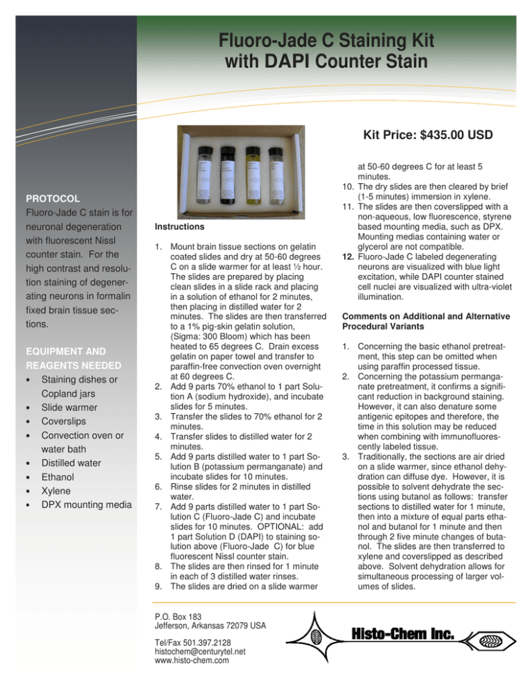
Fluoro-Jade
C Staining
Kit
Information
Technology
Solutions
with DAPI Counter Stain
Kit Price: $435.00 USD
PROTOCOL
Fluoro-Jade C stain is for
neuronal degeneration
with fluorescent Nissl
counter stain. For the
high contrast and resolution staining of degenerating neurons in formalin
fixed brain tissue sections.
EQUIPMENT AND
REAGENTS NEEDED
• Staining dishes or
Copland jars
• Slide warmer
• Coverslips
• Convection oven or
water bath
• Distilled water
• Ethanol
• Xylene
• DPX mounting media
Instructions
1. Mount brain tissue sections on gelatin
coated slides and dry at 50-60 degrees
C on a slide warmer for at least ½ hour.
The slides are prepared by placing
clean slides in a slide rack and placing
in a solution of ethanol for 2 minutes,
then placing in distilled water for 2
minutes. The slides are then transferred
to a 1% pig-skin gelatin solution,
(Sigma: 300 Bloom) which has been
heated to 65 degrees C. Drain excess
gelatin on paper towel and transfer to
paraffin-free convection oven overnight
at 60 degrees C.
2. Add 9 parts 70% ethanol to 1 part Solution A (sodium hydroxide), and incubate
slides for 5 minutes.
3. Transfer the slides to 70% ethanol for 2
minutes.
4. Transfer slides to distilled water for 2
minutes.
5. Add 9 parts distilled water to 1 part Solution B (potassium permanganate) and
incubate slides for 10 minutes.
6. Rinse slides for 2 minutes in distilled
water.
7. Add 9 parts distilled water to 1 part Solution C (Fluoro-Jade C) and incubate
slides for 10 minutes. OPTIONAL: add
1 part Solution D (DAPI) to staining solution above (Fluoro-Jade C) for blue
fluorescent Nissl counter stain.
8. The slides are then rinsed for 1 minute
in each of 3 distilled water rinses.
9. The slides are dried on a slide warmer
P.O. Box 183
Jefferson, Arkansas 72079 USA
Tel/Fax 501.397.2128
histochem@centurytel.net
www.histo-chem.com
at 50-60 degrees C for at least 5
minutes.
10. The dry slides are then cleared by brief
(1-5 minutes) immersion in xylene.
11. The slides are then coverslipped with a
non-aqueous, low fluorescence, styrene
based mounting media, such as DPX.
Mounting medias containing water or
glycerol are not compatible.
12. Fluoro-Jade C labeled degenerating
neurons are visualized with blue light
excitation, while DAPI counter stained
cell nuclei are visualized with ultra-violet
illumination.
Comments on Additional and Alternative
Procedural Variants
1. Concerning the basic ethanol pretreatment, this step can be omitted when
using paraffin processed tissue.
2. Concerning the potassium permanganate pretreatment, it confirms a significant reduction in background staining.
However, it can also denature some
antigenic epitopes and therefore, the
time in this solution may be reduced
when combining with immunofluorescently labeled tissue.
3. Traditionally, the sections are air dried
on a slide warmer, since ethanol dehydration can diffuse dye. However, it is
possible to solvent dehydrate the sections using butanol as follows: transfer
sections to distilled water for 1 minute,
then into a mixture of equal parts ethanol and butanol for 1 minute and then
through 2 five minute changes of butanol. The slides are then transferred to
xylene and coverslipped as described
above. Solvent dehydration allows for
simultaneous processing of larger volumes of slides.
Fluoro-Jade C Staining Kit
with DAPI Counter Stain—
Page 2
Troubleshooting
Q: The tissue wrinkles or falls off slides
when processing.
A: Use proper slide gelling procedure (see
processing procedure described above).
ST AI NI NG KI T S
Black Gold II Staining Kit
with Congo Red Counter
Stain
Black Gold II Staining Kit
with Toluidine Blue O
Counter Stain
Fluoro-Jade C Staining
Kit with DAPI Counter
Stain
SECURE ONLINE
ORDERING
Histo-Chem, Inc. adheres to international
PCI (payment card industry) compliance
standards for data security. Visit us at
www.histo-chem.com to
view our products.
Q: The staining is present, but has low contrast (high background).
A: Reduce dye concentration or increase
time in KMnO4.
Q: The staining is present, but faint.
A: Increase the FJ-C concentration or reduce time in KMnO4.
Q: The stain is present after final rinse, but
lost following coverslipping.
A: Air dry slides rather than ethanol dehydration and avoid mounting media that contain polar solvents (eg. water, ethanol, glycerin).
Q: What if there is no staining.
A: May be due to absence of neurodegeneration – verify by running positive control
(eg. kainic acid 10mg/kg i.p.) (Sigma). Fixative may consist of 10% formalin or 4%
paraformaldhyde dissolved in either neutral
phosphate buffer or physiological saline.
Survey view of the hippocampus of a rat exposed to
kainic acid. The section was triple labeled with FluoroJade C and DAPI staining combined with GFAP immunohistochemistry. The section reveals extensive
green Fluoro-Jade C positive neuronal degeneration
throughout the entire CA-1 region of the hippocampus.
The underlying blue viable positive granule cells of the
dentate gyrus are only DAPI positive. Both regions
exhibit red GFAP positive hypertrophied astrocytes.
Triple exposure combining ultraviolet, blue and green
light epi-fluorescent illumination. 10X mag.
P.O. Box 183
Jefferson, Arkansas 72079 USA
Copyright © 2011
Histo-Chem, Inc.
All Rights Reserved
View of the superficial layers of the cingulated cortex
of a rat exposed to kainic acid. Layer I contains conspicuous Fluoro-Jade C positive degenerating axon
terminals. Layer II contains densely packed DAPIpositive viable granule cells. Layer III contains a mixture of Fluoro-Jade C positive degenerating pyramidal
cells and DAPI positive viable pyramidal cells. Double
exposure using combined blue and ultraviolet epifluorescent illumination. 20X mag.
Tel/Fax 501.397.2128
histochem@centurytel.net
www.histo-chem.com

