The Effects of Positive End-Expiratory Pressure During Active
advertisement
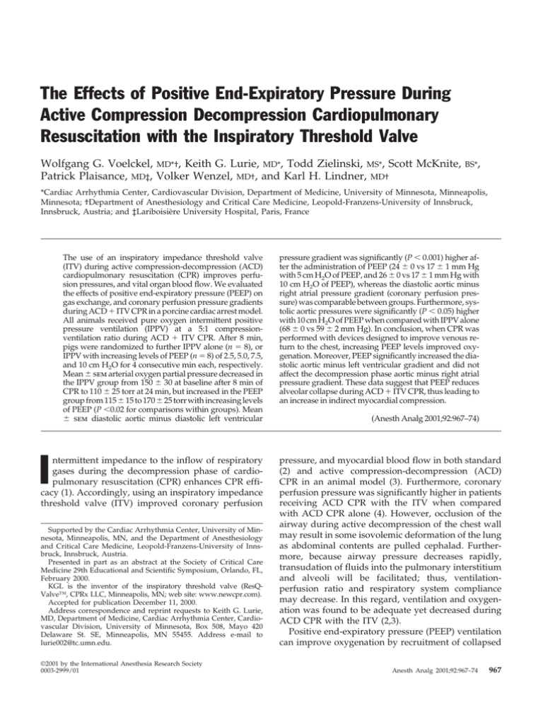
The Effects of Positive End-Expiratory Pressure During Active Compression Decompression Cardiopulmonary Resuscitation with the Inspiratory Threshold Valve Wolfgang G. Voelckel, MD*†, Keith G. Lurie, MD*, Todd Zielinski, MS*, Scott McKnite, Patrick Plaisance, MD‡, Volker Wenzel, MD†, and Karl H. Lindner, MD† BS*, *Cardiac Arrhythmia Center, Cardiovascular Division, Department of Medicine, University of Minnesota, Minneapolis, Minnesota; †Department of Anesthesiology and Critical Care Medicine, Leopold-Franzens-University of Innsbruck, Innsbruck, Austria; and ‡Lariboisière University Hospital, Paris, France The use of an inspiratory impedance threshold valve (ITV) during active compression-decompression (ACD) cardiopulmonary resuscitation (CPR) improves perfusion pressures, and vital organ blood flow. We evaluated the effects of positive end-expiratory pressure (PEEP) on gas exchange, and coronary perfusion pressure gradients during ACD ⫹ ITV CPR in a porcine cardiac arrest model. All animals received pure oxygen intermittent positive pressure ventilation (IPPV) at a 5:1 compressionventilation ratio during ACD ⫹ ITV CPR. After 8 min, pigs were randomized to further IPPV alone (n ⫽ 8), or IPPV with increasing levels of PEEP (n ⫽ 8) of 2.5, 5.0, 7.5, and 10 cm H2O for 4 consecutive min each, respectively. Mean ⫾ sem arterial oxygen partial pressure decreased in the IPPV group from 150 ⫾ 30 at baseline after 8 min of CPR to 110 ⫾ 25 torr at 24 min, but increased in the PEEP group from 115 ⫾ 15 to 170 ⫾ 25 torr with increasing levels of PEEP (P ⬍0.02 for comparisons within groups). Mean ⫾ sem diastolic aortic minus diastolic left ventricular I ntermittent impedance to the inflow of respiratory gases during the decompression phase of cardiopulmonary resuscitation (CPR) enhances CPR efficacy (1). Accordingly, using an inspiratory impedance threshold valve (ITV) improved coronary perfusion Supported by the Cardiac Arrhythmia Center, University of Minnesota, Minneapolis, MN, and the Department of Anesthesiology and Critical Care Medicine, Leopold-Franzens-University of Innsbruck, Innsbruck, Austria. Presented in part as an abstract at the Society of Critical Care Medicine 29th Educational and Scientific Symposium, Orlando, FL, February 2000. KGL is the inventor of the inspiratory threshold valve (ResQValve™, CPRx LLC, Minneapolis, MN; web site: www.newcpr.com). Accepted for publication December 11, 2000. Address correspondence and reprint requests to Keith G. Lurie, MD, Department of Medicine, Cardiac Arrhythmia Center, Cardiovascular Division, University of Minnesota, Box 508, Mayo 420 Delaware St. SE, Minneapolis, MN 55455. Address e-mail to lurie002@tc.umn.edu. ©2001 by the International Anesthesia Research Society 0003-2999/01 pressure gradient was significantly (P ⬍ 0.001) higher after the administration of PEEP (24 ⫾ 0 vs 17 ⫾ 1 mm Hg with 5 cm H2O of PEEP, and 26 ⫾ 0 vs 17 ⫾ 1 mm Hg with 10 cm H2O of PEEP), whereas the diastolic aortic minus right atrial pressure gradient (coronary perfusion pressure) was comparable between groups. Furthermore, systolic aortic pressures were significantly (P ⬍ 0.05) higher with 10 cm H2O of PEEP when compared with IPPV alone (68 ⫾ 0 vs 59 ⫾ 2 mm Hg). In conclusion, when CPR was performed with devices designed to improve venous return to the chest, increasing PEEP levels improved oxygenation. Moreover, PEEP significantly increased the diastolic aortic minus left ventricular gradient and did not affect the decompression phase aortic minus right atrial pressure gradient. These data suggest that PEEP reduces alveolar collapse during ACD ⫹ ITV CPR, thus leading to an increase in indirect myocardial compression. (Anesth Analg 2001;92:967–74) pressure, and myocardial blood flow in both standard (2) and active compression-decompression (ACD) CPR in an animal model (3). Furthermore, coronary perfusion pressure was significantly higher in patients receiving ACD CPR with the ITV when compared with ACD CPR alone (4). However, occlusion of the airway during active decompression of the chest wall may result in some isovolemic deformation of the lung as abdominal contents are pulled cephalad. Furthermore, because airway pressure decreases rapidly, transudation of fluids into the pulmonary interstitium and alveoli will be facilitated; thus, ventilationperfusion ratio and respiratory system compliance may decrease. In this regard, ventilation and oxygenation was found to be adequate yet decreased during ACD CPR with the ITV (2,3). Positive end-expiratory pressure (PEEP) ventilation can improve oxygenation by recruitment of collapsed Anesth Analg 2001;92:967–74 967 968 CRITICAL CARE AND TRAUMA VOELCKEL ET AL. PEEP DURING ACTIVE COMPRESSION DECOMPRESSION CPR WITH THE INSPIRATORY THRESHOLD VALVE alveolar spaces, thus reducing intrapulmonary shunting (5). However, an increase of alveolar volume can be associated with larger intrathoracic pressures, and impairment of venous return during CPR (6 – 8). The administration of PEEP has been previously described to decrease venous return and perfusion pressures in normovolemic animals undergoing standard chest compressions (8). With respect to pulmonary gas exchange, increasing PEEP levels resulted in increased oxygenation, carbon dioxide elimination, and subsequent restoration of pH to normal values (7,8). We hypothesized that the potential benefit of PEEP on forward blood flow and alveolar recruitment is dependent on several variables, including the amount of circulating blood volume (8) and the method of CPR. Given the fact that ACD ⫹ ITV CPR enhances cardiac output, and prevents inflow of respiratory gases, we postulated that increasing levels of PEEP may improve oxygenation during CPR. Accordingly, the purpose of the present investigation was to evaluate the effects of PEEP on oxygenation, and perfusion pressures during ACD CPR with the ITV. Methods Surgical Preparation This project was approved by the Committee on Animal Experimentation at the University of Minnesota, and the animals were managed in accordance with the American Physiological Society, institutional guidelines, and Position of the American Heart Association on Research Animal Use, as adopted on November 11, 1984. Animal care and use was performed by qualified individuals, supervised by veterinarians, and all facilities and transportation complied with current legal requirements and guidelines. Anesthesia was used in all surgical interventions, all unnecessary suffering was avoided, and research was terminated if unnecessary pain or fear resulted. Our animal facilities meet the standards of the American Association for Accreditation of Laboratory Animal Care. The study was performed according to Utstein-style guidelines (9) on 16 healthy, 4 – 6-wk-old female domestic farm pigs weighing 30 to 36 kg. The pigs were anesthetized with a single bolus dose of ketamine (20 mg/kg IM), pentobarbital (15 mg/kg IV bolus followed by 15 mg/kg per hour IV infusion), and morphine (2 mg bolus) given via an ear vein. The pigs were tracheally intubated during spontaneous respiration with a 7.5-mm cuffed endotracheal tube. During the preparation time, animals were mechanically ventilated at a volume controlled setting of 20 mL/kg, and respiratory frequency was adjusted at 10 –12 breaths/min to maintain the mean arterial carbon dioxide partial pressure at 35 torr; inspiratory oxygen concentration was 21%. Normal saline (10 mL · kg⫺1 · h⫺1) was administered continuously ANESTH ANALG 2001;92:967–74 throughout the preparation and study period with an infusion pump. Multiple catheters were used for hemodynamic monitoring and measurements. Left ventricular and ascending aortic arch blood pressures were monitored by using a single high-fidelity micromanometer-tipped catheter. It was positioned under fluoroscopic guidance by femoral cutdown. To monitor right atrial pressures, another micromanometer-tipped catheter was inserted through a right jugular vein sheath. Intratracheal pressure was obtained with a single high-fidelity micromanometertipped catheter, placed through the intratracheal tube, and positioned approximately 2 cm above the carina. The inlet of this micromanometer-tipped catheter was sealed to avoid any air leakage. Body temperature was maintained between 38.0° and 39.0°C with a heating blanket. Five minutes before induction of ventricular fibrillation, 5000 U IV of sodium heparin was administered to prevent intracardiac clot formation. Pressure tracings obtained from the high-fidelity micromanometer-tipped catheters were continuously monitored with a data acquisition and recording system. Digitized data were analyzed electronically to provide hemodynamic measurements. Accordingly, the arteriovenous pressure difference (time-coincident difference between aortic and right atrial pressure), and arterio-left ventricular pressure difference (timecoincident difference between aortic and left ventricular pressure) were calculated during end diastole (peak of active decompression of the chest wall). Intratracheal pressure changes and the time course of ACD CPR were continuously recorded, and stored on representative data files for each sequence of the experiment. These data were further analyzed, and mean ⫾ sem airway pressures were plotted for each sequence of the experimental protocol. Expired gas volume was measured with a calibrated waterseal spirometer. Plateau airway pressure was assessed with an intratracheal airway pressure transducer during pressure-controlled ventilation with an automatic ventilator. Static compliance (mL/cm H2O) was calculated subsequently according to standard formulas (exhaled tidal volume ⫼ [plateau airway pressure ⫺ PEEP]). Arterial and mixed venous blood was sampled, and analyzed with a blood gas machine every 4 min. Ventricular fibrillation was induced with a single 5-s administration of alternating current via an indwelling bipolar pacing catheter, and ventilation was discontinued. After 4 min of cardiac arrest, ACD ⫹ ITV CPR was performed with a 9.0-cm silicon suction cup positioned fluoroscopically over the right and left ventricles. It was attached to a pneumatically driven automatic piston device as described previously (3). The compression rate was 80/min with a 50% duty cycle, a depth of 25% of the anterior-posterior diameter of the chest wall, and a velocity of 7.5 in./s. Active ANESTH ANALG 2001;92:967–74 CRITICAL CARE AND TRAUMA VOELCKEL ET AL. PEEP DURING ACTIVE COMPRESSION DECOMPRESSION CPR WITH THE INSPIRATORY THRESHOLD VALVE decompression was performed with a 9.0-cm silicon suction cup to produce a sternal displacement of 10% more than the resting anterior-posterior diameter. Compression and decompression excursions were continuously monitored and adjusted with the control module, as necessary, during the experiment. During the first 8 min of CPR, all pigs received pressurecontrolled pure oxygen (Fio2 ⫽ 1.0) ventilation at a constant flow rate (160 L/min) with the aforementioned semiautomatic ventilator, which was manually controlled to achieve a 5:1 compression-ventilation ratio. After 8 min of ACD ⫹ ITV CPR, animals were randomly assigned to receive either further intermittent positive pressure ventilation alone (n ⫽ 8) or increasing levels of PEEP with 2.5, 5.0, 7.5, and 10 cm H2O for 4 consecutive min each (n ⫽ 8), respectively. PEEP was delivered by using a PEEP valve, which was manually adjusted every 4 min. The inspiratory threshold valve (ResQ-Valve™; CPRx, Minneapolis, MN) used in this experiment is a small, single-use one-way valve which is placed between the endotracheal tube and the ventilator, thus becoming part of the respiration circuit. While providing no resistance to active ventilation or expiration, the impedance valve functions to occlude inspiratory gas exchange during the decompression phase of CPR (Fig. 1). This results in a decrease of intrathoracic pressure, thereby promoting blood return to the thorax during the decompression phase of CPR. Given the possibility that the animals will regain their own ventilatory drive during the experiment, or that decompression force may generate a negative airway pressure of more than 21 cm of water, a safety check valve is attached to the side of the ITV. Accordingly, the safety check valve allows air to bypass the diaphragm system by entering a separate side port when pressure more than 21 cm of water is applied to the patient port (Fig. 1). Immediately after acquiring the last blood sample during CPR (i.e., after a total of 28 min of CPR, including 4 min of ventricular fibrillation), we attempted to restore spontaneous circulation with three successive 200-J DC shocks. The return of spontaneous circulation was defined as an unassisted pulse with a systolic blood pressure of at least 50 mm Hg lasting at least 5 min. After finishing the experimental protocol, the animals were euthanized with an overdose of pentobarbital and potassium chloride; all pigs were then necropsied to check correct positioning of the catheters, damage to the rib cage, and internal organs. All values were expressed as mean ⫾ sem. All data were stored on a computer system (Power Macintosh 7100/66), and statistical assessments were performed by using GB Stat™ software (Scolari, Sage Publications, London, UK; web site: www.statistics.com). The comparability of weight and baseline data was tested 969 with the t-test for continuous variables. One way analysis of variance was used to determine statistical significance between groups, and paired Student’s t-test (two-tailed) was used for comparisons within treatment groups. Statistical significance was considered to be at P ⬍ 0.05. Results Before the induction of cardiac arrest, there were no statistically significant differences in weight, temperature, hemodynamic variables, and blood gases between groups (Table 1). With the administration of PEEP, arterial partial pressure of oxygen significantly improved when compared with baseline values obtained after 8 min of ACD ⫹ ITV CPR. When intermittent positive pressure ventilation was maintained without PEEP, arterial oxygen pressure significantly decreased at corresponding time points when compared with baseline. Although tidal volume decreased after the application of 7.5 and 10 cm H2O of PEEP, static compliance significantly improved from baseline values at Minute 8 when 7.5 cm H2O of PEEP was administered (Table 1). Figures 2 and 3 demonstrate the changes in intratracheal pressure after a single ventilation, and four subsequent compression-decompression cycles. In these representative experiments, intratracheal pressure decreased below atmospheric pressures during both the compression and decompression phases of ACD ⫹ ITV CPR within two compression cycles after each breath. Airway pressures in the trachea, at a level just above the carina, remained subatmospheric with PEEP levels below 5 cm H2O. When 10 cm H2O of PEEP was applied, intratracheal pressure changes were significantly (P ⬍ 0.05) lower during ventilation in the PEEP group (24 ⫾ 1 vs 30 ⫾ 2 cm H2O), but significantly (P ⬍ 0.01) higher during the fourth compression-decompression phase when compared with intermittent positive pressure ventilation (IPPV) alone (15 ⫾ 1 vs 11 ⫾ 2 cm H2O). Moreover, with each incremental increase in PEEP, both maximal and minimal airway pressures shifted by the degree of PEEP applied such that the differences in intratracheal pressures in the IPPV group alone versus the IPPV with PEEP group were defined by the level of PEEP. The coronary perfusion pressure (CPP), defined as the aortic diastolic minus right atrial diastolic pressure gradient was comparable in both groups during the study period. In contrast, when the CPP was defined as the aortic diastolic minus left ventricular diastolic pressure gradient, it was significantly (P ⬍ 0.001) higher when using PEEP (Fig. 4). When 10 cm H2O of PEEP was applied, aortic and left ventricular pressures were significantly higher at maximal compression during CPR when compared with intermittent 970 CRITICAL CARE AND TRAUMA VOELCKEL ET AL. PEEP DURING ACTIVE COMPRESSION DECOMPRESSION CPR WITH THE INSPIRATORY THRESHOLD VALVE ANESTH ANALG 2001;92:967–74 Figure 1. Components and function of the inspiratory threshold valve. The valve consists of a clear polycarbonate plastic hard shell containing a silicon rubber diaphragm, which occludes the airflow through the patient port, but only when a negative pressure is applied at the patient port (i.e., during the decompression phase of cardiopulmonary resuscitation); and a safety check valve, which allows air to bypass the diaphragm system by entering a separate port when pressure more than ⫺20 cm H2O is applied to the patient port (i.e., during active inspiration). Arrows indicate the airflow through the inspiratory impedance threshold valve during chest compression, active ventilation, chest decompression, and spontaneous ventilation. CPR ⫽ cardiopulmonary resuscitation. Table 1. Blood Gases, Tidal Volume, and Static Compliance at Prearrest, and During Cardiopulmonary Resuscitation Cardiopulmonary resuscitation (min) Variable pHa (U) IPPV IPPV ⫹ PEEP Pao2 (torr) IPPV IPPV ⫹ PEEP Paco2 (torr) IPPV IPPV ⫹ PEEP pHv (U) IPPV IPPV ⫹ PEEP Pvo2 (torr) IPPV IPPV ⫹ PEEP Pvco2 (torr) IPPV IPPV ⫹ PEEP VT (mL) IPPV IPPV ⫹ PEEP CRS (mL/cm H2O) IPPV IPPV ⫹ PEEP Prearrest 0–8 0 PEEP 9–12 2.5 PEEP 13–16 5 PEEP 17–20 7.5 PEEP 21–24 10 PEEP 7.40 ⫾ .01 7.47 ⫾ .01 7.36 ⫾ .01 7.30 ⫾ .02 7.31 ⫾ .02 7.29 ⫾ .03 7.29 ⫾ .02 7.26 ⫾ .03 7.27 ⫾ .03 7.24 ⫾ .03 7.26 ⫾ .03 7.23 ⫾ .04 80 ⫾ 2 80 ⫾ 2 150 ⫾ 30 115 ⫾ 15 140 ⫾ 35 135 ⫾ 25 120 ⫾ 25* 155 ⫾ 25† 120 ⫾ 30† 160 ⫾ 25† 110 ⫾ 25* 170 ⫾ 25‡ 34 ⫾ 1 36 ⫾ 1 41 ⫾ 2 48 ⫾ 3 40 ⫾ 3 46 ⫾ 3 41 ⫾ 2 46 ⫾ 3 42 ⫾ 3 50 ⫾ 4 41 ⫾ 3 49 ⫾ 4 7.44 ⫾ .00 7.43 ⫾ .01 7.26 ⫾ .02 7.26 ⫾ .01 7.20 ⫾ .01 7.20 ⫾ .01 7.18 ⫾ .03 7.13 ⫾ .02 7.18 ⫾ .04 7.17 ⫾ .02 7.14 ⫾ .04 7.16 ⫾ .02 40 ⫾ 1 40 ⫾ 2 25 ⫾ 2 28 ⫾ 2 25 ⫾ 3 26 ⫾ 1 25 ⫾ 2 26 ⫾ 2 25 ⫾ 2 27 ⫾ 2 24 ⫾ 2 26 ⫾ 2 41 ⫾ 2 42 ⫾ 2 54 ⫾ 3 56 ⫾ 3 57 ⫾ 3 62 ⫾ 3 59 ⫾ 4 64 ⫾ 3 62 ⫾ 4 64 ⫾ 4 60 ⫾ 5 64 ⫾ 3 660 ⫾ 25 640 ⫾ 20 375 ⫾ 25 385 ⫾ 30 360 ⫾ 25 370 ⫾ 30 350 ⫾ 25 360 ⫾ 30 350 ⫾ 20 355 ⫾ 30† 360 ⫾ 20 345 ⫾ 10* 43 ⫾ 2 41 ⫾ 2 22 ⫾ 5 16 ⫾ 2 21 ⫾ 6 20 ⫾ 4 18 ⫾ 4 18 ⫾ 4 20 ⫾ 5 28 ⫾ 5† 15 ⫾ 2 21 ⫾ 3 All variables are given as mean ⫾ sem. Prearrest indicates measurements before induction of cardiac arrest. IPPV ⫽ intermittent positive pressure ventilation, IPPV ⫹ PEEP ⫽ intermittent positive pressure ventilation with incremental PEEP from Minute 8, PEEP ⫽ positive end-expiratory pressure given in cm H2O, pHa ⫽ arterial pH, Pao2 ⫽ arterial oxygen partial pressure, Paco2 ⫽ arterial carbon dioxide partial pressure; pHv ⫽ mixed venous pH, Pvo2 ⫽ mixed venous oxygen partial pressure, Pvco2 ⫽ mixed venous carbon dioxide partial pressure, VT ⫽ tidal volume administered with a 5:1 synchronized compression-ventilation ratio, CRS ⫽ calculated static respiratory system compliance. Fio2 was 0.21 at prearrest, and 1.0 during cardiopulmonary resuscitation. * P ⬍ 0.02 versus values before intervention at Minute 8 for comparison within groups; † P ⬍ 0.05 versus values before intervention at Minute 8 for comparison within groups; ‡ P ⬍ 0.005 versus values before intervention at Minute 8 for comparison within groups. ANESTH ANALG 2001;92:967–74 CRITICAL CARE AND TRAUMA VOELCKEL ET AL. PEEP DURING ACTIVE COMPRESSION DECOMPRESSION CPR WITH THE INSPIRATORY THRESHOLD VALVE Figure 2. Mean ⫾ sem intratracheal pressure during ACD ⫹ ITV CPR with either intermittent positive pressure ventilation (□) or the administration of increasing levels of PEEP given in cm H2O (䉫) with a 5:1 synchronized ventilation-compression ratio. ACD ⫹ ITV CPR indicates active compression-decompression cardiopulmonary resuscitation with the inspiratory impedance threshold valve. IPPV ⫽ intermittent positive pressure ventilation, PEEP ⫽ continuous positive expiratory pressure; *P ⬍ 0.05 for intratracheal pressure change defined as the difference between airway pressure at maximal decompression and at peak compression between groups; †P ⬍ 0.01 for intratracheal pressure change between groups. The time course of ACD CPR includes four compressions, and four decompressions. The time between each compression is 0.75 s. positive pressure ventilation at corresponding time points (P ⬍ 0.05 between groups) (Table 2). Seven of eight animals in the IPPV ⫹ PEEP group, and five of eight animals in the IPPV alone group were successfully defibrillated (data not shown). Necropsy confirmed appropriate catheter positions, and revealed no signs of pulmonary edema or injuries of the rib cage or intrathoracic organs in any animals; however, we did not perform histology studies to determine possible presence of pulmonary edema. Discussion Previous studies have suggested that PEEP decreases CPP when applied during performance of CPR (6,7), 971 Figure 3. Mean ⫾ sem intratracheal pressure during ACD ⫹ ITV CPR with either intermittent positive pressure ventilation (□) or the administration of increasing levels of PEEP given in cm H2O (䉫) with a 5:1 synchronized ventilation-compression ratio. ACD ⫹ ITV CPR indicates active compression-decompression cardiopulmonary resuscitation with the inspiratory impedance threshold valve. IPPV ⫽ intermittent positive pressure ventilation, PEEP ⫽ continuous positive expiratory pressure; *P ⬍ 0.05 for intratracheal pressure change defined as the difference between airway pressure at maximal decompression and at peak compression between groups; †P ⬍ 0.01 for intratracheal pressure change between groups. The time course of ACD CPR includes four compressions, and four decompressions. The time between each compression is 0.75 s. except when the experimental preparation is hypervolemic before arrest (8). We hypothesized that the combination of ACD and the ITV would significantly enhance blood return to the chest, resulting in an increased intrathoracic blood volume. With this relative “intrathoracic hypervolemia,” we reassessed oxygenation and cardiopulmonary circulation after application of positive expiratory airway pressure during ACD ⫹ ITV CPR. Results from this study demonstrate that the combination of ACD CPR with the ITV, and PEEP ventilation maintained CPP during prolonged CPR in a porcine cardiac arrest model. Levels of 5, 7.5, and 10 cm H2O of PEEP improved oxygenation, and static respiratory system compliance. Moreover, PEEP did not decrease the CPP when defined as the diastolic arterio-right atrial pressure difference. However, when defined as the diastolic aortic-left ventricular pressure gradient (10), PEEP significantly increased CPP. Furthermore, defibrillation was successful in 972 CRITICAL CARE AND TRAUMA VOELCKEL ET AL. PEEP DURING ACTIVE COMPRESSION DECOMPRESSION CPR WITH THE INSPIRATORY THRESHOLD VALVE Figure 4. Mean ⫾ sem calculated pressure gradients between diastolic aortic and diastolic right atrial pressure; and between diastolic aortic and diastolic left ventricular pressure during each sequence of ACD ⫹ ITV CPR at increasing levels of PEEP (□) versus intermittent positive pressure ventilation (䡵). ACD ⫹ ITV CPR indicates active compression-decompression cardiopulmonary resuscitation with the inspiratory impedance threshold valve, IPPV ⫽ intermittent positive pressure ventilation, PEEP ⫽ positive end-expiratory pressure. *P ⬍ 0.001 between PEEP and IPPV. Time during CPR is given in minutes. seven of eight animals in the PEEP group after 28 minutes of cardiac arrest. These results from the addition of PEEP during ACD CPR with the ITV underscore the importance of both positive and negative intrathoracic pressure changes during CPR. ACD CPR generates a negative intrathoracic pressure gradient, thus enhancing blood return to the thorax, and vital organ perfusion in animals and humans (11–14). However, when inflow of respiratory gases is further limited with an inspiratory threshold valve, equilibration of the negative intrathoracic pressure generated by active chest wall expansion occurs to a greater extent, and blood return to the thorax is further enhanced. Accordingly, coronary perfusion and vital organ blood flow was subsequently improved in a porcine model of cardiac arrest using ITV during ACD CPR (3). A physiological consequence of intermittent inspiratory impedance is that lungs may be driven to the residual volume during compression because no air is allowed to enter during ANESTH ANALG 2001;92:967–74 decompression unless a negative pressure of 20 cm H2O is exceeded. Although pleural pressures are most likely to be positive during the compression phase, we found airway pressures to become negative after the second compression phase of ACD CPR. We speculate that the combination of negative airway pressure and positive intrathoracic pressure may cause alveolar collapse, thus decreasing the ventilation/perfusion ratio. Moreover, transudation of fluids into the alveoli may be facilitated, which may cause pulmonary edema. As such, during ACD ⫹ ITV CPR there is a physiological tradeoff between coronary perfusion and decreased oxygenation. It is noteworthy that arterial Po2 values exceeded a critical threshold within the oxygenhemoglobin dissociation curve in most cases; accordingly, we speculate that arterial oxygen content was comparable between groups. However, the decrease in oxygenation without further intervention is evident, and impaired oxygen delivery may subsequently affect outcome. Use of PEEP appears to minimize the decrease in oxygenation and may increase blood flow to the heart. Previous studies have shown that with PEEP levels more than 10 cm H2O, increased frequency of ventilation, or continuous positive airway pressure during standard CPR have all improved oxygenation and elimination of carbon dioxide. However, carotid blood flow and CPP were significantly impaired (6,7). The effects of PEEP ventilation on forward blood flow may be closely related to the amount of the circulating blood volume. In this regard, Ido et al. (8) found that PEEP levels more than 5 cm H2O decreased venous return between chest compressions, and subsequently, forward blood flow in normovolemic animals during standard CPR. In hypervolemic animals (i.e., after transfusion of 25 mL/kg blood), higher levels of up to 20 cm H2O of PEEP further improved common carotid artery blood flow. The results in the hypervolemic animals are similar to our current findings. As the use of the ITV in combination with ACD CPR improves blood return to the thorax, and subsequently forward blood flow, the central volume status of these animals may be considered as “hypervolemic.” These data are also consistent with a study focusing on alternative ventilation during CPR (15). In this study, constantflow insufflation in the trachea generated a positive airway pressure of approximately 10 cm H2O with concomitant chest compressions, which was found to maintain adequate gas exchange, and furthermore, to improve systolic aortic pressure and common carotid blood flow (15). Accordingly, the current results suggest that even 10 cm H2O of PEEP improved aortic blood pressure during the compression phase. The diastolic arterio-right atrial pressure difference, which is typically used to calculate CPP (16), was not influenced by PEEP in our study. Interestingly, the diastolic aortic-left ventricular pressure gradient, which ANESTH ANALG 2001;92:967–74 CRITICAL CARE AND TRAUMA VOELCKEL ET AL. PEEP DURING ACTIVE COMPRESSION DECOMPRESSION CPR WITH THE INSPIRATORY THRESHOLD VALVE 973 Table 2. Aortic, Right Atrial, Left Ventricular, and Right Atrial Pressures During Active Compression Decompression Cardiopulmonary Resuscitation with the Inspiratory Threshold Valve Cardiopulmonary resuscitation (min) Variable Ao compression (mm Hg) IPPV IPPV ⫹ PEEP Ao decompression (mm Hg) IPPV IPPV ⫹ PEEP MAP (mm Hg) IPPV IPPV ⫹ PEEP LV compression (mm Hg) IPPV IPPV ⫹ PEEP LV decompression (mm Hg) IPPV IPPV ⫹ PEEP RA decompression (mm Hg) IPPV IPPV ⫹ PEEP 0–8 0 PEEP 9–12 2.5 PEEP 13–16 5 PEEP 17–20 7.5 PEEP 21–24 10 PEEP 61 ⫾ 2 64 ⫾ 2 70 ⫾ 1 67 ⫾ 1 71 ⫾ 1 66 ⫾ 0 67 ⫾ 1 67 ⫾ 1 59 ⫾ 2* 68 ⫾ 0 27 ⫾ 1 27 ⫾ 1 30 ⫾ 0 28 ⫾ 0 32 ⫾ 0 29 ⫾ 0 32 ⫾ 0 29 ⫾ 0 28 ⫾ 1 29 ⫾ 0 38 ⫾ 1 39 ⫾ 1 43 ⫾ 0 41 ⫾ 1 45 ⫾ 0 41 ⫾ 0 44 ⫾ 1 42 ⫾ 0 37 ⫾ 1 42 ⫾ 0 69 ⫾ 4 73 ⫾ 3 83 ⫾ 1 78 ⫾ 1 80 ⫾ 2 76 ⫾ 0 74 ⫾ 1 79 ⫾ 0 64 ⫾ 3* 80 ⫾ 1 6⫾1 5⫾0 13 ⫾ 1† 5⫾0 14 ⫾ 0† 5⫾0 12 ⫾ 1† 4⫾0 8 ⫾ 0† 3⫾0 1⫾0 2⫾0 1⫾0 2⫾1 0 ⫾ 0* 1⫾0 0 ⫾ 0* 2⫾0 0 ⫾ 0* 2⫾0 All variables are given as mean ⫾ sem. Ao ⫽ aortic pressure, MAP ⫽ mean arterial pressure, LV ⫽ left ventricular pressure, RA ⫽ right atrial pressure, IPPV ⫽ intermittent positive pressure ventilation, IPPV ⫹ PEEP ⫽ intermittent positive pressure ventilation with incremental PEEP from Minute 8, PEEP ⫽ positive end-expiratory pressure. * P ⬍ 0.05 between IPPV and PEEP; † P ⬍ 0.001 between IPPV and PEEP. has been considered as an alternative to estimate left ventricular diastolic coronary blood flow (10), was significantly higher when PEEP was administered. In the fibrillating heart, the area between the aortic and left ventricular pressure curves, which reflects the pressure gradient across the left ventricle, provides a reasonable estimate of endomyocardial blood flow (17). We are aware that the functional significance of the observed increase in diastolic aortic-left ventricular pressure gradient remains speculative without knowledge of coronary blood flow. Nevertheless, the hemodynamic findings from our study suggest that PEEP may be helpful to transmit the compression force to the myocardium, thus improving forward blood flow, and subsequently to reduce left ventricular afterload during ventricular fibrillation. Accordingly, PEEP may play an important role when CPR devices, designed to enhance blood flow to the thorax, are used during CPR. This may be supported by the fact that in our pigs, CPP remained at levels that normally cannot be maintained without a vasopressor during prolonged CPR (18). When further analyzing airway pressures in this study, we observed that the first peak airway pressure with a synchronized compression-ventilation ratio of 5:1 was followed by a second similar high peak during the second compression phase (Fig. 2). These findings suggest that a portion of the inspired tidal volume was trapped during the initial compression after ventilation, but completely exhaled during the following compression phase. Air trapping in smaller airways seems to be of significant value for transmitting the force of chest compressions to pulmonary vessels and the myocardium, thus generating a forward blood flow during CPR. In this regard, Halperin et al. (19) demonstrated in a dog model of vest CPR that this air trapping occurs in small airways distal of the carina. Without inspiratory gas exchange at this point, intratracheal pressure rapidly becomes negative. Because the ITV prevents inflow of respiratory gases during the decompression phase, but allows exhalation, airway pressures further remained negative even at maximal compression during continuing ACD ⫹ ITV CPR. With increasing levels of PEEP, we found intratracheal pressures shifted toward positive values, but PEEP did not interfere with the benefits of ACD ⫹ ITV CPR. This new observation demonstrates that there are marked regional differences in pressure within different parts of the thorax that, when manipulated by adjunctive CPR devices, can significantly improve forward blood flow out of the chest. In this regard, PEEP increases the content of respiratory gases within the lungs, which may be beneficial to convert external chest compressions into forward blood flow out of the chest. Moreover, because intratracheal pressure changes were even more pronounced during the fifth chest compression when 10 cm H2O of PEEP were applied, the “bellows-like” pumping action of the thorax may be further improved with the concept of PEEP ventilation. 974 CRITICAL CARE AND TRAUMA VOELCKEL ET AL. PEEP DURING ACTIVE COMPRESSION DECOMPRESSION CPR WITH THE INSPIRATORY THRESHOLD VALVE We observed a decrease in respiratory system compliance during the first eight minutes before intervention, which may indicate the occurrence of alveolar and/or small airway collapse. Accordingly, the nonsignificant difference in arterial oxygen partial pressure between groups at this point may be explained by individual factors such as the quantity of functional residual capacity, and degree of intrapulmonary shunting. Lung compliance diminishes as the duration of cardiac inactivity continues, and static compliance may be as low as 22 mL/cm H2O, especially at low lung volumes (20). With higher volumes, static compliance was found to be approximately 50 mL/cm H2O in patients immediately after termination of CPR efforts (21). We observed compliance values as low as 15 mL/cm H2O in our study, which is in good relation to the results of other investigators using a comparable pig model (22). The etiology of the changes in lung compliance is undoubtedly multifactorial, and vascular congestion as well as alveolar collapse before and during CPR are likely to contribute to the observed decrease in compliance (21). However, we found that the application of 7.5 and 10 cm H2O of PEEP improved calculated static compliance. Some limitations of the present study should be noted. First, we did not measure vital organ blood flow, thus leaving the question unanswered whether the improved diastolic aortic-left ventricular pressure gradient, which we observed in the PEEP group, resulted in higher left ventricular perfusion. Second, we did not measure cardiac output because of the questionable accuracy of the thermodilution technique during extreme low-flow states. Accordingly, we are not able to report on oxygen delivery, and oxygen consumption data. We did not assess the effect of higher values of PEEP during ACD ⫹ ITV CPR, and are not able to state whether higher PEEP levels may have yielded different results. Finally, we incrementally increased PEEP and did not compare this stepwise increase to beginning with a higher amount of PEEP from the start of the study. In conclusion, when CPR was performed with devices designed to improve venous return to the chest, increasing PEEP levels improved oxygenation. Moreover, PEEP significantly increased the diastolic aortic minus left ventricular gradient and did not affect the decompression phase aortic minus right atrial pressure gradient. These data suggest that PEEP reduces alveolar collapse during ACD ⫹ ITV CPR, thus leading to an increase in indirect myocardial compression. References 1. Lurie KG, Voelckel W, Plaisance P, et al. Use of an inspiratory impedance threshold valve during cardiopulmonary resuscitation: a progress report. Resuscitation 2000;44:219 –30. ANESTH ANALG 2001;92:967–74 2. Lurie KG, Mulligan KA, McKnite S, et al. Optimizing standard cardiopulmonary resuscitation with an inspiratory impedance threshold valve. Chest 1998;113:1084 –90. 3. Lurie KG, Coffen P, Shultz J, et al. Improving active compressiondecompression cardiopulmonary resuscitation with an inspiratory impedance valve. Circulation 1995;91:1629 –32. 4. Plaisance P, Lurie KG, Payen D. Inspiratory impedance during active compression decompression cardiopulmonary resuscitation: a randomized evaluation in patients in cardiac arrest. Circulation 2000;101:989 –94. 5. Colgan FJ, Nichols FA, DeWeese JA. Positive end-expiratory pressure, oxygen transport, and the low output state. Anesth Analg 1974;53:538 – 43. 6. Hevesi ZG, Thrush DN, Downs JB, Smith RA. Effect of CPAP on gas exchange during chest compressions. Anesthesiology 1999; 90:1078 – 83. 7. Hodgkin BC, Lambrew CT, Lawrence FH, Evangelos T. Effects of Peep and of increased frequency of ventilation during CPR. Crit Care Med 1980;8:123– 6. 8. Ido Y, Goto H, Lavin MJ, et al. Effects of positive end-expiratory pressure on carotid blood flow during closed-chest cardiopulmonary resuscitation in dogs. Anesth Analg 1982;61:557– 60. 9. Idris AH, Becker LB, Ornato JP, et al. Utstein-style guidelines for uniform reporting of laboratory CPR research. Resuscitation 1996;33:69 – 84. 10. Aversano T, Klocke FJ, Mates RE, Canty JM. Preload-induced alterations in capacitance-free diastolic pressure-flow relationship. Am J Physiol 1984;246(3 Pt 2):H410 –7. 11. Cohen TJ, Tucker KL, Lurie KG, et al. Active compressiondecompression: a new method of cardiopulmonary resuscitation. JAMA 1992;267:2916 –23. 12. Lindner KH, Pfenninger E, Lurie KG, et al. Effects of active compression-decompression resuscitation on myocardial and cerebral blood flow in pigs. Circulation 1993;88:1254 – 63. 13. Plaisance P, Adnet F, Vicaut E. Benefit of active compressiondecompression cardiopulmonary resuscitation as a prehospital advanced cardiac life support: a randomized multicenter study. Circulation 1997;95:955– 61. 14. Plaissance P, Lurie KG, Vicaut E, et al. A comparison of standard cardiopulmonary resuscitation and active compressiondecompression resuscitation for out-of-hospital cardiac arrest. N Engl J Med 1999;341:569 –75. 15. Brochard L, Boussignac G, Adnot S, et al. Efficacy of cardiopulmonary resuscitation using intratracheal inflation. Am J Respir Crit Care Med 1996;154:1323–9. 16. Ditchey RV, Winkler JV, Rhodes CA. Relative lack of coronary blood flow during closed-chest resuscitation in dogs. Circulation 1982;66:297–302. 17. Livesay JJ, Folette DM, Fey KH, et al. Optimizing myocardial supply/demand balance with ␣-adrenergic drugs during cardiopulmonary resuscitation. J Thorac Cardiovasc Surg 1978;76: 244 –51. 18. Wenzel V, Lindner KH, Krismer AC, et al. Survival with full neurological recovery, and no cerebral pathology after prolonged cardiopulmonary resuscitation with vasopressin in pigs. J Am Coll Cardiol 2000;35:527–33. 19. Halperin HR, Brower R, Weisfeldt ML, et al. Air trapping in the lungs during cardiopulmonary resuscitation in dogs: a mechanism for generating changes in intrathoracic pressure. Circ Res 1989;65:946 –54. 20. Ornato JP, Bryson BL, Donovan PJ. Measurement of ventilation during cardiopulmonary resuscitation. Crit Care Med 1983;11: 79 – 82. 21. Davis K, Johannigmann JA, Johnson RC, Branson RD. Lung compliance following cardiac arrest. Acad Emerg Med 1995;2: 874 – 8. 22. Wenzel V, Idris AH, Banner MJ, et al. Respiratory system compliance decreases after cardiopulmonary resuscitation and stomach inflation: impact of large and small tidal volumes on calculated peak airway pressure. Resuscitation 1998;38:113– 8.
