Arrhythmias
advertisement
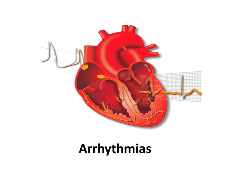
Arrhythmias Arrhythmias - Objectives • • • • • Definition Clinical presentation Diagnosis - ECG analysis Classification Management Sinus rhythm - SA node controls the ventricle on a 1 :1 ratio Definition of arrhythmia Ventricular activity (QRS) is not regulated by SA node on a one to one conduction Interruption in conduction – Heart block Abnormal focus - Atrial tachycardia, VT Re entry circuit - SVT, Atrial flutter, VT Classification of Arrhythmias • Narrow complex tachycardia (NCT) • Broad complex tachycardia (BCT) • Bradycardias and A-V Block Clinical presentations • • • • • • • • • Cardiac arrest - VT,VF, Ventricular asystole Breathlessness Chest pain Dizziness Embolic episode Falls Low GCS Hypotension Syncope Suspected Arrhythmia • Immediate assessment –A –B –C • Establish intravenous access • Attach cardiac monitor • Classify arrhythmia Diagnosis and classification of arrhythmia 1. ? Sinus rhythm (SR) – – P preceding QRS and PR interval equal Assess in lead II or V1 If not in SR 2. Heart rate > 100- Tachyarrhythmia < 60 – Bradyarrhythmia 3. QRS duration < 120 ms - Narrow Complex Tachycardia > 120 ms - Broad Complex Tachycardia 4. Assess whether Regular or Irregular NCT Arrythmia Tachyarrhythmia Narrow QRS Regular Irregular Bradyarrythmia Broad QRS Regular Irregular Regular narrow-complex tachycardia • • Atrial flutter with regular AV block Re entrant tachycardia • AV nodal (AVNRT) • Atrio ventricular(AVRT) • Atrial tachycardia • Abnormal P wave Atrial flutter Diagnosed only in the presence of flutter waves on the ECG Management of Atrial flutter (<48 hrs) • DC Cardioversion – Haemodynamic compromise…. – Normal echo – No underlying cause • Drugs (less effective) – Flecainide better avoided – Amiodarone - drug of choice to revert to SR (50 %) – Ibutilide may reduce the need for DC shock . Re - entrant Tachycardia No P wave present – hidden in ST or T waves Management of Re - entrant tachycardia (AVNRT / AVRT) Vagal manoeuvre • • Carotid sinus massage Valsalva manoeuvre Adenosine test • • NOT in acute asthmatics (bronchospasm) Warn patient of symptoms (chest pain, SOB,flushing) Flecainide • 2mg/Kg ( 30-60) min if adenosine fails. DC cardioversion • • If haemodynamic instability and no response to adenosine or flecainide DC seldom required in Re-entrant tachycardia Management of Re - entrant tachycardia (AVNRT/ AVRT) To prevent recurrence: • Flecainide or B-blocker • Consider Ablation for all Re-entrant tachycardias Irregular narrow-complex tachycardia • Atrial fibrillation • Atrial flutter • Multifocal atrial tachycardia Atrial Fibrillation Atrial fibrillation Classification of Atrial fibrillation • • Acute Atrial fibrillation (within 7 days) Chronic Atrial fibrillation – Paroxysmal: • – Spontaneous termination < 7 days Persistent: • – Continues indefinitely unless cardioverted Permanent: • Cannot restore sinus rhythm Cardiac causes of AF • • • • • • Hypertension Valvular heart disease Coronary Heart disease Cardiomyopathy Post cardiac surgery Pericardial disease Non cardiac causes of Atrial Fibrillation • • • • • • Alcohol Lung disease – Pneumonia, COPD Thyroid disease Sepsis Stroke Post surgery Echocardiography in AF • Identify the cause - Mitral valve, myocardial or pericardial • Assess LV size and Function - H/T, IHD, cardiomyopathy • Assess atrial size /clot Management of acute Atrial fibrillation • If < 48 hrs of onset – – • DC cardioversion if haemodynamic compromise IV Flecainide 2mg/kg over 30-60 min if echocardiography normal. If > 48 hrs Rate control Early Cardioversion after TOE or – Delayed Cardioversion after 4 weeks of warfarin – – Drugs to achieve rate control • Verapamil / Diltiazem – Avoid if IHD, LV impairment • Beta blocker – Usual CI should be observed • Digoxin – often ineffective rate control on exertion hence combined with beta or Ca channel blocker • Amiodarone Rate or Rhythm control in AF? Acute AF Rhythm control Rate control Persistent AF Rhythm control Rate control Permanent AF ? Ablation Paroxysmal AF Rhythm/Ablation Rate-control in AF Try rate control first for patients with Persistent AF: • • • • • > 65 years Duration > 1 year Hypertension with LVH Dilated LA > 5 cm Unsuitable for cardioversion Rhythm control in AF • • • • • Acute AF AF < 6 months < 65 years Structurally normal heart ? Non ischaemic LV dysfunction Maintaining Sinus Rhythm in AF Normal Heart /No CHD Flecanide/Propafenone Disopyramide Mild LVH/CHD Sotalol Significant LVH/HF Beta blocker/ Amiodarone Digoxin not useful for maintaining SR Broad Complex Tachycardia • Clinically Any cardiac rhythm >100 /min, with QRS duration of >0.12s • Electrophysiologically Mostly ventricular in origin, involving automatic focus or re-entry circuit within the ventricles Broad Complex Tachycardia Causes: • Ventricular Tachycardia (VT) • Supraventricular arrythmia with aberrant conduction (BBB, rate related BBB) • WPW with antidromic pathway Default diagnosis is VT. Ventricular Tachycardia Ventricular Tachycardia • Monomorphic Ventricular Tachycardia (>120/min) Accelerated idioventricular rhythm (< 120/min) • – • Polymorphic Ventricular Tachycardia – • AMI most common cause - Rx not necessary Acute MI or ischaemia Long QT interval (Torsades) VT • NSVT- >3 VEs (rate>120) lasting <30s – Prognostically significant in HCM, DCM, post MI with low EF – Rarely in structurally N heart- not associated with risk • VT in structurally normal heart – RVOTT and Fascicular tachycardia – Good prognosis, good response to RFA if symptomatic VT • • • • • • P/H IHD,drugs VA dissociation QRS>140ms QRS>160ms Left axis with RBBB Concordance in chest leads • Fusion or Capture beat “SVT” • No cardiac history Ventricular tachycardia AF with LBBB Assessment of patient with BCT • Immediate assessment –A –B –C • Establish intravenous access • Attach cardiac monitor (ideally with printout) Management of VT If patient compromised (low BP, decreased conscious level, heart failure, chest pain): – Urgent electrical cardio-version If patient not compromised: – – LV not impaired - IV lignocaine If evidence of cardiac failure or poor LV function - IV amiodarone Management of VT Second-Line drugs: – – – – Beta-blockers (avoid in heart failure) Flecanide (avoid if LV impaired or IHD) Mexiletine (do not use in cardiogenic shock) Procainamide (can cause torsade) Causes of VT • • • • • IHD Cardiomyopathy Arrythmogenic RV dysplasia Brugada syndrome Congenital Heart disease Causes of Torsades de pointes (TDP) • Congenital long QT syndromes – Jervell and Lange-Nielsen syndrome – Romano-Ward syndrome • Acquired long QT syndromes – Bradycardia – Electrolytes – Hypokalaemia, Hypomagnesaemia – Drugs Drugs causing long QT Syndromes • • • • • • • Antiarrythmics – Sotalol, Amiodarone, disopyramide H1-receptor antagonists - Terfenadine, astemizole Cholinergic antagonists – Cisapride Antibiotics - Erythromycin, clarithromycin Antifungals - Ketoconazole, itraconazole Psychotropic agents - Haloperidol, phenothiazines Tricyclic and tetracyclic antidepressants Long QT Syndrome ECG in Torsades de pointes • Paroxysms of 5-20 beats, with a heart rate of 200/min • Sustained episodes occasionally can be seen and can degenerate in VF • Complete 180° twist of QRS complexes within 10-12 beats • Torsade occurring in the setting of acquired long QT is preceded by pauses in almost all cases Torsades de points Management of Torsades de points • Give all patients IV magnesium even when Mg normal: – 8mmol stat followed by 2.5mmol/hr infusion • Isoprenaline infusion can help suppress TDP • Overdrive atrial pacing (if no AV block) at rate of 100 bpm is treatment of choice • DC cardioversion if sustained • Stop antiarrhythmic drugs Ventricular Fibrillation Ventricular Fibrillation • Uncoordinated contraction (fibrillation) of the ventricles • Most common arrhythmia in cardiac arrest • Manifests as completely irregular activity on ECG Bradyarrythmias • SA node disease • AV block • First degree • Second degree – Mobitz 1 or Mobitz 11 • Third degree or Complete block • Intraventricular block – Bifasicular, Trifasicular Heart Block First degree AV block First degree AV block • • • • Prolonged PR interval > 200ms Every P wave followed by a QRS complex Can be normal Rarely causes symptoms Mobitz type1/ Wenckebach AV block Mobitz type 1 AV block • Progressive prolongation of PR interval until a QRS complex is “dropped” • Usually benign and requires no treatment Mobitz type 2 Mobitz type 2 block • Intermittently non-conducted P waves • No prolongation of P-R interval • Block occurs in the His-Purkinje system, and therefore the QRS complex is often wide • May progress to complete heart block • Requires pacemaker Complete heart block Complete heart block • Complete dissociation of atrial and ventricular activity • Ventricular escape rhythm of 40 bpm • Many causes • Requires pacemaker insertion Indications for temporary pacing • Cardiac arrest with ventricular asystole or bradycardia • Asystole due to eletrolyte imbalance • Mobitz II or CHB in anterior infarct • CHB in Inferior MI if HR < 40, Ventricular arrythmia Intracardiac defibrillator (ICD) Indications for ICD • Impaired LV with sustained or non sustained VT • Resuscitated VF/VT arrests not due to reversible cause • Patients with Previous MI , LVEF<35%,QRS >120 • Brugada syndrome, ARVD, Long QT syndrome What the Guidelines say…. ICD indications Yes No No Comment Secondary Prevention Absolute redn in mortality with ICD (per annum) 3.5% (VF,unstable VT, VT with poor LV) Primary Prevention - Post MI, EF<0.36, 8% NSVT,+ve EPS -Post MI, QRS>120ms, EF<0.30-0.35 5% - High risk inherited conditions (HOCM, Brugada, LQTS) 2-3% - Post MI, EF<0.30-0.35 2.7% - DCM, EF<0.30-0.35 1.3 - 3% NICE ACC/AHA/ESC ICD –Delivering Shock Any questions?

