Applications for the MACSQuant® VYB
Analysis of fluorescent proteins
Background
The use of flow cytometric techniques for the analysis of cell
populations enables scientists to evaluate cells based on both
intrinsic and extrinsic properties. Using fluorescent markers
such as dyes, antibodies, or fluorescent reporters multiple
characteristics of thousands of cells can be investigated
within a matter of minutes. Fluorescent protein reporters are
used for many applications, including analysis of transcription
factors, cell differentiation, cell expression patterns, protein
localization, and transfection efficiency. However, the wide
array of available fluorescent reporters calls for high-capacity
flow cytometers offering the simultaneous detection
of even closely related fluorescent spectra (table 1). The
MACSQuant® VYB is equipped with three excitation lasers
with wavelengths of 405 nm, 488 nm, and 561 nm and
can detect up to eight different fluorescent colors. This
configuration allows for simultaneous detection of multiple
fluorescent proteins such as GFP, YFP, mCherry, and CFP.
ZsGreen
tdTomato
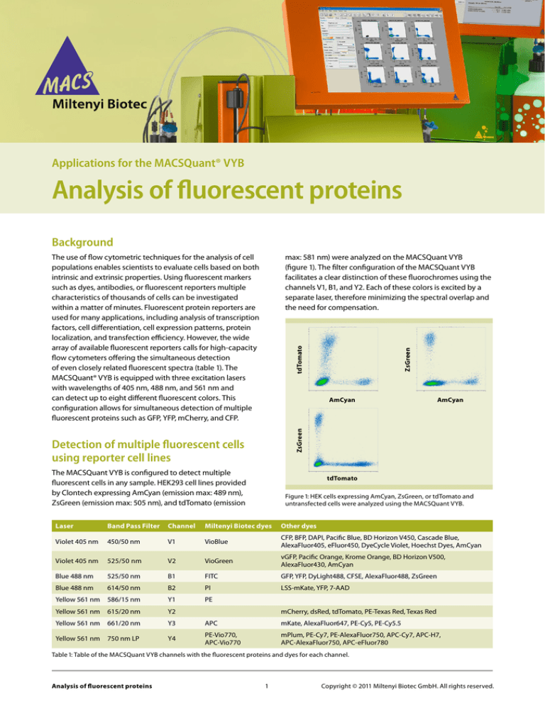
max: 581 nm) were analyzed on the MACSQuant VYB
(figure 1). The filter configuration of the MACSQuant VYB
facilitates a clear distinction of these fluorochromes using the
channels V1, B1, and Y2. Each of these colors is excited by a
separate laser, therefore minimizing the spectral overlap and
the need for compensation.
AmCyan
ZsGreen
AmCyan
Detection of multiple fluorescent cells
using reporter cell lines
The MACSQuant VYB is configured to detect multiple
fluorescent cells in any sample. HEK293 cell lines provided
by Clontech expressing AmCyan (emission max: 489 nm),
ZsGreen (emission max: 505 nm), and tdTomato (emission
tdTomato
Figure 1: HEK cells expressing AmCyan, ZsGreen, or tdTomato and
untransfected cells were analyzed using the MACSQuant VYB.
Laser
Band Pass Filter
Channel
Miltenyi Biotec dyes
Other dyes
Violet 405 nm
450/50 nm
V1
VioBlue
CFP, BFP, DAPI, Pacific Blue, BD Horizon V450, Cascade Blue,
AlexaFluor405, eFluor450, DyeCycle Violet, Hoechst Dyes, AmCyan
Violet 405 nm
525/50 nm
V2
VioGreen
vGFP, Pacific Orange, Krome Orange, BD Horizon V500,
AlexaFluor430, AmCyan
Blue 488 nm
525/50 nm
B1
FITC
GFP, YFP, DyLight488, CFSE, AlexaFluor488, ZsGreen
Blue 488 nm
614/50 nm
B2
PI
LSS-mKate, YFP, 7-AAD
Yellow 561 nm
586/15 nm
Y1
PE
Yellow 561 nm
615/20 nm
Y2
Yellow 561 nm
661/20 nm
Y3
APC
mKate, AlexaFluor647, PE-Cy5, PE-Cy5.5
Yellow 561 nm
750 nm LP
Y4
PE-Vio770,
APC-Vio770
mPlum, PE-Cy7, PE-AlexaFluor750, APC-Cy7, APC-H7,
APC-AlexaFluor750, APC-eFluor780
mCherry, dsRed, tdTomato, PE-Texas Red, Texas Red
Table 1: Table of the MACSQuant VYB channels with the fluorescent proteins and dyes for each channel.
Analysis of fluorescent proteins 1
Copyright © 2011 Miltenyi Biotec GmbH. All rights reserved.
Simultaneous detection of GFP and YFP
A
before compensation
after compensation
YFP
YFP
The simultaneous detection of GFP and YFP can be
challenging with many flow cytometers due to similar
emission wavelengths of the two proteins. However, only
GFP will be excited at 405 nm. Combining this signal and
the YFP signal obtained with a 488 nm laser, discrimination
of both signals is possible (figure 2).
GFP
405 nm
488 nm
525/50
GFP
B
100
CFP
80
60
40
20
mCherry
0
400
500
exitation GFP
emission GFP
exitation YFP
emission YFP
Figure 3: CHO cells transfected with GFP, YFP, mCherry, or CFP were
analyzed with the MACSQuant VYB. GFP and YFP were detected using
the V2 and B1 channels. (A) Dot plot data of GPF and YFP before and
after compensation. (B) Dot plot data of mCherry and CFP.
Conclusions
Figure 2: The excitation and emission spectra of GFP (blue) and YFP
(green). Shaded area represents the bandpass filter used for collection
of the emission spectra of GFP and YFP. The V2 and B1 channels of the
MACSQuant VYB both use the 525/50 filter which enables detection of
GFP and YFP separately.
The MACSQuant VYB is a versatile 3 laser, 10-parameter
flow cytometer that has enhanced capabilities for
detection of fluorescent protein reporters. As shown
here using reporter cell lines, there are a number of
fluorescent markers that can be distinguished using this
instrument. Virtually any cell can be analyzed based
on a number of fluorescent parameters as well as by
size and granularity. The power of flow cytometry to
analyze thousands of cells per second combined with the
ability of the MACSQuant VYB to detect a large range of
fluorescent proteins makes this a powerful tool for a wide
range of research fields, such as stem cell detection and
differentiation, neuroscience, development, cell biology,
cancer research, plant, and marine research.
Using the MACSQuant Analyzer 10 or the MACSQuant VYB,
GFP and YFP can be distinguished by utilizing the 405 nm
laser and the channel V2 to detect GFP fluorescence. At
the 405 nm excitation, only GFP is detectable within this
channel and can therefore be distinguished from the YFP
signal by applying compensation to the B1 channel of
the 488 nm laser. When proper compensation has been
applied, the two cell populations expressing GFP or YFP
can be clearly distinguished (figure 3A).
Further, along with GFP and YFP, many other fluorescent
proteins can be used simultaneously. In the experiment
shown here, GFP, YFP, mCherry, and CFP reporter cell lines
were analyzed in one experiment (figure 3).
Miltenyi Biotec provides products and services worldwide. Visit www.miltenyibiotec.com/local to find your nearest Miltenyi Biotec contact.
MACS, MACSQuant and VioBlue are registered trademarks and VioGreen and Vio770 are trademarks of Miltenyi Biotec GmbH. Cy is a trademark of GE Healthcare
companies. All other trademarks mentioned in this publication are the property of their respective owners and are used for identification purposes only. Unless
otherwise specifically indicated, Miltenyi Biotec products and services are for research use only and not for therapeutic or diagnostic use.
Copyright © 2011 Miltenyi Biotec GmbH. All rights reserved.
Analysis of fluorescent proteins 2
Copyright © 2011 Miltenyi Biotec GmbH. All rights reserved.

