L 1 Human Gametogenesis Example Lab Report...Incomplete
advertisement
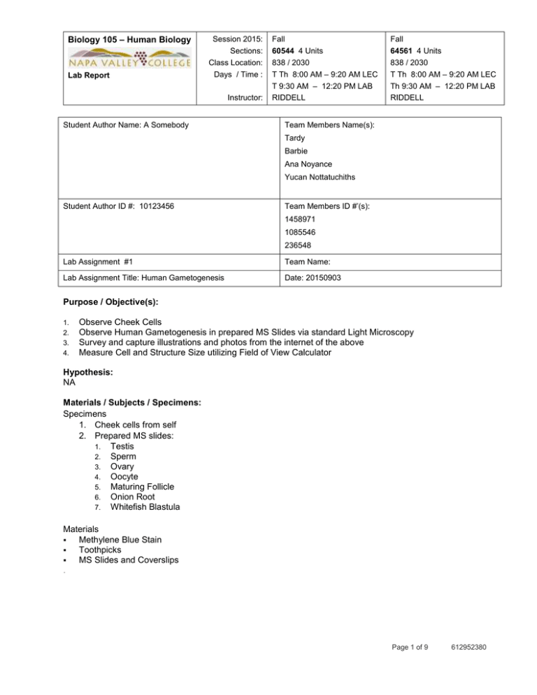
Biology 105 – Human Biology Lab Report Session 2015: Sections: Class Location: Days / Time : Instructor: Student Author Name: A Somebody Fall 60544 4 Units Fall 64561 4 Units 838 / 2030 T Th 8:00 AM – 9:20 AM LEC T 9:30 AM – 12:20 PM LAB RIDDELL 838 / 2030 T Th 8:00 AM – 9:20 AM LEC Th 9:30 AM – 12:20 PM LAB RIDDELL Team Members Name(s): Tardy Barbie Ana Noyance Yucan Nottatuchiths Student Author ID #: 10123456 Team Members ID #’(s): 1458971 1085546 236548 Lab Assignment #1 Team Name: Lab Assignment Title: Human Gametogenesis Date: 20150903 Purpose / Objective(s): 1. 2. 3. 4. Observe Cheek Cells Observe Human Gametogenesis in prepared MS Slides via standard Light Microscopy Survey and capture illustrations and photos from the internet of the above Measure Cell and Structure Size utilizing Field of View Calculator Hypothesis: NA Materials / Subjects / Specimens: Specimens 1. Cheek cells from self 2. Prepared MS slides: 1. Testis 2. Sperm 3. Ovary 4. Oocyte 5. Maturing Follicle 6. Onion Root 7. Whitefish Blastula Materials Methylene Blue Stain Toothpicks MS Slides and Coverslips . Page 1 of 9 612952380 Biology 105 – Human Biology Lab Report Session 2015: Sections: Class Location: Days / Time : Instructor: Fall 60544 4 Units Fall 64561 4 Units 838 / 2030 T Th 8:00 AM – 9:20 AM LEC T 9:30 AM – 12:20 PM LAB RIDDELL 838 / 2030 T Th 8:00 AM – 9:20 AM LEC Th 9:30 AM – 12:20 PM LAB RIDDELL Methods / Tools / Instrumentation / Procedures: Instruments Standard Binocular Light Microscope Brand and model Number…see Photo 1 in Attachments Procedures 1. Prepare Human Cheek cell MS slide using standardized methylene blue stain 2. View Human Cheek cell all magnifications 3. Prepare Human hair MS Slide 4. Viewed Human hair at all magnifications 5. View Human sperm smear, and view ovary at all magnifications 6. Viewed Letter e, Colored Threads as MS practice specimens 7. Viewed Mitosis in Onion Root and White fish Blastula Measurements and Calculations Used the Cell Size FOV On Line Calculator to estimate the sizes of (diameters) of all specimens See Figure 1 in Attachments Results Viewing the target specimens and structures with the standard light microscopes required skill and patience. Making photographs of the specimens using my Cell Phone Camera required skill beyond my ability. Gametogenesis tissues and cells are presented in Table 1 o Male and Female structures Sample measurements from the FOV Calculator are presented in Table 2 Table 3 lists the size measurements for the cells and structures of Gametogenesis and Mitosis Figure 1 compares the cells and structure sizes of Gametogenesis and Mitosis Specimens Analysis / Discussion: Compare and Contrast … …. …. ….. Page 2 of 9 612952380 Biology 105 – Human Biology Lab Report Session 2015: Sections: Class Location: Days / Time : Instructor: Fall 60544 4 Units Fall 64561 4 Units 838 / 2030 T Th 8:00 AM – 9:20 AM LEC T 9:30 AM – 12:20 PM LAB RIDDELL 838 / 2030 T Th 8:00 AM – 9:20 AM LEC Th 9:30 AM – 12:20 PM LAB RIDDELL Research Phases of Mitosis http://www.biology.iupui.edu/biocourses/N100/images/8mitosiscropped.jpg Phases of Meiosis Page 3 of 9 612952380 Biology 105 – Human Biology Lab Report Session 2015: Sections: Class Location: Days / Time : Instructor: Fall 60544 4 Units Fall 64561 4 Units 838 / 2030 T Th 8:00 AM – 9:20 AM LEC T 9:30 AM – 12:20 PM LAB RIDDELL 838 / 2030 T Th 8:00 AM – 9:20 AM LEC Th 9:30 AM – 12:20 PM LAB RIDDELL Conclusions/Further Considerations: Take Away The Microscope is a valuable essential device for the study of cells and histology. ……….. …………. References 1. http://webnt.calhoun.edu/distance/internet/natural/anatomy/EndoRepro/ovary4X.html 2. http://faculty.baruch.cuny.edu/jwahlert/bio1003/mitosis.html Page 4 of 9 612952380 Biology 105 – Human Biology Lab Report Session 2015: Sections: Class Location: Days / Time : Instructor: Fall 60544 4 Units Fall 64561 4 Units 838 / 2030 T Th 8:00 AM – 9:20 AM LEC T 9:30 AM – 12:20 PM LAB RIDDELL 838 / 2030 T Th 8:00 AM – 9:20 AM LEC Th 9:30 AM – 12:20 PM LAB RIDDELL ATTACHMENTS: Table 1: Illustrations and Photomicrographs of cells- Human Gametogenesis, (Testis, Sperm, and Ovary) and Human Development Table 1 Human Gametogenesis Specimen Key Events Testis Spermatogenesis Meiosis Web Reference Notes / Description /Illustration Low Magnification Photo Medium Magnification Photo High Magnification Photo http://www.bing.com/images/search?q=Testis+M odel&vie w=detailv2&&id=FAD0D6375C7F1CBDF8FEC27A94A3F 4A91CACA823&selectedIndex=40&ccid=uFxvgOai&simid http://www.bing.com/images/search?q=Testis+M odel&vie w=detailv2&id=6AABF80730719E94F749F699174AEAE B6C746A45&selectedindex=38&ccid=%2BTEhpdiO&simid http://www.bing.com/images/search?q=human+testis+med ium+magnification+histology&view=detailv2&&id=346B81 0E391D82012C85F162172A3994BEA93759&selectedInd http://www.bing.com/images/search?q=human+testis+med ium+magnification+histology&view=detailv2&id=DA3C78B 78196F1733D03E3FB7A7C374046CC0A09&selectedind Very High Magnification Photo Cell Size Sperm Maturation Discharge into Lumen of Seminiferous Tubuls Web Reference http://bigthink.com/ideafeed/how-sequencing-spermcould-help-fight-cancer Cell Size Ovary Oogenesis by Meiosis Web Reference Cell Size Oocytes Mature from Primary to Secondary Web Reference http://www.firstscience.com/home/images/stories/egg.jpg Cell Size Mature Follicle Web Reference Cell Size Page 5 of 9 612952380 Biology 105 – Human Biology Lab Report Session 2015: Sections: Class Location: Days / Time : Instructor: Fall 60544 4 Units Fall 64561 4 Units 838 / 2030 T Th 8:00 AM – 9:20 AM LEC T 9:30 AM – 12:20 PM LAB RIDDELL 838 / 2030 T Th 8:00 AM – 9:20 AM LEC Th 9:30 AM – 12:20 PM LAB RIDDELL Table 2: Specimens of Gametogenesis and estimated sizes in mm and microns Average Size mm Average Size μm Human Sperm 0.00 2.8 Onion Root Cell 0.01 14.6 Blastula Cell 0.02 15.2 My Hair 0.02 22.9 Cheek Cell 0.06 55.9 Colored Threads 0.08 76.1 Human Oocyte 0.15 145.8 White Fish Blastula 0.70 700.0 Letter e 1.05 1053.5 Corpus Luteum 3.50 3500.0 Specimen Page 6 of 9 612952380 Biology 105 – Human Biology Lab Report Session 2015: Sections: Class Location: Days / Time : Instructor: Fall 60544 4 Units Fall 64561 4 Units 838 / 2030 T Th 8:00 AM – 9:20 AM LEC T 9:30 AM – 12:20 PM LAB RIDDELL 838 / 2030 T Th 8:00 AM – 9:20 AM LEC Th 9:30 AM – 12:20 PM LAB RIDDELL Figure 1 Estimated cell Size in mm Average Size mm 3.50 3.00 2.50 Human Sperm Onion Root Cell Blastula Cell 2.00 My Hair Cheek Cell Colored Threads 1.50 Human Oocyte White Fish Blastula Letter e 1.00 Corpus Luteum 0.50 0.00 Human Sperm Onion Root Cell Blastula Cell My Hair Cheek Cell Colored Threads Human Oocyte White Fish Blastula Letter e Corpus Luteum Page 7 of 9 612952380 Biology 105 – Human Biology Session 2015: Sections: Class Location: Days / Time : Lab Report Instructor: Fall 60544 4 Units Fall 64561 4 Units 838 / 2030 T Th 8:00 AM – 9:20 AM LEC T 9:30 AM – 12:20 PM LAB RIDDELL 838 / 2030 T Th 8:00 AM – 9:20 AM LEC Th 9:30 AM – 12:20 PM LAB RIDDELL Illustration 1 Example of Cell Size Calculator FOV Field of View Check / Set Specimen Size Therefore OCULAR MAG Therefore The Field of View Diameter is Magnification Name your specimens here Therefore OBJECTIVE MAG TOTAL MAG 1,000 Specimen Estimated Count * Pi times radius Put your estimate here squared - How many specimens (S) could be placed "shoulder to shoulder?" in a straight line along the diameter of the FOV? For a Specimen with a Round Form Ratio The Specimen Diameter = FOV Area in μm squared FOV / Specimen units in mm 1,000,000 1 The Field of View Area is FOV FOV Area in FOV Diameter Diameter in mm in μm mm squared Size for Round and Spherical Set Ocular and Objective Powers here Count = units in microns 1,000 mm μm Cheek Cell 10 4 40 3.50 3500 9.62 9,621,150 59.0 0.017 0.06 59 Cheek Cell 10 10 100 1.40 1400 1.54 1,539,384 24.0 0.042 0.06 58 Cheek Cell 10 40 400 0.35 350 0.10 96,212 7.0 0.143 0.05 50 10 63 630 0.22 222 0.04 38,785 292.5 1.4 1368.1 2.8 Cheek Cell AVERAGE Specimen 2,823,883 Set Ocular and Objective Powers here 30.0 0.1 Count = 0.056 55.9 mm μm Colored Threads 10 4 40 3.50 3500 9.62 9,621,150 35.0 0.029 0.10 100 Colored Threads 10 10 100 1.40 1400 1.54 1,539,384 20.0 0.050 0.07 70 Colored Threads 10 40 400 0.35 350 0.10 96,212 6.0 0.167 0.06 58 Colored Threads 10 63 630 0.22 222 0.04 38,785 292.5 1.4 1368.1 2.8 AVERAGE 2,823,883 20.3 0.1 0.1 Page 8 of 9 76.1 612952380 Biology 105 – Human Biology Lab Report Session 2015: Sections: Class Location: Days / Time : Instructor: Fall 60544 4 Units Fall 64561 4 Units 838 / 2030 T Th 8:00 AM – 9:20 AM LEC T 9:30 AM – 12:20 PM LAB RIDDELL 838 / 2030 T Th 8:00 AM – 9:20 AM LEC Th 9:30 AM – 12:20 PM LAB RIDDELL Photo 1 Microscope Jkosdfnglnvlfvdflpvpsdfkvpfv Page 9 of 9 612952380
