Lab 4 Cell Structure
advertisement
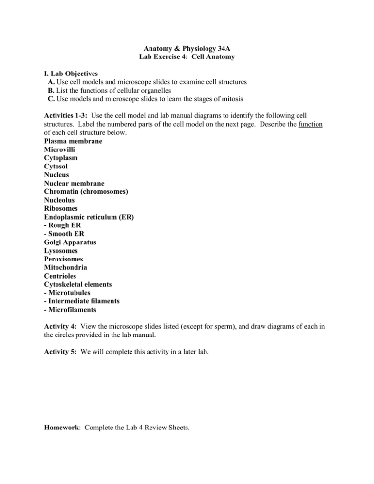
Anatomy & Physiology 34A Lab Exercise 4: Cell Anatomy I. Lab Objectives A. Use cell models and microscope slides to examine cell structures B. List the functions of cellular organelles C. Use models and microscope slides to learn the stages of mitosis Activities 1-3: Use the cell model and lab manual diagrams to identify the following cell structures. Label the numbered parts of the cell model on the next page. Describe the function of each cell structure below. Plasma membrane Microvilli Cytoplasm Cytosol Nucleus Nuclear membrane Chromatin (chromosomes) Nucleolus Ribosomes Endoplasmic reticulum (ER) - Rough ER - Smooth ER Golgi Apparatus Lysosomes Peroxisomes Mitochondria Centrioles Cytoskeletal elements - Microtubules - Intermediate filaments - Microfilaments Activity 4: View the microscope slides listed (except for sperm), and draw diagrams of each in the circles provided in the lab manual. Activity 5: We will complete this activity in a later lab. Homework: Complete the Lab 4 Review Sheets. Anatomy 30 Lab 3: Cells Label the numbered parts of the cell model below and include in your lab notebook.


