A CFD STUDY OF THE EFFECTS OF PHYSIOLOGICAL... OXYGEN TRANSPORT IN CORONARY ARTERIES
advertisement
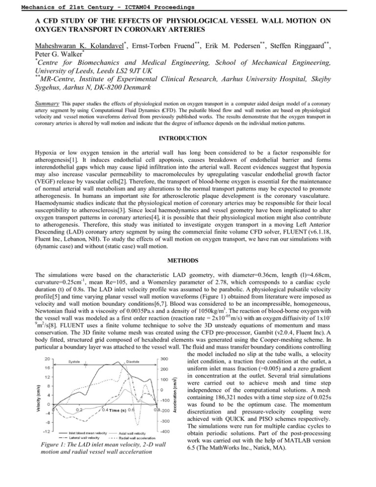
Mechanics of 21st Century - ICTAM04 Proceedings A CFD STUDY OF THE EFFECTS OF PHYSIOLOGICAL VESSEL WALL MOTION ON OXYGEN TRANSPORT IN CORONARY ARTERIES Maheshwaran K. Kolandavel*, Ernst-Torben Fruend **, Erik M. Pedersen**, Steffen Ringgaard**, Peter G. Walker* * Centre for Biomechanics and Medical Engineering, School of Mechanical Engineering, University of Leeds, Leeds LS2 9JT UK ** MR-Centre, Institute of Experimental Clinical Research, Aarhus University Hospital, Skejby Sygehus, Aarhus N, DK-8200 Denmark Summary This paper studies the effects of physiological motion on oxygen transport in a computer aided design model of a coronary artery segment by using Computational Fluid Dynamics (CFD). The pulsatile blood flow and wall motion are based on physiological velocity and vessel motion waveforms derived from previously published works. The results demonstrate that the oxygen transport in coronary arteries is altered by wall motion and indicate that the degree of influence depends on the individual motion patterns. INTRODUCTION Hypoxia or low oxygen tension in the arterial wall has long been considered to be a factor responsible for atherogenesis[1]. It induces endothelial cell apoptosis, causes breakdown of endothelial barrier and forms interendothelial gaps which may cause lipid infiltration into the arterial wall. Recent evidences suggest that hypoxia may also increase vascular permeability to macromolecules by upregulating vascular endothelial growth factor (VEGF) release by vascular cells[2]. Therefore, the transport of blood-borne oxygen is essential for the maintenance of normal arterial wall metabolism and any alterations to the normal transport patterns may be expected to promote atherogenesis. In humans an important site for atherosclerotic plaque development is the coronary vasculature. Haemodynamic studies indicate that the physiological motion of coronary arteries may be responsible for their local susceptibility to atherosclerosis[3]. Since local haemodynamics and vessel geometry have been implicated to alter oxygen transport patterns in coronary arteries[4], it is possible that their physiological motion might also contribute to atherogenesis. Therefore, this study was initiated to investigate oxygen transport in a moving Left Anterior Descending (LAD) coronary artery segment by using the commercial finite volume CFD solver, FLUENT (v6.1.18, Fluent Inc, Lebanon, NH). To study the effects of wall motion on oxygen transport, we have run our simulations with (dynamic case) and without (static case) wall motion. METHODS The simulations were based on the characteristic LAD geometry, with diameter=0.36cm, length (l)=4.68cm, curvature=0.25cm-1, mean Re=105, and a Womersley parameter of 2.78, which corresponds to a cardiac cycle duration (t) of 0.8s. The LAD inlet velocity profile was assumed to be parabolic. A physiological pulsatile velocity profile[5] and time varying planar vessel wall motion waveforms (Figure 1) obtained from literature were imposed as velocity and wall motion boundary conditions[6,7]. Blood was considered to be an incompressible, homogeneous, Newtonian fluid with a viscosity of 0.0035Pa.s and a density of 1050kg/m3. The reaction of blood-borne oxygen with the vessel wall was modeled as a first order reaction (reaction rate = 2x10-05m/s) with an oxygen diffusivity of 1x10⁹ 2 m /s[8]. FLUENT uses a finite volume technique to solve the 3D unsteady equations of momentum and mass conservation. The 3D finite volume mesh was created using the CFD pre-processor, Gambit (v2.0.4, Fluent Inc). A body fitted, structured grid composed of hexahedral elements was generated using the Cooper-meshing scheme. In particular a boundary layer was attached to the vessel wall. The fluid and mass transfer boundary conditions controlling the model included no slip at the tube walls, a velocity inlet condition, a traction free condition at the outlet, a uniform inlet mass fraction (=0.005) and a zero gradient in concentration at the outlet. Several trial simulations were carried out to achieve mesh and time step independence of the computational solutions. A mesh containing 186,321 nodes with a time step size of 0.025s was found to be the optimum case. The momentum discretization and pressure-velocity coupling were achieved with QUICK and PISO schemes respectively. The simulations were run for multiple cardiac cycles to obtain periodic solutions. Part of the post-processing work was carried out with the help of MATLAB version Figure 1: The LAD inlet mean velocity, 2-D wall 6.5 (The MathWorks Inc., Natick, MA). motion and radial vessel wall acceleration Mechanics of 21st Century - ICTAM04 Proceedings RESULTS AND DISCUSSION Figure 2 shows the time-averaged surface deposition rate (SDR) (rate of oxygen consumption by the arterial wall) for the static and dynamic cases. It is evident from these contour plots that the wall motion has led to alterations in the SDR of oxygen. In the static model the hypoxic regions (low SDR) are mainly located along the inner wall; whereas in the dynamic model the SDR of oxygen is not only higher than that of the static case but it is almost similar along both the outer and inner walls. Upon inspection, it is evident that this is caused by secondary flows as shown in Figure 3 for an axial plane along the bend. In the static model the overall patterns of the secondary velocity vectors are typical of a curved tube flow situation in which two symmetric counter rotating vortices (Dean type) form in the cross sectional plane having a tendency to move towards the outer wall. In the dynamic model the vessel wall acceleration led to stronger Dean type vortices when compared to the static case. It seems apparent that the stronger vortices promote SDR Kg.m2/s Figure 2: Time-averaged oxygen SDR in static (left) and dynamic (right) models. The vertical axis gives the circumferential location (900 = inner wall & 2700 = outer wall). The horizontal axis indicates the position along the length of the tube in degrees. cross mixing and as a result the oxygen rich blood is carried to the arterial wall more effectively. That is why the dynamic case demonstrates little regional differences in the SDR while the static case predicts a low SDR along the inner wall. This is interesting as it is often thought that mass transport in a curved artery is affected along the inner wall due to the influence of local haemodynamics. In contrast our results suggest that the inner wall may not be as vulnerable to atherosclerotic plaque formation as one would have thought it to be. In the cases presented here the SDR was greater in the dynamic model Figure 3: In-plane velocity vector plots in static although this will of course depend upon the motion patterns (left) and dynamic (right) models at t=0.225s at used in this study. l=2.34cm. CONCLUSIONS Taken together, these results suggest that the vessel wall motion may play an important role as far as oxygen transport in coronary arteries is concerned. However, it should be noted that its degree of influence would almost certainly be dependent on the individual motion patterns and reaction mechanisms. This warrants future studies on more physiologic models over a wide range of population to gain a fundamental understanding on wall / blood interactions, hypoxia and consequently on conditions such as atherosclerosis. References [1] [2] [3] [4] [5] [6] [7] [8] << session Crawford D.W., Blankenhorn D.H.: Arterial Wall Oxygenation, Oxyradicals, and Atherosclerosis . Atherosclerosis 89:97-108, 1991. Tarbell J.M.: Mass Transport in Arteries and the Localization of Atherosclerosis. Annu. Rev. Biomed. Eng. 5:79-118, 2003. Kolandavel M.K., Fruend E.T., Pedersen E.M., et al.: Three Dimensional Numerical Simulations of Wall Shear Stress Patterns in the Coronary Arteries: A Preliminary Study of the Effects of Wall Motion. IPEM Conference on Physical, Mathematical and Numerical Modelling of Blood Flow in Cardiovascular Diseases, York, UK, 2003. Kaazempur-Mofrad M.R., Ethier C.R.: Mass Transport in an Anatomically Realistic Human Right Coronary Artery. Ann. Biomed. Eng. 29:121-127, 2001. Marcus J.T, Smeenk H.G, Kuijer J.P.A., et al.: Flow Profiles in the Left Anterior Descending and the Right Coronary Artery Assessed by MR Velocity Quantification: Effects of Through-Plane and In-Plane Motion of the Heart. J. Comp Asst Tomography 23:567-576; 1999. Mackay S.A., Potel M.J., Rubin J.M.: Graphics Method for Tracking Three-Dimensional Heart Wall Motion. Comp. Biomed. Res. 15:455-473, 1982. Kozerke S., Scheidegger M.B., Pederson E.M., Boesiger P.: Heart Motion Adapted Cine Phase-Contrast Flow Measurements Through the Aortic Valve. MRM 42:970-978, 1999. Stangeby D.K., Ethier C.R.: Computational Analysis of Coupled Blood-Wall Arterial LDL Transport. J. Biomech. Eng. 124:1-8, 2002. << start
