Your objectives today!
advertisement
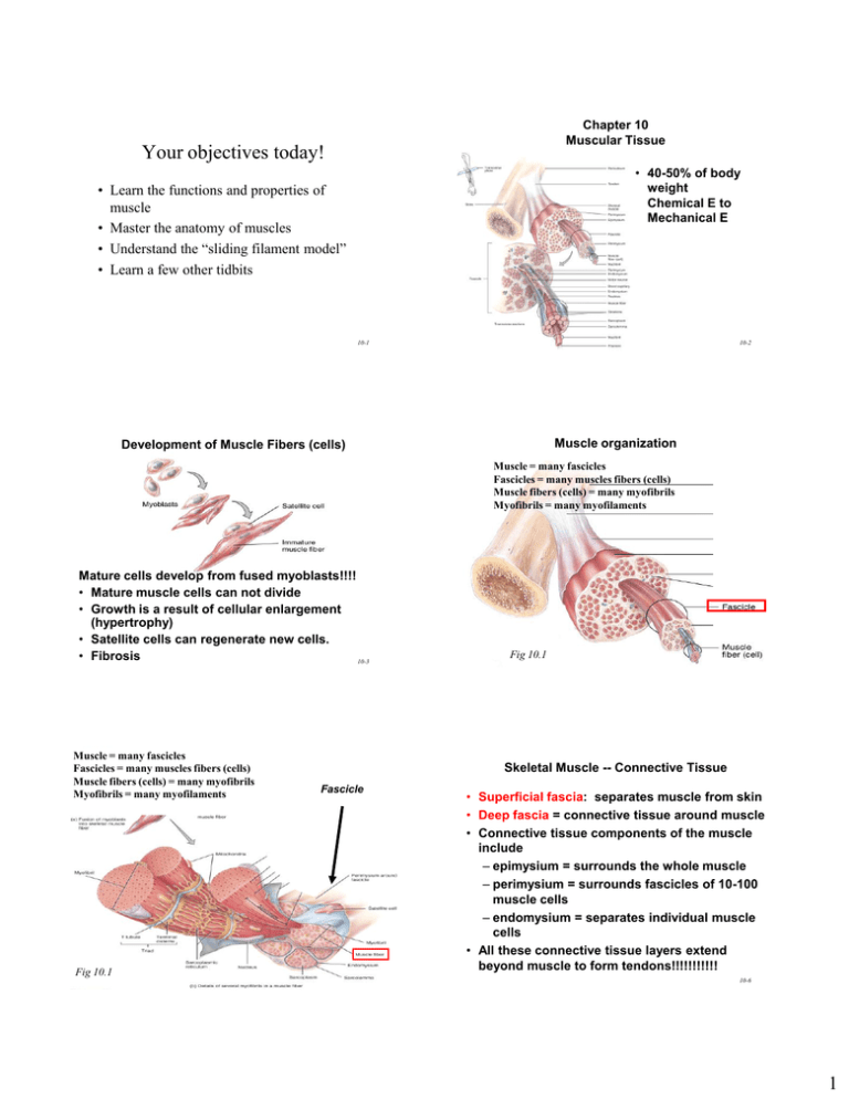
Chapter 10 Muscular Tissue Your objectives today! • 40-50% of body weight Chemical E to Mechanical E • Learn the functions and properties of muscle • Master the anatomy of muscles • Understand the “sliding filament model” • Learn a few other tidbits 10-1 10-2 Muscle organization Development of Muscle Fibers (cells) Muscle = many fascicles Fascicles = many muscles fibers (cells) Muscle fibers (cells) = many myofibrils Myofibrils = many myofilaments Mature cells develop from fused myoblasts!!!! • Mature muscle cells can not divide • Growth is a result of cellular enlargement (hypertrophy) • Satellite cells can regenerate new cells. • Fibrosis 10-3 Muscle = many fascicles Fascicles = many muscles fibers (cells) Muscle fibers (cells) = many myofibrils Myofibrils = many myofilaments Fig 10.1 10-4 Skeletal Muscle -- Connective Tissue Fascicle Fig 10.1 10-5 • Superficial fascia: separates muscle from skin • Deep fascia = connective tissue around muscle • Connective tissue components of the muscle include – epimysium = surrounds the whole muscle – perimysium = surrounds fascicles of 10-100 muscle cells – endomysium = separates individual muscle cells • All these connective tissue layers extend beyond muscle to form tendons!!!!!!!!!!! 10-6 1 Connective Tissue Components Muscle = many fasciles Fascicles = many muscles fibers (cells) Muscle fibers (cells) = many myofibrils Myofibrils = many myofilaments 10-7 10-8 Myofibrils & Myofilaments Muscle Fiber (Cell) • Sarcolemma = muscle cell membrane • Sarcoplasm = cytoplasm • Myofibrils Myofibrils are made up of myofilaments • Myofilaments are contractile proteins 1. Thin myofilaments = actin 2. Thick myofilaments = myosin 10-9 10-10 Other components of Muscle cell Sarcoplasmic Reticulum (SR) Sarcoplasmic reticulum • T-tubules are tubes from surface into the center of the cell (interstitial fluid) 10-11 • • • • System of tubular sacs Stores Ca+ in a relaxed muscle Release Ca+ - - contraction Let’s return to the Myofibril 10-12 2 Thick & Thin Myofilaments Myosin Structural Proteins Actin • The M line (myomesin) connects to titin and adjacent thick filaments. • Titin filaments • Z-discs (Z-lines) • The contracting proteins of muscle 10-13 10-14 Myofilaments and the Sarcomere!!!! • Thick and thin filaments overlap each other in a pattern that creates striations • They are arranged in compartments called sarcomeres!!!!!!! 10-15 Overlap of Thick & Thin Myofilaments within a Myofibril 10-17 10-16 Myosin myofilaments • Thick filaments = Myosin • myosin heads Actin myofilaments = thin filaments 10-18 3 Actin • Associated with actin: troponin, & tropomyosin AKA = T-T Complex Motor Unit: a motor neuron AND all the mm fibers it supplies 10-19 10-20 9.13a Figure Structures of NMJ Region Neuromuscular Junction (NMJ) or Synapse • Motor neurons • End bulbs of motor neurons release acetylcholine (ACh) (neurotransmitter) • NMJ = myoneural junction 10-21 10-22 The Big Picture Neuromuscular Junction Axon terminal of somatic motor neuron Motor nerve fiber Myelin Synaptic knob Schwann cell 1 Muscle fiber Synaptic vesicles (containing ACh) Basal lamina Sarcolemma Synaptic cleft 2 Motor end plate RyR Nucleus T-tubule ACh receptor DHP Z disk Actin Sarcoplasm Myofilaments Troponin Tropomyosin M line Myosin head Mitochondria Nucleus Sarcoplasmic reticulum Ca2+ Junctional folds Figure 11.7b 11-23 (b) ACh Na+ Myosin thick filament (a) Initiation of muscle action potential KEY DHP = dihydropyridine L-type calcium channel RyR = ryanodine receptor-channel Figure 12-11a 4 1. Chemical step Muscle contraction: “Sliding filament model” • motor nerve stimulates muscle (acetyocholine) - this causes an electrical change in the sarcolema (depolarization) - electrical impulse moves along sarcolema T-tubules Sarcoplasmic reticulum • Functional unit of muscle contraction is the sarcomere • Two steps to muscle contraction: 1. Chemical 2. Mechanical 10-25 10-26 Figure 38-8 • In response to electrical impulse sarcoplasmic reticulum releases Ca+ - Released Ca+ causes Actin and Myosin filaments to slide past each other Impulse • (causes the sarcomere to shorten) Ca+ Ca+ Ca+ 10-27 2. Mechanical step 10-28 Fig. 38-9 2. Mechanical step (cross bridges) - Myosin filaments have little heads Myosin Actin = binding sites How does this happen? 10-29 10-30 5 At rest binding sites on Actin are covered by •Active sites are exposed Troponin Tropomyosin Tropomyosintroponin complex Ca+ released from sarcoplasmic reticulum - Myosin heads reach down and bind “Cross bridges” - Requires ATP - binds to troponin - causes TT complex to change shape and move off of the binding site 10-31 10-32 10-33 10-34 Binding sites Muscle Contraction & Relaxation • four major phases of contraction and relaxation 1. excitation • firing nerve leads to muscle excitation 2. excitation-contraction coupling • Wave of depolarization of sarcolemma to activation of myofilaments 3. contraction • step in which the muscle fiber develops tension and may shorten 4. Relaxation • work is done, muscle fiber relaxes and returns to its resting length Cross bridge 11-36 10-35 6 Excitation-Contraction Coupling Axon terminal of somatic motor neuron Terminal cisterna of SR 1 Muscle fiber 2 T tubule T tubule ACh Sarcoplasmic reticulum Na+ Ca2+ Motor end plate RyR Ca2+ T-tubule Sarcoplasmic reticulum Ca2+ DHP Z disk 6 Action potentials propagated down T tubules Troponin Actin Tropomyosin Myosin thick filament (a) Initiation of muscle action potential 7 Calcium released from terminal cisternae Voltage-gated Ca+ channels on Transverse Tubules –change shape and cause Ca+ release channels on S.R. to release Ca+ M line Myosin head KEY DHP = dihydropyridine L-type calcium channel 11-38 RyR = ryanodine receptor-channel Figure 12-11a Contraction Excitation-Contraction Coupling Process in which APs initiate Ca+ signals that activate a contraction-rleaxation cycle Troponin Ca2+ Tropomyosin Active sites Troponin Tropomyosin Actin Thin filament ADP Pi Myosin 10 Myosin Ca2+ 8 Binding of calcium to troponin 9 Hydrolysis of ATP to ADP + Pi; activation and cocking of myosin head Shifting of tropomyosin; exposure of active sites on actin Cross-bridge: Actin Myosin 11 Formation of myosin–actin cross-bridge 11-39 Contraction ATP 13 Binding of new ATP; breaking of cross-bridge ADP ADP PPi i 12 Power stroke; sliding of thin filament over thick filament Please note that due to differing operating systems, some animations will not appear until the presentation is viewed in Presentation Mode (Slide Show view). You may see blank slides in the “Normal” or “Slide Sorter” views. All animations will appear after viewing in Presentation Mode and playing each animation. Most animations will require the latest version of the Flash Player, which is available at http://get.adobe.com/flashplayer. 42 10-42 7 Relaxation • Nerve Impulse stops •No electrical charge (depolorization) over sarcolemma – t-tubules – sarcoplasmic ret. •Ca+ moves back into sarcoplasmic reticulum •Myosin and actin binding prevented Please note that due to differing operating systems, some animations will not appear until the presentation is viewed in Presentation Mode (Slide Show view). You may see blank slides in the “Normal” or “Slide Sorter” views. All animations will appear after viewing in Presentation Mode and playing each animation. Most animations will require the latest version of the Flash Player, which is available at http://get.adobe.com/flashplayer. Also Acetylcholinesterase – breaks down Ach remaining in synapse • Muscle fiber relaxes 43 10-43 •Rigor Mortis!!!!!!!!!!!! 44 10-44 8
