Cell Membrane Structure
advertisement
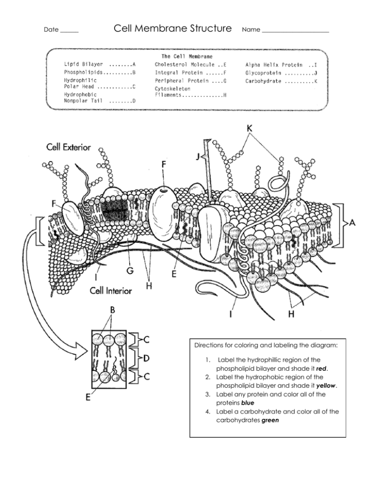
Date ______ Cell Membrane Structure Name ______________________ Directions for coloring and labeling the diagram: 1. Label the hydrophillic region of the phospholipid bilayer and shade it red. 2. Label the hydrophobic region of the phospholipid bilayer and shade it yellow. 3. Label any protein and color all of the proteins blue 4. Label a carbohydrate and color all of the carbohydrates green Date ______ Cell Membrane Structure Name ______________________ 1. Looking at the diagram, describe the locations of the hydrophilic and the hydrophobic regions on the cell membrane. 2. What is the difference between the hydrophilic molecules and hydrophobic molecules? 3. Looking at the diagram, how can you tell if the carbohydrates on the cell membrane are monosaccharides, disaccharides or polysaccharides? What is the function of the carbohydrates on the cell membrane? 4. Looking at the diagram, how does the structure of the integral protein allow it to perform its function? 5. How are the functions of the integral protein and the alpha helix protein similar? 6. Differentiate between peripheral proteins and integral protein? 7. Explain why this structure of the cell membrane is referred to as the fluid mosaic model.
