Document 13310556
advertisement
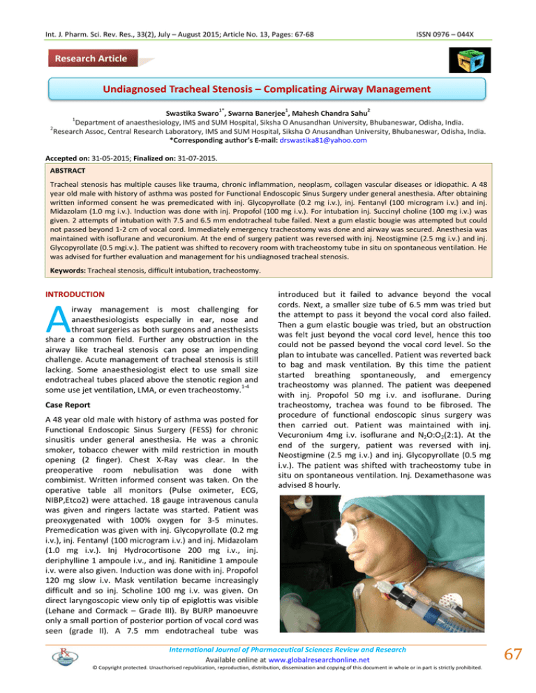
Int. J. Pharm. Sci. Rev. Res., 33(2), July – August 2015; Article No. 13, Pages: 67-68 ISSN 0976 – 044X Research Article Undiagnosed Tracheal Stenosis – Complicating Airway Management 1* 1 2 Swastika Swaro , Swarna Banerjee , Mahesh Chandra Sahu Department of anaesthesiology, IMS and SUM Hospital, Siksha O Anusandhan University, Bhubaneswar, Odisha, India. 2 Research Assoc, Central Research Laboratory, IMS and SUM Hospital, Siksha O Anusandhan University, Bhubaneswar, Odisha, India. *Corresponding author’s E-mail: drswastika81@yahoo.com 1 Accepted on: 31-05-2015; Finalized on: 31-07-2015. ABSTRACT Tracheal stenosis has multiple causes like trauma, chronic inflammation, neoplasm, collagen vascular diseases or idiopathic. A 48 year old male with history of asthma was posted for Functional Endoscopic Sinus Surgery under general anesthesia. After obtaining written informed consent he was premedicated with inj. Glycopyrollate (0.2 mg i.v.), inj. Fentanyl (100 microgram i.v.) and inj. Midazolam (1.0 mg i.v.). Induction was done with inj. Propofol (100 mg i.v.). For intubation inj. Succinyl choline (100 mg i.v.) was given. 2 attempts of intubation with 7.5 and 6.5 mm endotracheal tube failed. Next a gum elastic bougie was attempted but could not passed beyond 1-2 cm of vocal cord. Immediately emergency tracheostomy was done and airway was secured. Anesthesia was maintained with isoflurane and vecuronium. At the end of surgery patient was reversed with inj. Neostigmine (2.5 mg i.v.) and inj. Glycopyrollate (0.5 mgi.v.). The patient was shifted to recovery room with tracheostomy tube in situ on spontaneous ventilation. He was advised for further evaluation and management for his undiagnosed tracheal stenosis. Keywords: Tracheal stenosis, difficult intubation, tracheostomy. INTRODUCTION A irway management is most challenging for anaesthesiologists especially in ear, nose and throat surgeries as both surgeons and anesthesists share a common field. Further any obstruction in the airway like tracheal stenosis can pose an impending challenge. Acute management of tracheal stenosis is still lacking. Some anaesthesiologist elect to use small size endotracheal tubes placed above the stenotic region and some use jet ventilation, LMA, or even tracheostomy.1-4 Case Report A 48 year old male with history of asthma was posted for Functional Endoscopic Sinus Surgery (FESS) for chronic sinusitis under general anesthesia. He was a chronic smoker, tobacco chewer with mild restriction in mouth opening (2 finger). Chest X-Ray was clear. In the preoperative room nebulisation was done with combimist. Written informed consent was taken. On the operative table all monitors (Pulse oximeter, ECG, NIBP,Etco2) were attached. 18 gauge intravenous canula was given and ringers lactate was started. Patient was preoxygenated with 100% oxygen for 3-5 minutes. Premedication was given with inj. Glycopyrollate (0.2 mg i.v.), inj. Fentanyl (100 microgram i.v.) and inj. Midazolam (1.0 mg i.v.). Inj Hydrocortisone 200 mg i.v., inj. deriphylline 1 ampoule i.v., and inj. Ranitidine 1 ampoule i.v. were also given. Induction was done with inj. Propofol 120 mg slow i.v. Mask ventilation became increasingly difficult and so inj. Scholine 100 mg i.v. was given. On direct laryngoscopic view only tip of epiglottis was visible (Lehane and Cormack – Grade III). By BURP manoeuvre only a small portion of posterior portion of vocal cord was seen (grade II). A 7.5 mm endotracheal tube was introduced but it failed to advance beyond the vocal cords. Next, a smaller size tube of 6.5 mm was tried but the attempt to pass it beyond the vocal cord also failed. Then a gum elastic bougie was tried, but an obstruction was felt just beyond the vocal cord level, hence this too could not be passed beyond the vocal cord level. So the plan to intubate was cancelled. Patient was reverted back to bag and mask ventilation. By this time the patient started breathing spontaneously, and emergency tracheostomy was planned. The patient was deepened with inj. Propofol 50 mg i.v. and isoflurane. During tracheostomy, trachea was found to be fibrosed. The procedure of functional endoscopic sinus surgery was then carried out. Patient was maintained with inj. Vecuronium 4mg i.v. isoflurane and N2O:O2(2:1). At the end of the surgery, patient was reversed with inj. Neostigmine (2.5 mg i.v.) and inj. Glycopyrollate (0.5 mg i.v.). The patient was shifted with tracheostomy tube in situ on spontaneous ventilation. Inj. Dexamethasone was advised 8 hourly. International Journal of Pharmaceutical Sciences Review and Research Available online at www.globalresearchonline.net © Copyright protected. Unauthorised republication, reproduction, distribution, dissemination and copying of this document in whole or in part is strictly prohibited. 67 © Copyright pro Int. J. Pharm. Sci. Rev. Res., 33(2), July – August 2015; Article No. 13, Pages: 67-68 ISSN 0976 – 044X DISCUSSION REFERENCES Risk factors for adult tracheal stenosis include a history of prolonged intubation (>24 hours), history of traumatic intubation, unrecognised cases of malignancy, infections, 5 inflammation or collagen vascular disorders . 1. Saito S, Dohi S, Tajima K. Failure of double lumen tube placement: congenital tracheal Stenosis in an adult. Anesthesiology. 66, 1987, 83-85. 2. Esener Z, Tiir A, Diren 8. Difficulty in endotracheal intubation due to congenital tracheal stenosis: a case report. Anesthesiology. 69, 1988, 279-281. 3. Baraka A. Oxygen-jet ventilation during tracheal reconstruction in patients with tracheal stenosis. Anesth Analg, 56, 1977, 429-432. 4. Macnaughton FI. Catheter inflation ventilation and tracheal stenosis. Br J Anaesth, 47, 1975, 12257. 5. Ernst A, Feller-Kopman D, Becker HD, Mehta AC. Central airway obstruction. Am J Respir Crit Care Med, 169, 2004, 1278. 6. Whited RE. A prospective study of laryngotracheal sequelae in long-term intubation. Laryngoscope, 94, 1984, 367-377. 7. Wain JC Postintubation tracheal stenosis. Chest Surg Clin N Am. 13(2), 2003 May, 231-246. In 6-21% of cases of prolonged intubation tracheal stenosis develops. History of prolonged intubation (>24 hours) is the most common cause of tracheal stenosis6,7. Asymptomatic patients pose a real challenge for the anaesthesiologist. Idiopathic tracheal stenosis is an uncommon form of tracheal stenosis usually involving the subglottic area. Our patient did not have any respiratory symptoms and was misdiagnosed to have bronchial asthma 2 years back. There was no history of any subglottic stenosis suggesting possibility of idiopathic tracheal stenosis. Treatment includes laser, balloons, biopsy forcep, steroid injection, mitomycin ointment, tracheal transplant or resection. Source of Support: Nil, Conflict of Interest: None. International Journal of Pharmaceutical Sciences Review and Research Available online at www.globalresearchonline.net © Copyright protected. Unauthorised republication, reproduction, distribution, dissemination and copying of this document in whole or in part is strictly prohibited. 68 © Copyright pro
![Chester _Royals_FCOT[1]](http://s3.studylib.net/store/data/006762869_1-d0409422479a6dd3b4d6752fba5dcd6e-300x300.png)
