Cten, a COOH-Terminal Tensin-like Protein with Prostate Restricted Expression, Is
advertisement
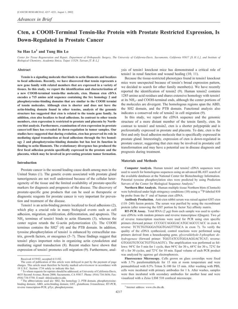
[CANCER RESEARCH 62, 4217– 4221, August 1, 2002] Advances in Brief Cten, a COOH-Terminal Tensin-like Protein with Prostate Restricted Expression, Is Down-Regulated in Prostate Cancer Su Hao Lo1 and Tung Bin Lo Center for Tissue Regeneration and Repair, Department of Orthopaedic Surgery, The University of California-Davis, Sacramento, California 95817 [S. H. L.], and Institute of Biological Chemistry, Academia Sinica, Taipei 11529, Taiwan [T. B. L.] Abstract Tensin is a signaling molecule that binds to actin filaments and localizes to focal adhesions. Recently, we have discovered that tensin represents a new gene family with related members that are expressed in a variety of tissues. In this study, we report the identification and characterization of a new COOH-terminal tensin-like molecule, cten. Human cten cDNA encodes a 715 amino acid sequence containing the Src homology 2 and phosphotyrosine-binding domains that are similar to the COOH termini of tensin molecules. Although cten is shorter and does not have the actin-binding domain found in other tensins, analysis of the genomic structure has suggested that cten is related to the tensin gene family. In addition, cten also localizes to focal adhesions. In contrast to other tensin members, cten expression is restricted to prostate and placenta by Northern blot analysis. Furthermore, examination of cten expression in prostate cancer/cell lines has revealed its down-regulation in tumor samples. Our studies have suggested that during evolution, cten has preserved its role in mediating signal transduction at focal adhesions through the Src homology 2 and phosphotyrosine-binding domains but has lost its function in binding to actin filaments. The evolutionary divergence has produced the first focal adhesion protein specifically expressed in the prostate and the placenta, which may be involved in preventing prostate tumor formation. ysis of tensin1 knockout mice has demonstrated a critical role of tensin1 in renal function and wound healing (10, 11). Because the tissue-restricted phenotypes found in tensin1 knockout mice were unexpected because of tensin’s broad expression pattern, we decided to search for other family member(s). We have recently reported the identification of tensin2 (9). Human tensin2 contains 1285 amino acid residues and shares extensive homology with tensin1 at its NH2- and COOH-terminal ends, although the center portions of the molecules are divergent. The homologous regions span the ABD, the SH2 domain, and the PTB domain. Functional analysis also indicates a conserved role of tensin2 in cell migration (9). In this study, we report the cDNA sequence and the genomic structure of a more distant member of the tensin family, cten. In contrast to tensin1 and tensin2, cten is a shorter polypeptide and is preferentially expressed in prostate and placenta. To date, cten is the first and only focal adhesion molecule that is specifically expressed in prostate gland. Interestingly, expression of cten is down-regulated in prostate cancer, suggesting that cten may be involved in prostatic cell transformation and may have a potential use in disease diagnosis and prognosis during treatment. Materials and Methods Introduction Prostate cancer is the second leading cause death among men in the United States (1). The genetic events associated with prostate gland tumorigenesis are not well understood because of the cellular heterogeneity of the tissue and the limited availability of prostate-specific markers for diagnosis and prognosis of the disease. The discovery of prostate-specific gene products that can be used as therapeutic or diagnostic reagents for prostate cancer is very important for prevention and treatment of the disease. Tensin1 is an actin-binding protein localized to focal adhesions (2), which play a crucial role in many biological events such as cell adhesion, migration, proliferation, differentiation, and apoptosis. The NH2 terminus of tensin1 binds to actin filaments (3), whereas the center region retards the G-actin polymerization (3). The COOH terminus contains the SH22 (4) and the PTB domain. In addition, tyrosine phosphorylation of tensin1 is enhanced by extracellular matrix, growth factors, or oncogenes (5–7). These findings suggest that tensin1 plays important roles in organizing actin cytoskeleton and mediating signal transduction (8). Recent studies have shown that expression of tensin1 promotes cell migration (9). Furthermore, analReceived 4/19/02; accepted 6/11/02. The costs of publication of this article were defrayed in part by the payment of page charges. This article must therefore be hereby marked advertisement in accordance with 18 U.S.C. Section 1734 solely to indicate this fact. 1 To whom requests for reprints should be addressed, at University of California-Davis, 4635 Second Avenue, Room 2000, Sacramento, CA 95817. Phone: (916) 734-3656; Fax: (916) 734-5750; E-mail: shlo@ucdavis.edu. 2 The abbreviations used are: SH2, Src homology 2; PTB domain, phosphotyrosinebinding domain; ABD, actin-binding domain; GST, glutathione S-transferase; RT-PCR, reverse transcription-PCR; pTyr, phosphotyrosine. Computer Analysis. Human tensin1 and tensin2 cDNA sequences were used to search for homologous sequences using an advanced BLAST search of the available databases at the National Center for Biotechnology Information. Potential tyrosine phosphorylation sites were predicted by using Prediction Servers of the Center for Biological Sequence Analysis.3 Northern Blot Analysis. Human multiple tissue Northern blots (Clontech) were hybridized under high stringency conditions (10) using a 32P-labeled 810 bp probe from the 5⬘ end of human cten cDNA. Antibody Production. Anti-cten rabbit serum was raised against GST-cten (118 –240) fusion protein. The serum was purified by using the recombinant protein (after removing the GST portion by factor Xa) affinity matrix. RT-PCR Assay. Total RNA (2 g) from each sample was used to synthesize cDNAs with random primers and reverse transcriptase (Qiagen). Two l of reverse transcription reactions were used for PCR using cten specific primers (forward primer: CCCGCCAGGGACATGCAGCCCACC in exon 4; reverse: TCTCTGTGGGATGGTGAGTTTGCA in exon 7). To verify the quality of the cDNA synthesized, control reactions were performed using primers derived from a housekeeping gene, glyceraldehyde-3-phosphate dehydrogenase (forward primer: TGGTATCGTGGAAGGACTCAT; reverse: GTGGGTGTCGCTGTTGAAGTC). The amplification was performed as follows: 94°C for 5 min for 1 cycle, then 94°C for 30 s, 68°C for 30 s, 72°C for 45 s for 30 cycles, and 72°C for 10 min. Equal volume of each PCR product was analyzed by agarose gel electrophoresis. Fluorescence Microscopy. Cells grown on glass coverslips were fixed with 3.7% paraformaldehyde for 15 min at room temperature and were permeabilized with 0.5% Triton X-100 for 15 min. After washing with PBS, cells were incubated with primary antibodies for 1 h. After washes, samples were then incubated with secondary antibodies for another hour and were visualized with a Zeiss LSM 510 confocal microscope. 4217 3 Internet address: www.cbs.dtu.dk. CTEN IN PROSTATE CANCER Results Identification, Cloning, and Sequence Analysis of Cten. By using human tensin1 and tensin2 cDNA sequences as probes, we have searched the available database for related genes, and a candidate cDNA clone (FLJ14950) was identified. The partial open reading frame of this clone encodes an SH2 domain followed by a PTB domain. Additional sequence of this clone was obtained by performing rapid amplification of cDNA ends. The total composed cDNA is 4015 bp with an open reading frame encoding 715 amino acid residues (GenBank access no. AF417488; Fig. 1A). There is an in-frame stop codon 27 bp upstream of the predicted initiation site. The predicted molecule mass is 76,930 Da with an estimated isoelectric point of 6.95. Analysis of the predicted amino acid sequence has revealed that residues 418 –715 are very similar with the COOH termini of tensin1 (50% identity or 68% similarity) and tensin2 (45% identity or 56% similarity; Fig. 1, B and C). In addition, six potential tyrosine phosphorylation sites were found in cten (Fig. 1A). Because this gene product is a shorter polypeptide and lacks the NH2-terminal homologous regions found in tensin1 and tensin2, it is likely to be a distant member of the tensin family. We have named it cten for the COOH-terminal tensin-like molecule. A search of the available human genomic sequence databases using the human cten cDNA revealed that the length of human cten is about 21 kb on the Hs17-25057 clone, which was derived from chromosome 17q12-21. Analysis of the sequences revealed 12 exons in human cten gene (Fig. 1D). All splice acceptor/donor sites for these exons conform to the GT-AG rule. The putative start codon is in exon 1. Exon 12 is the largest exon with 1883 bp, but only 142 bp comprise the coding sequence, whereas the rest is the 3⬘ untranslated region. Exon 11 is the smallest exon with 27 bp, and the sizes of exons 4 –11 are very similar to other tensin genes (9), supporting the idea that cten is related to tensin family. Restricted Expression of Cten in Human Normal Tissues. The distribution of cten mRNA in normal human tissues was examined by Fig. 1. Analysis of cten amino acid sequence. A, the cDNA-derived amino acid sequence of human cten. The potential tyrosine phosphorylation sites are in bold. B, alignment of cten amino acid sequence with the COOH termini of tensin1 and tensin2. The shaded and unshaded areas in the boxes represent identity and similarity, respectively. C, domain structures of tensin1, tensin2, and cten. ABD I interacts with actin filaments. The same region also contains the PTEN homologous sequence. ABD II retards G-actin polymerization rate. SH2 and PTB domains are two binding molecules for pTyr. The center regions of these genes show no sequence homology. D, organization of human cten gene. Exon/intron boundaries were determined by comparison of sequences of genomic DNA and cDNA. In the splice site, exon sequences are indicated by uppercase letters, and intron sequences are indicated by lowercase letters. Codon phase refers to the codon split at the splice acceptor. Introns that do not split codon triplets are indicated by phase 0, interruption after the first nucleotide is indicated by codon phase I, and interruption after the second nucleotide is indicated by codon phase II. N indicates noncoding region. Numbers in the brackets indicate the sizes of the corresponding exons in human tensin1 and tensin2, respectively. 4218 CTEN IN PROSTATE CANCER Fig. 2. Tissue distribution of cten mRNAs. Northern blot analysis of cten (top panel) in human tissues: Lane 1: brain; Lane 2: heart; Lane 3: skeletal muscle; Lane 4: colon; Lane 5: thymus; Lane 6: spleen; Lane 7: kidney; Lane 8: liver; Lane 9: small intestine; Lane 10: placenta; Lane 11: lung; Lane 12: peripheral blood leukocyte; Lane 13: prostate; Lane 14: testis; and Lane 15: ovary. The blots were stripped and probed with actin cDNA probe (bottom panel). Northern blot analysis. The 5⬘ 810-bp cDNA fragment was used as a probe to avoid cross-hybridizing to other tensin genes. With 15 human normal tissues examined, a 4.4-kb message was readily detected in the prostate and placenta (Fig. 2). In contrast, tensin1 and tensin2 are expressed widely (9, 12), suggesting that cten plays a specific role in the prostate and the placenta. Molecular Mass and Subcellular Localization of Cten Protein. To examine the expression of protein encoded by human cten cDNA, 35 S-methionine labeled recombinant cten was generated by in vitro transcription and translation and analyzed by SDS-PAGE. Although the predicted molecular mass was 77 kDa, the recombinant cten migrated as a 90-kDa protein on a 10% gel (Fig. 3A). To examine whether the endogenous cten also migrated at a similar rate, we raised antibodies against GST-cten (118 –240) fusion protein. This anti-cten antibody was used to detect cten expression in an immortalized, nontransformed human prostate epithelial line, MLC (13), because cten was only detected in the prostate and the placenta by Northern blotting. A single band ⬃90 kDa was recognized by anti-cten antibodies, indicating that we have cloned the full-length cten cDNA. To determine subcellular localization of the protein, MLC cells were analyzed by immunofluorescent staining. As shown in Fig. 3C, cten was colocalized with pTyr-containing proteins at focal adhesions in MLC cells. In addition, recombinant cten fused to green fluorescence protein also targeted focal adhesions in transfected MLC cells (data not shown). These results demonstrated that both endogenous and recombinant cten target to focal adhesions, and focal adhesion localization is a common feature of the tensin family members. Cten Expression in Human Prostate Cancer/Cell Lines. To explore the potential role of cten in prostate cancer, we have examined the mRNA levels of cten in cancer cell lines. Human prostate cancer cell lines LnCap, DU145, and PC3 (prostate cancer cell lines derived from lymph node, brain, and bone metastases, respectively) and nontransformed MLC were analyzed by RT-PCR assays (Fig. 4). Although every cell line expressed considerable amounts of a housekeeping gene, glyceraldehyde-3-phosphate dehydrogenase, we detected a strong signal of cten in MLC, a weak band in DU145, and no signal in LnCap and PC3. To evaluate whether mRNA levels detected by RT-PCR assay correlate with protein levels, we examined protein expressions in these cell lines by Western blotting and immunostaining. The Western blot analysis showed that DU145 cells expressed a lower amount of cten when compared with MLC cells, whereas LnCap and PC3 cells had no detectable cten (Fig. 3B). As Fig. 3. The apparent molecular mass and subcellular localization of cten. A, [35S]Methioninelabeled tensin1, tensin2, and cten were generated by TnT in vitro transcription/translation (Promega) and separated on 10% SDS-PAGE. The gel was dried on filter paper and analyzed by autoradiograpy. B, equal amounts of cell lysate protein (30 g) from MLC, LnCap, DU145, and PC3 cell lines were separated on 10% SDS-PAGE, transferred to membrane, and then probed with anti-cten antibody. C, MLC, DU145, and PC3 cells grown on coverslips were fixed, permeabilized, and then labeled with rabbit anti-cten and mouse anti-pTyr antibodies, followed by Texas Red-conjugated goat antirabbit and fluorescein-conjugated goat antimouse secondary antibodies. Cells were then visualized with a Zeiss LSM 510 laser scanning microscope. Arrows indicated focal adhesions labeled by both cten and pTyr antibodies. Arrowheads showed focal adhesions detected by only pTyr but not cten antibodies. 4219 CTEN IN PROSTATE CANCER Fig. 4. Analysis of cten expressions in human prostate cancer/ cell lines. cDNAs were synthesized with random primers and reverse transcriptase from 2 g of total RNA prepared from each sample. Two l of reverse transcription reactions were used for PCR with primers specific for indicated molecules. Equal volume (15 l) of each PCR product was loaded. T1, T2, and T3 were three prostate tumor tissue samples. Additional four pairs of matched tumor/normal prostate tissues were analyzed. shown in Fig. 3C, although all prostate cancer cells showed focal adhesion staining by anti-pTyr antibodies, no focal adhesion staining was detected in PC3 and LnCap (data not shown) cells by anti-cten antibodies. Within DU145 cells, most of the cells showed no focal adhesion staining and only a small population was labeled by cten antibodies (Fig. 3C). These findings were consistent with the RT-PCR result, indicating that cten mRNA levels detected by RT-PCR assay correlate with protein levels. Therefore, we analyzed human patient samples using RT-PCR assay. Characterization of three prostate cancer patient samples (T1, T2, and T3) has detected no or very weak cten expression. Additional analysis of four pairs of matched tumor/ normal prostate samples (Fig. 4) has shown that cten expression was lost in tumor in one pair (pair 1), reduced in two pairs (pairs 3 and 4), and similar in one pair (pair 2). These results showed that cten expression was down-regulated in the majority of prostate cancer samples tested. Discussion Here we report the identification of a distant member of the tensin family with a tissue expression pattern restricted to the prostate gland in normal tissues, whereas its expression is markedly reduced in a large fraction of tumor samples. The homology of cten to the tensin genes is intriguing. Tensin proteins are composed of three regions with different structural roles: the NH2-terminal, middle, and the COOH-terminal regions (Fig. 1C). The NH2 and COOH termini were conserved between tensin1 and tensin2, whereas the middle regions were divergent. Interestingly, cten did not contain the NH2-terminal region, which includes the ABD and sequence similar to the tumor suppressor, phosphatase and tensin homologue deleted from chromosome 10. Therefore, we predicted that cten is unlikely to interact with actin cytoskeleton. Nonetheless, cten contains the SH2 and PTB domains. Both domains are binding modules for pTyr, although the PTB domain can also interact with molecules via non-pTyr interaction. The SH2 domain is found among many cytoplasmic proteins, which functions mainly by regulating various cellular events, including enzyme activity, substrate recruitment, and protein localization. SH2 domains bind ligands containing pTyr residues within a specific sequence (14). High affinity binding is provided by the pTyr residue itself and by subsequent residues toward the COOH-terminal (14). The optimal phosphopeptide-binding specificity for tensin1’s SH2 domain is pY (E or D), N (I, V, or F [Ref. 15]; pY, phosphotyrosine; E, glutamic acid; D, aspartic acid; N, aspargine; I, isoleucine; V, valine; and F, phenylalanine). Because tensin1 and cten’s SH2 domains are 75% identical in amino acid sequence (or 87% homologous), we expect that cten’s SH2 domain binds to a similar motif. PTB domains also bind to tyrosine-phosphorylated peptides, but in contrast to the SH2 domain, binding specificity is determined by residues preceding the pTyr toward the NH2 terminus (16). However, it has recently been demonstrated that PTB domains participate in pTyrindependent interactions (17). In either case, PTB domains normally function as protein-protein interaction modules. Analysis of genomic structures of tensin genes has shown that the sizes and arrangement of exons coding for the SH2 and PTB domains of tensin1, tensin2, and cten are almost identical (Fig. 1D), indicating that these functional domains have been conserved through evolution. However, during evolution cten has lost actin-binding function while gaining a more restricted expression pattern limiting to the prostate and placenta. Prostate tissue specificity of cten expression prompted us to evaluate whether cten expression was altered during tumorigenesis. Our initial studies by using RT-PCR assay have shown that prostate cancers exhibit significantly decreased expression of cten. These results indicate that cten expression may be critical in preventing prostatic cell transformation. Tumor suppressor genes are believed to be strong contributors to the progression of prostate cancers as are other epidemiological factors. Chromosome 17 is one of the most frequently lost chromosomes in human malignancies (18). Previous studies have shown that one or more deleted regions were found in prostate tumors close to BRCA1 (the breast and ovarian cancer susceptibility gene) on chromosome 17. Gao et al. (19) showed loss of heterozygosity on chromosome 17q in a 2-cM region that is centromeric to BRCA1, whereas data from Dai et al. (20) suggested that a region distal to BRCA1 may contain a prostate-specific suppressor gene, suggesting that there may be unidentified tumor suppressor genes near 17q21 that play a pivotal role in prostate cancer progression. An attractive candidate for this function is cten, which localizes at 17q12-21, although whether it functions as a tumor suppressor for prostate cancer remains to be investigated. At present, it is not yet clear how cten under expression may play a role in tumor progression. Intriguingly, although all prostate cell lines examined in this study contain pTyr-positive focal adhesions, only nontransformed MLC and a small population in DU145 cells express cten at focal adhesions, suggesting that loss of cten expression might be an early event during prostatic cell transformation. It is possible that its function as a signaling molecule is required for maintaining normal growth and differentiation events in the prostate. Future experiments will focus on the biological function of cten in normal and tumorigenic prostate tissue. Acknowledgments We thank Dr. Liz Allen for critical reading and discussion of the manuscript. We also thank Dr. Huaiyang Chen for preparing the cten antibody. References 4220 1. Parker, S. L., Tong, T., Bolden, S., and Wingo, P. A. Cancer statistics. CA Cancer J. Clin., 47: 5–27, 1997. 2. Lo, S. H., An, Q., Bao, S., Wong, W. K., Liu, Y., Janmey, P. A., Hartwig, J. H., and Chen, L. B. Molecular cloning of chick cardiac muscle tensin. Full-length cDNA sequence, expression, and characterization. J. Biol. Chem., 269: 22310 –22319, 1994. 3. Lo, S. H., Janmey, P. A., Hartwig, J. H., and Chen, L. B. Interactions of tensin with actin and identification of its three distinct actin-binding domains. J. Cell Biol., 125: 1067–1075, 1994. 4. Davis, S., Lu, M. L., Lo, S. H., Lin, S., Butler, J. A., Druker, B. J., Roberts, T. M., An, Q., and Chen, L. B. Presence of an SH2 domain in the actin-binding protein tensin. Science (Wash. DC), 252: 712–715, 1991. 5. Bockholt, S. M., and Burridge, K. Cell spreading on extracellular matrix proteins induces tyrosine phosphorylation of tensin. J. Biol. Chem., 268: 14565–14567, 1993. 6. Jiang, B., Yamamura, S., Nelson, P. R., Mureebe, L., and Kent, K. C. Differential effects of platelet-derived growth factor isotypes on human smooth muscle cell proliferation and migration are mediated by distinct signaling pathways. Surgery (St. Louis), 120: 427– 431, 1996. 7. Salgia, R., Brunkhorst, B., Pisick, E., Li, J. L., Lo, S. H., Chen, L. B., and Griffin, J. D. Increased tyrosine phosphorylation of focal adhesion proteins in myeloid cell lines expressing p210BCR/ABL Oncogene, 11: 1149 –1155, 1995. CTEN IN PROSTATE CANCER 8. Lo, S. H., Weisberg, E., and Chen, L. B. Tensin: a potential link between the cytoskeleton and signal transduction. Bioessays, 16: 817– 823, 1994. 9. Chen, H., Duncan, I. C., Bozorgchami, H., and Lo, S. H. Tensin1 and a previously undocumented family member, tensin2, positively regulate cell migration. Proc. Natl. Acad. Sci. USA, 99: 733–738, 2002. 10. Lo, S. H., Yu, Q. C., Degenstein, L., Chen, L. B., and Fuchs, E. Progressive kidney degeneration in mice lacking tensin. J. Cell Biol., 136: 1349 –1361, 1997. 11. Ishii, A., and Lo, S. H. A role of tensin in skeletal muscle regeneration. Biochem. J., 356: 737–745, 2001. 12. Chen, H., Ishii, A., Wong, W. K., Chen, L. B., and Lo, S. H. Molecular characterization of human tensin. Biochem. J., 351: 403– 411, 2000. 13. Lee, M. S., Garkovenko, E., Yun, J. S., Weijerman, P. C., Peehl, D. M., Chen, L. S., and Rhim, J. S. Characterization of adult human prostatic epithelial cells immortalized by polybrene-induced DNA transfection with a plasmid containing an origindefective SV40 genome. Int. J. Oncol., 4: 821– 830, 1994. 14. Songyang, Z., Shoelson, S. E., Chaudhuri, M., Gish, G., Pawson, T., Haser, W. G., King, F., Roberts, T., Ratnofsky, S., Lechleider, R. J., et al. SH2 domains recognize specific phosphopeptide sequences. Cell, 72: 767–778, 1993. 15. Auger, K. R., Songyang, Z., Lo, S. H., Roberts, T. M., and Chen, L. B. Plateletderived growth factor-induced formation of tensin and phosphoinositide 3-kinase complexes. J. Biol. Chem., 271: 23452–23457, 1996. 16. Zhou, M. M., Ravichandran, K. S., Olejniczak, E. F., Petros, A. M., Meadows, R. P., Sattle, M., Harlan, J. E., Wade, W. S., Burakoff, S. J., and Fesik, S. W. Structure and ligand recognition of the phosphotyrosine binding domain of Shc. Nature (Lond.), 378: 584 –592, 1995. 17. Zambrano, N., Buxbaum, J. D., Minopoli, G., Fiore, F., De Candia, P., De Renzis, S., Faraonio, R., Sabo, S., Cheetham, J., Sudol, M., and Russo, T. Interaction of the phosphotyrosine interaction/phosphotyrosine binding-related domains of Fe65 with wild-type and mutant Alzheimer’s -amyloid precursor proteins. J. Biol. Chem., 272: 6399 – 6405, 1997. 18. Seizinger, B. R., Klinger, H. P., Junien, C., Nakamura, Y., Le Beau, M., Cavenee, W., Emanuel, B., Ponder, B., Naylor, S., Mitelman, F., Louis, D., Menon, A., Newsham, I., Decker, J., Kaelbling, M., Henry, I., and von Deimling, A. Cytogenet. Cell Genet., 58: 1080 –1096, 1991. 19. Gao, X., Zacharek, A., Grignon, D. J., Sakr, W., Powell, I. J., Porter, A. T., and Honn, K. V. Localization of potential tumor suppressor loci to a ⬍ 2 Mb region on chromosome 17q in human prostate cancer. Oncogene, 11: 1241–1247, 1995. 20. Dai, Q., Deubler, D. A., Maxwell, T. M., Zhu, X. L., Cui, J., Rohr, L. R., Stephenson, R. A., and Brothman, A. R. A common deletion at chromosomal region 17q21 in sporadic prostate tumors distal to BRCA1. Genomics, 71: 324 –329, 2001. 4221
