Cleavage of cten by caspase-3 during apoptosis Su-Shun Lo
advertisement
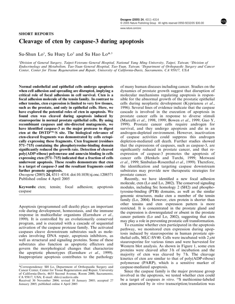
Oncogene (2005) 24, 4311–4314 & 2005 Nature Publishing Group All rights reserved 0950-9232/05 $30.00 www.nature.com/onc SHORT REPORTS Cleavage of cten by caspase-3 during apoptosis Su-Shun Lo1, Su Huey Lo2 and Su Hao Lo*,3 1 Division of General Surgery, Taipei-Veterans General Hospital, National Yang Ming University, Taipei, Taiwan; 2Division of Endocrinology and Metabolism, Tao-Yuan General Hospital, Tao-Yuan, Taiwan; 3Department of Orthopaedic Surgery and Cancer Center, Center for Tissue Regeneration and Repair, University of California-Davis, Sacramento, CA 95817, USA Normal endothelial and epithelial cells undergo apoptosis when cell adhesion and spreading are disrupted, implying a critical role of focal adhesions in cell survival. Cten is a focal adhesion molecule of the tensin family. In contrast to other tensins, cten expression is limited to very few tissues, such as the prostate, and only in epithelial cells. Here, we have explored the potential roles of cten in apoptosis. We found cten was cleaved during apoptosis induced by staurosporine in normal prostate epithelial cells. By using recombinant caspases and site-directed mutagenesis, we have identified caspase-3 as the major protease to digest cten at the DSTD570kS site. The biological relevance of cten-cleaved fragments was demonstrated by cells ectopically expressing these fragments. Cten fragment (residues 571–715) containing the phosphotyrosine-binding domain significantly reduced the growth rate. Detection of cleaved poly(ADP-ribose) polymerase and annexin binding in cells expressing cten (571–715) indicated that a fraction of cells underwent apoptosis. These results demonstrate that cten is a target of caspase-3 and the resultant fragments could further promote apoptosis. Oncogene (2005) 24, 4311–4314. doi:10.1038/sj.onc.1208571 Published online 4 April 2005 Keywords: cten; caspase tensin; focal adhesion; apoptosis Apoptosis (programmed cell death) plays an important role during development, homeostasis, and the immune response in multicellular organisms (Earnshaw et al., 1999). It is controlled by an evolutionarily conserved program, and is executed with a cascade of sequential activation of the caspase protease family. The activated caspases cleave downstream substrates such as molecules involving DNA repair, apoptosis inhibitors, as well as structural and signaling proteins. Some of these substrates also function as apoptotic effectors and govern the morphological changes that characterize the apoptotic phenotypes (Earnshaw et al., 1999). Inappropriate apoptosis contributes to the pathology *Correspondence: SH Lo, Department of Orthopaedic Surgery and Cancer Center, Center for Tissue Regeneration and Repair, University of California-Davis, 4635 Second Avenue, Room 2000, Sacramento, CA 95817, USA; E-mail: shlo@ucdavis.edu Received 30 November 2004; revised 18 January 2005; accepted 27 January 2005; published online 4 April 2005 of many human diseases including cancer. Studies on the dynamics of prostate growth suggest that disruption of molecular mechanisms regulating apoptosis is responsible for the abnormal growth of the prostatic epithelial cells during neoplastic development (Kyprianou et al., 1996). Several lines of evidence indicate that the caspase cascade is involved in the execution of apoptosis in prostate cancer cells in response to diverse stimuli (Marcelli et al., 1998, 1999; Bowen et al., 1999; Guo Y, 1999). Prostate cancer cells require androgen for survival, and they undergo apoptosis and die in an androgen-depleted environment. However, inactivation of caspase activities could suppress this androgen depletion-mediated cell death. Further studies showed that the expressions of caspases, such as caspase-3, are significantly reduced in prostate cancer, and that reexpression of caspase-3 promotes the apoptosis of cancer cells (Henkels and Turchi, 1999; Motwani et al., 1999; Simbulan-Rosenthal et al., 1999). Therefore, the identification and targeting caspase downstream substrates may provide new therapeutic strategies for prostate cancer. Recently, we have identified a new focal adhesion molecule, cten (Lo and Lo, 2002). The conserved signaling modules, including Src homology 2 (SH2) and phosphotyrosine-binding (PTB) domains, as well as the similar genomic structures, make cten a member of the tensin family (Lo, 2004). However, cten protein is shorter than other tensins and cten expression pattern is more restricted. It is concentrated in the prostate gland and the expression is downregulated or absent in the prostate cancer patients (Lo and Lo, 2002), suggesting that cten may play a role in preventing prostatic cell transformation. To examine whether cten participated in the apoptosis pathway, we monitored cten expression during apoptosis induced by staurosporine in human prostate epithelial cells, MLC-SV40. Cells were incubated with 2 mM staurosporine for various times and were harvested for Western blot analysis. As shown in Figure 1, some cten proteins were cleaved after 3 h of incubation and the majority of cten was cleaved by 7 h. The cleavage kinetics of cten are similar to that of poly(ADP-ribose) polymerase (PARP), which is a sensitive marker of caspase-mediated apoptosis. Since the caspase family is the major protease group involved in the apoptosis, we tested whether cten could be a target of caspases in vitro. 35S methionine-labeled cten generated by in vitro transcription/translation was Cten in apoptosis S-S Lo et al 4312 Figure 1 Cleavage of cten during apoptosis induced by staurosporine. Subconfluent MLC-SV40 cells (a kind gift from Dr Johng Rhim at Uniform Services University) incubated with 2 mM staurosporine for 0, 1, 3, 5, and 7 h were harvested. Equal amount of proteins were separated by SDS–PAGE and immunoblotted with anti-cten antibodies. The same blot was reprobed with antiPARP antibodies. Arrow indicates intact cten. Arrowhead shows the cten fragment incubated with recombinant caspase-1, -2, -3, -6, -7, -8, -9, and -10. Figure 2 shows that only caspase-3 was able to cleave cten and generate a proteolytic fragment similar to the one detected in staurosporine-induced apoptosis. Two DXXD consensus sites, preferred by caspase-3, were found in cten: DSND506kL and DSTD570kS (Figure 3a). To investigate whether these were the cleavage sites for caspase-3, we mutated each of the two critical aspartic acid residues to alanine (cten D506A and cten D570A) and examined the effects of these mutations on cten sensitivity to caspase-3 digestion. As shown in Figure 3b, cten D570A but not cten D506A was resistant to caspase-3, indicating that cten D570 is the cleavage site for the enzyme. Cten was specifically cleaved by caspase-3 among 10 tested caspases, suggesting that cten is not just a bystander and the cleavage of cten is biologically relevant. Interestingly, D570 localizes between the SH2 and PTB domains, such that the cleavage generates one cten fragment (1–570) without the PTB domain and the other fragment (571–715) with the PTB domain alone (Figure 3a). We hypothesized that these cleaved cten fragments might be involved in promoting apoptosis by virtue of their deregulated binding activities. To test this possibility, we generated transfection constructs fused wild-type cten (1–715), cten (1–570), or cten (571–715) to the C-terminus of GFP. These constructs were initially transfected into MLC-SV40 cells for establishing stable cell lines. While we were able to generate stable MLC-SV40 cell lines expressing either GFP-cten or GFP-cten (1–570), we failed to establish any stable line expressing GFP-cten (571–715) in numerous attempts. The cells appeared to express GFP-cten (571–715) fusion proteins confirmed by Western blot and green fluorescence microscopy when transfected (not shown), but started to detach and die during drug selection, suggesting that cten 571–715 might, indeed, be involved in promoting apoptosis. Since MLC-SV40 is a Oncogene Figure 2 In vitro cleavage of cten by caspase-3. 35S-labeled recombinant proteins were generated by using the TNTs T7coupled in vitro transcription/translation system (Promega) according to the manufacturer’s instruction. 35S-labeled samples (2 ml) were incubated without ( ) or with 1 ml of recombinant caspase-1, -2, -3, -6, -7, -8, -9, or -10 (Cal Biochem.) in cleavage buffer (25 mM HEPES (pH 7.4), 0.1% chaps, 10% glycerol, 1 mM EDTA, 10 mM DTT) at 371C for 1 h. The samples were then analysed by SDS– PAGE and autoradiography. Arrow indicates full-length cten. Arrowhead shows the cten fragment generated by caspase-3 Figure 3 DSTD570kS of cten is the cleavage site for caspase-3. (a) Schematic structure of cten. Ag is the antigen region for making cten antibodies used in this study. (b) 35S methionine-labeled cten wt, cten D506A, and cten D570A were incubated with or without caspase-3 for 30 min at 371C, and then analysed by SDS–PAGE and autoradiography. Cten D570A was not cleaved by caspase-3 normal epithelial cell line expressing endogenous cten, it might be more sensitive to cten fragments. We then used MDA-MB-468 cells, a breast cancer cell line that does not express endogenous cten, with the thought that cancer cells are more resistant to apoptosis and cten fragments will not function as ‘dominant negative’ to endogenous intact cten. With this cell line, we were able to establish stable MDA-MB-468 cell lines expressing GFP, GFP-cten, GFP-cten (1–570), and, most importantly, GFP-cten (571–715). The Western blot analysis has confirmed the expression of GFP or GFP fusion proteins (Figure 4a). Cell numbers were counted to examine the effects of cten and its fragments on cell growth. As shown in Figure 4b, while GFP, GFP-cten, and GFP-cten (1–570) cells grew at the same rate, the growth rate of GFP-cten (571–715) was significantly Cten in apoptosis S-S Lo et al 4313 slower than the others. However, the ability in reducing tetrazolium compounds was the same among viable transfectants (Figure 4c), indicating that proliferation rates are the same. In addition, we have observed more floating cells in GFP-cten (571–715) cells than others during routine culturing. This phenomenon was more significant when transfectants were seeded and cultured in the absence of serum. To investigate whether cells underwent apoptosis, cells were collected and analysed for the presence of PARP. While all samples contained intact PARP, significant amount of cleaved PARP was detected in GFP-cten (571–715) cells (Figure 4d). Figure 4 Cten fragments affected cell growth and death. (a) GFP-cten plasmids were transfected into MDA-MB-468 cells by SuperFect reagents (Qiagen). Cells were selected against 800 mg/ml G418 in the medium for 10 days. The survival colonies were isolated by cloning filter prewetted with trypsin and transferred to 24-well plates. The positive clones were verified by GFP labeling and Western blotting. Protein lysates prepared from MDA-MB-468 cells expressing GFP (lane 1), GFP-cten (lane 2), GFP-cten 1–570 (lane3), and GFP-cten 571–715 (lane 4) were immunoprecipitated with goat anti-GFP antibodies (Rockland), and then detected by Western blotting using mouse anti-GPF antibodies (Zymed). (b) Cells (50 000) from indicated lines were seeded at day 1 in six-well plates and counted every day. The figure represented data from triplicate experiments of at least two independent clones of each GFP transfectant. *Po0.005. (c) Transfectants (1 105) were seeded in duplicate six-well plates. After 24 h, one set of plates was used to count cell numbers, and the other was analysed by Aqueous One Solution Cell Proliferation assay (Promega). The absorbance readings were normalized to cell number counted. No significant difference was found. (d) Cells (both attached and floating) expressing GFP (lane 1), GFP-cten (lane 2), GFP-cten 1–570 (lane3), and GFP-cten 571–715 (lane 4) were harvested. Equal amount of proteins were separated by SDS–PAGE and immunoblotted with anti-PARP antibodies. The same blot was reprobed with anti-GAPDH antibodies for loading and transferring control. Arrow indicates the cleaved PARP. (e) Transfectants grown on coverslips were washed in cold PBS twice, and then in annexin-binding buffer (10 mM HEPES, 140 mM NaCl, 2.5 mM CaCl2 (pH 7.4)). Cells were incubated with biotin-X-conjugated annexin V (Molecular probes) for 15 min at room temperature followed by 10 min Alexa Fluor 594-labeled streptavidin (Molecular probes) treatment. After extensive washes, cells were fixed in 4% paraformaldehyde, mounted, and observed under Ziess axiovert 200 microscope. Arrowheads indicate strong annexin V membrane staining (red). Arrows show focal adhesion sites Oncogene Cten in apoptosis S-S Lo et al 4314 Furthermore, there was significantly higher degree of annexin V labeling on the surface of GFP-cten (571– 715) cells (Figure 4e). Both results indicated that some GFP-cten (571–715) cells underwent apoptosis. Interestingly, both GFP-cten and GFP-cten (1–570) proteins localized to focal adhesion sites (Figure 4d, arrows), whereas GFP-cten (571–715) and GFP proteins were in the cytoplasm and nuclei (Figure 4e). Taken together, we have demonstrated that cten is cleaved by caspase-3 at DSTD570kS site. One of the resulting fragments, cten 571–715, is able to reduce cell growth by inducing apoptosis, as judged by detecting cleaved PARP in the transfectants and annexin V binding on the cell surface. These effects are probably under-rated because cancer cells are more resistant to apoptosis and in favor of cell growth. The mechanism involved is currently not clear, but is likely due to deregulated binding activity of cten PTB domain in the cleaved fragment 571–715. The PTB domain was originally identified as a binding motif for tyrosinephosphorylated peptides. However, many PTB domains bind to proteins in a nonphosphorylated manner. Tensin’s PTB domain does not interact with tyrosinephosphorylated proteins (Chen and Lo, 2003). Instead, it binds to the NPXY motifs in the b integrin cytoplasmic tails (Calderwood et al., 2003). Since the PTB domains of tensin members are highly conserved, we would predict that they interact with similar targets. If so, cten 571–715 fragments could bind to the b integrin tails and disrupt the links between integrins and their associated molecules. This disruption might facilitate the breakdown of focal adhesions and lead to cell detachment. It is known that cells undergo apoptosis (or anoikis) when cell adhesions were compromised (Meredith et al., 1993; Frisch and Francis, 1994; Re et al., 1994; Boudreau et al., 1995). Cten is specifically expressed in the prostate epithelial cells and the expression is reduced/loss in the prostate cancer. In addition, cten gene locates at human chromosome 17q21, a region often deleted in prostate cancer (Lo and Lo, 2002), suggesting that cten may be a tumor suppressor and play a role in preventing prostatic cell transformation. Present studies have explored a new function of cten in regulating cell death, which may be critical in balancing cell numbers in normal prostate. The reduced/loss of cten expression may lead to uncontrolled epithelial cell growth and result in cell transformation. Acknowledgements This work was supported in part by the CRCC award from University of California and CA102537 from the NIH (SHL). References Boudreau N, Sympson CJ, Werb Z and Bissell MJ. (1995). Science, 267, 891–893. Bowen C, Voeller HJ, Kikly K and Gelmann EP. (1999). Cell Death Differ., 6, 394–401. Calderwood DA, Fujioka Y, de Pereda JM, Garcia-Alvarez B, Nakamoto T, Margolis B, McGlade CJ, Liddington RC and Ginsberg MH. (2003). Proc. Natl. Acad. Sci. USA, 100, 2272–2277. Chen H and Lo SH. (2003). Biochem. J., 370, 1039–1045. Earnshaw WC, Martins LM and Kaufmann SH. (1999). Annu. Rev. Biochem., 68, 383–424. Frisch SM and Francis H. (1994). J. Cell Biol., 124, 619–626. Guo YKN. (1999). Cancer Res., 59, 1366–1371. Henkels KM and Turchi JJ. (1999). Cancer Res., 59, 3077–3083. Kyprianou N, Tu H and Jacobs SC. (1996). Hum. Pathol., 27, 668–675. Oncogene Lo SH. (2004). Int. J. Biochem. Cell Biol., 36, 31–34. Lo SH and Lo TB. (2002). Cancer Res., 62, 4217–4221. Marcelli M, Cunningham GR, Walkup M, He Z, Sturgis L, Kagan C, Mannucci R, Nicoletti I, Teng B and Denner L. (1999). Cancer Res., 59, 382–390. Marcelli ML, Cunningham GR, Haidacher SJ, Padayatty SJ, Sturgis L, Kagan C and Denner L. (1998). Cancer Res., 58, 76–83. Meredith JEJ, Fazeli B and Schwartz MA. (1993). Mol. Cell. Biol., 4, 953–961. Motwani M, Delohery TM and Schwartz GK. (1999). Clin. Cancer Res., 5, 1876–1883. Re F, Zanetti A, Sironi M, Polentarutti N, Lanfrancone L, Dejana E and Colotta F. (1994). J. Cell Biol., 127, 537–546. Simbulan-Rosenthal CM, Rosenthal DS, Luo R and Smulson ME. (1999). Cancer Res., 59, 2190–2194.
