.. . . tn .............presented on................. Ttl ....for
advertisement

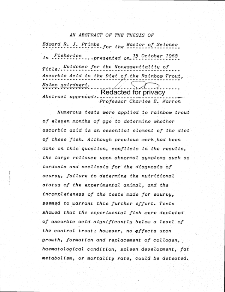
AN ABSTRACT OF THE THESIS OF' Edward P. J. Prirnbs .............*.e. ....for e Master of Science Fisheries 15 October 1968 tn .............presented on................. Evidence for the Nonessentiality of Ttl Q....................................,.... L Ascorbic Acid in. the Diet of the Rainbow Trout, S S S 7. Salmo gairdneri. I -1 /.s . . . . .')_t . . . Redacted for privacy Abs t rac I approved: . . . . . . . . . .. . . . . . . Professor Charles E. Warren . . . . . . . S S I S S S S S S S S S S S S S S S S -.. . . . I S I . . . Numerous tests were applied to rainbow trout of eleven months of qge to determine whether ascorbic acid is an essential element of the diet of these fish. Although previous work had been done on this question, conflicts in the results, the large reliance upon abnormal symptoms such as lordosis and .scoliosis for the diagnosis of scurvy, failure to determine the rwtritional status of the experimental animal, and the tricontpleteness of the tests made for scurvy, seemed to warrant this further effort. Tests showed that the experimental fish were deoleted of ascorbic acid significantly below a level of the control trout, however, no affects upon collagen, haematological condition, soleen development, fat growth, formation and replaceriiertt of metabolts'n, or mortality rate, could be detected. planned. are therefrom rising hypotheses the and experiment this of findings the verify to work Further vitamin. another with acid ascorbic of interrelationship an by caused be may salpionoids the by acid ascorbic of requirement dietetic the show that others of results the that hypothesized is It hundred. three of population total a of out scoliosis of case one .of exception the with months, two of age initial an of fish in also but months eleven of age initial an of fish in only not develop to failed scoliosis and lordosis Specifically, 2 Evidence for the Nonessentiality of Ascorbic Acid in the Diet of the Rainbow Trout, Salmo gjel. Edward Rudolph Joseph Primbs A THESIS submitted to Oregon State University in partial fulfillment of the requirements for the iegree of Master of Science June 1969 555 is thesis Date Graduate of Dean School 5SS5S5 S. ..S...U... S.U.....S.. 5,5.5.55 Redacted for privacy 5 5 Wildlife & Fisheries 55 S S S ofS Department of lead 4'. 5 5 S S S S S S U S S S S S . . . Redacted for privacy major of charge in Fisheries of Professor ItISeI S .SS. ItSS S.. S S . .. . . ... . .. .. Redacted for privacy APPROVED: Acknowledgments In. a very factual sense, the author of an experiment is but a coordinator of the contributions of many. Thus, this experiment would riot have been possible without the fellowship granted by the Bureau of Commercial Fisheries, nor without the laboratory facilities, equipment, fish, arid feed generously provided by the Department of Food Science and Technology of Oregon State University. More subtle, perhaps, but of no less value, has been the stimulating envzrorurient and the readiness to inquire that prevails under Dr. Harold W. Schultz's leadership. More specifically, Professor Russell 0. Sirinhuber has been of inestimable value in all of the following work, and its success is due to him more than to any other. Credit must also be given to Professor Joseph H. Wales for his suggestions, criticisms, and advice, and to Mrs. June L. Hunter for her assistance. Dr. Jorz.g Lee translated articles written in Japanese, a contribution of no small value. George B. Putman has been a helpful colleague, and to Theodore Wales and Richard Foster credit for the preparation of the diets and daily feeding of the fish must go. Finally, thanks to my wife, Julia, are expressed for her patience, encouragements, and suggestions. Evidence for the IVoriessentiality of Ascorbic Acid in the Diet of the Rainbow Trout, Saln'io qairdrieri. Introduction In the decade 1947-1958 some effort was made to determine whether ascorbic acid was a dietetic requirement of the salmonoids. The work in 1947 of ftfcLaren (NcLaren, 1947), which actually had been preceded by an experiment on ascorbic acid requirements of trout of 1924 even before ascorbic acid had been isolated (Davis and James, 1924) and by an experiment of 1937 (Hewitt, 193?) during a period of active investigation of vitamin C, evidenced not only that ascorbic aci-' is a dietetic requirement but also that an excess of the vitamin had depressing effects on growth. McLaren's work undoubtedly had been suggested by Elvehjem's suspicions that the trout is dependent on a dietary intake of ascorbtc acid. (Field, 1943) However, later experiments of Wolf (Wolf, 1951), Halver (Halver, 1957), and Coates (Coates and Halver, 1958) clearly contradicted that of HcLaren and of Hewitt, but the results of these efforts are questionable, because of (1) the failure to appreciate algae as a source of ascorbic acid, (2) the termination of the 2 experimental period short of PicLaren's time, (3) the interference with the established criteria by disease, arid (4) the failure to test diets for the presence of ascorbic acid. In 1965 and 1967 a Japanese team reported the results of experiments that suggested that rainbow trout require 2áscorbic acid in the diet. (Kitamura, 1965, 1967) The symptoms accepted as the criteria for the development of scurvy were accompanying scoliosis, lordosis, abnormal mortalities, a decrease in growth rate, hemorrhages, arid an incomplete development of the operculum. Following these findings, Halver recently with a diet deficient in ascorbic acid also developed curvatures of the spinal column of silver salmon, as well as change in shin pigmentation, susceptibility to subcutaneous and intramuscular hemorrhage, loss of coordination, a deficient cartilage development in gill and eye structures, and hyperplasia of adrenal cortical cells. (Halver, 1967) (Ashlgy, 1967) (Halver, 1968) The Cort land laboratory also reversed their previous conclusions on the need of ascorbic acid by trout after the Japanese publications: Poston. found lordosis arid scoliosis, tritrnaZ hemorrhaging, and a 3 higher condition factor among brook trout fed a diet lacking ascorbic acid (Poston, 1966) These experiments previously performed, however, have been incomplete tests for the scurvy syndrome. Moreover, with the exception of Hewitt, none have mentioned any effort to test whether their subject animals actually were in a state of ascorbic acid deficiency. Hence this experiment was undertaken to attempt to reach a conclusion that could be applicable to hatchery diets and that could provide necessary data as a basis for future research into the needs for and function of ascorbic acid in fish in general and other animals. 4 Nat ertals and Methods The water used was that of a well (40 feet deep) at the Food Toxicology and Nutrition Laboratory of Oregon State University. After an examiriatiorz. of the water for the type of endemic organisms, an assay of this bioniass for ascorbic acid concentration was made by the method of Roe, Kuether, Oesterlirig, and Mills (Roe, 1954), as reported arid modified by the Association of Vitamin Chemists. (Freed, 1966) Light in the indoor fish tanks was reduced below the level at which endemic organisms were collected, arid the tanks were scrubbed whenever any slight growth was apparent and cleaned at least once a week. Two hundred rainbow trout (Salrn.o çirdneri), approximately 11 months of age and 43 grams in weight, of a Mt. Shasta strain were randomly taken from the population of the Food Toxicology and Nutrition Laboratory arid randomly divided into four lots of 50 each, and four hundred trout, approximately two months of age and 2.30 grams in weight, from the population of the following year were randomly divided into four lots of 100 each. From time of segregation into lots, two lots from each age group were fed ad libi. turn the Oregon Test Diet No 1 (Table No 1) which had the 5 cidditarnent of 1.2 mg. of ascorbic acid per gm of diet, and two lots were fed the same diet without the ascorbic acid. Table I Oregon Test Diet No 1 The caseiri used was VitaminNutrient % of Diet free Casein, a product of NBC, which had rreviousl been Salt Nix No 2 USP XII Calcium Carbonate Carboxyinethylcellulose Cellulose (NBC) tested in Choline Chloride (7) ascorbic acid Vitamin Nix Vitamin A Vitanurt , Cone (11OIU/g) 4.0 0.9 1.3 6.4 1.0 0.1 0.6 2.0 deficient diets. (NBC, 1967) The diet in toto and specific suspected components were assayed for ascorbic acid concentration by the method of Roe Table 2 (Roe, 1954), as (mg/kg diet) modified by the Aiphacel 1,200 Ascorbic Acid 15 Association of Butylated Flydroxyanisole ii Vitamin Chemists. (Freed, 1966) The concentration of ascorbic acid in the blood plasma was measured by the method of Roe for Butylated Hydroxytoluene Supp. BThtjri B Ca Pantotheri.ate Ioljc Acid Inositol Nenadione Niacin P-Aminobenzoie Acid Pyridoxine Riboflavin Thiamine Vitamin D 15 53 2 288 19 2,500 16 512 400 48 144 64 8 6 indicating iomediate intake of the vitamin. Tests for the state of ascorbic acid deficiency were riicide and the degree of deficiency measured by determination of ascorbic acid concentrations in. the liver, brain, and spleen by the Roe method. J'rom the 24th to the 35th week after initiation of the feeding trial, samples of fish from the 43-gram age group were examined for scurvy symptoms as defined by the following criteria 1. Decrease in growth Total fish were weighed biweekly, and calculations of contht ion and conversion factors were made. 2. Decrease in formation and replacement of collagen (i) Bone abnormalities. Plural ribs were extracted and examined for (a) "ground glass appearance", (b) the "white line of Frarikel", (c) the "corner sign", and (d) fractures. (Li) Fat lure of wound healing process Vertical wounds, ir'medtately above the lateral line and directly vertical to anterior margin of dorsal fin, were made with scalpel on samples of each lot under anesthesia MS-222). Wound length was ten mm, and depth was six mm. After 15 days wGurids were sutured, and subsequently excised on the 23rd day after wounding, preserved in Bouin's solution, sectioned with a 7 niicrotOnie, stained with friasson trichrome connective tissue stain, and compared at 20X, 128X, and 800X. (iii) Weakness in collagenous structures. Gas bladders of samples of all lots were removed and tested for relative strength by application of air pressure, which was measured by a manometer. (iv) Hemorrhagic tissue. Gross examination of liver, intestine, and epidermis was made. (v) Development of lordosis and scotiosis. Gross examination of all fish, including fish of the 2.30-gram age group, was made. Dorsal and lateral radiograms were taken of all fish apparently deformed. 3. Development of anemia. Erythrocytes were counted by use of a Fisher autocytometer and the B. D. Unopette diluting technique. (ii) Herncztocrit readings were determined by a clinical centrifuge. Ci) (iii) Erythrocytes were measured with a micrometer (800X) after stairiirLg with Wright's. (iv) Hemoglobin gm. % was determined by use of Hycel Cyanmethemoglobin reagent and standard solutions with a Beckman D. U. spectrophotometer at 540 m. u. wavelength. 4. Enlargement and congestion of spleen. A spleen index was computed by determination of the 8 ratio of the weight of a spleen to the weight of the subject animal. 5. Pat deposits in liver. Extracted livers were fixed in Boutri's solution and sectioned with a microtome. Samples were stained with Sudan IV and compared at 800X. 6. Mortalities. All mortalities were examined for abnormalities and recorded. The sources of ascorbic acid for the test lots were irwesttgated. Ninety to ninety-five percent of the biomass in the water consisted of diatoms (Naviculu minima GrünOw: and Achnonthes minutissinia Kutzirtg) (Mclnttre, 1967), which contained approximately 34 mg. of ascorbic acid per 100 g of algae (dry basis). This determination compares favorably with that of Pratt, who found 39 nig per 100 g in Chiorella (dry). (Pratt, 1967) However, the growth present in any one tank could be estimated at no more than a few milligrams, and this was promptly eliminated when the growth became apparent. The concentration of ascorbic acid in the Oregon Diet without added vitamin C was also suspected to be insignificant dte the was found to have on the average Table 3 Ascorbic Acid in Diet Components Component ug/g Diet seven ug per g of diet with a range of five to eight ug. Vitamin Mix 1.3 Pive of the major components of the diet contributed at least half of this amount. (Table 3) Prom the time of initiation to the time of 10 terin.in.atioa of the experiment 66,740 grams of the Oregon Diet was fed to the test grouc. Thus, the diet provided but 467 mg to 100 fish over a period of approximately six months, or approximately 26 ug per fish per day. Measurements of concentrations of ascorbic acid in the blood plasma of the test and control groups indicated a Table 4 Ascorbic Acid Concentration in Blood Plasma SigrLij ican difference in intakes of the utanitn. Test Mean (mg/100 cc) Standard Deviation Number of Samples Control 1.02 4.57 1.42 .69 19 19 (P<.005) (Table 4) Two zero concentrations were found among the test lots. Figure 1 shows the distribution ft . graphically. / L Tes \...eoNiroL A distinctly defined ,, : state of ascorbic acid deficiency in the test 0 groups was found by the measurements of ascorbic acid concentrations in \ 1 1.1 7 3.7 f7 5.7 M(Asorbi h.7 AcJ//cc cc Fig. 1. Frequency of Plasma Ascorbic Acid Levels in Ascorbic Acid Deprived (Test) and Ascorbic Acid Fed (Control) Groups. 11 the liver, brain, and spleen. The differences between test groups and control groups were significant in each case. Table 5 Ascorbic Acid Concentrations in Organs Liver Mean (mg/100 g liver) Standard Deviation Number of Samples Test Control 1.43 18.00 6.85 12 .85 14 P< .005 Brain Mean (mg/100 g brain) Standard Deviation Number of Samples 9.00 1.60 8 P< .005 Spleen Méin (mg/100 g spleen) Standard Deviation Nunber of Samples 6.75 P<.005 36.75 12.20 8 1.50 45.75 8.26 4 4 Of particular interest were the 1 differences between Te standard deviations. Whereas the It distributions of values within the test groups lie within a narrow range, the distributions of values within the control groups encompass '' t II. .o Iy AccJ//eo Liver Fig. 2. Frequency of Liver Ascorbic Acid Levels in Ascorbic Acid Deprived (Test) and Ascorbic Acid led (Control) Groups. . 12 considerable scope. This is seen in I figures 2, 3, and 4. 4" 1 Te Of equal : interest was the large gradual 0, Cd,virc'J increase in the ascorbic acid / concentration in the o ç Ia Ic .lo e 3ç ç \, lic 5a $ 0 M(i45coYhc ,4Ld//oo ?. $rqV liver of the control Pig. 3. Frequency of Brain Ascorbic Acid Levels in Ascorbic Acid groups over a period Deprived (Test) and Ascorbic Acid Fed (Control) Groups. of 50 days, while the ascorbic acid concentration in the liver of the test groups remained fairly constant over the entire period with the exception of a slight decline within the last ten days. (See figure 5.) Both, the large differences in the sI ci standard deviations of'c the data of the three organs exarnineu anu i ; 'i I , s Co#Vvo i ! the differerwe in C ange as or ic , 3 3c 90 MAcoi'6ic Acd//cia 4f fe (f .gp)eew acid concentrations in Pig. Frequency of Spleen Ascorbic Acid Levels in Ascorbic the liver with time, Acid Deprived (Test) and Ascorbic Acid Fed (Control) Groups. suggest that the major . 13 portion of the ascorbic acid in the 2J organs is deposited n these tissues only for storage. .35 . 510 9$ 50 Pig. 5. Chcirtge of Liver Ascorbic Acid with Time in Ascorbic Acid Deprived (Test) and Ascorbic Acid Fed (Control) Groups. (0 day of 26th week of experimental period.) day is 1st Upon examtnatLon of growth data and calculatton of the tndex food conuerston factor no sLgnLftcant Table6 H Comparison of Growth between Ascorbic Acid Deprived and Ascorbic Acid Fed Groups Test Total Fish 100 Total Weight Increment (g) 23,216 Final Average Wt. (g) (58 fish) 300 Food Conversion Factor 1.01 Control 99 22,895 308 0.98 differences were found between the control and test groups. (Table 6) fiforeover, no stgrttftcant dtfference was found between the condttLon factors calculated for the test and control groups, although the condttion factors of 14 both groups were larger than either of the two reported by Poston as indicating ascorbic acid deficiency. (Poston, 1966) The mean condition factor for 50 test fish is 1.62 arid the mean condition factor for 50 control fish is 1.65. (P>.i0) No distinction up to 40X could be made between the bone structures and composition of the pleural ribs of the test and control groups, and none of the traditional scurvy symptoms of the cost ochoridral junctions could be discerned. Statistical measurement showed a high probability (P > .25) of no difference between the mean air pressures requt red to break uie g as bladders o 1 the Table 7 Comparison of Gas Bladder Breaking Points of Ascorbic Acid Deprived and Ascorbic Acid Fed Trout test and control Test result sugg ests Nean (mJY.9 Standard Deviation 105 that there was P> .25 groups. This Number of Samples Control 34 i3T 116 35 no interruption in the formation and replacement of collagen in the ascorbic acid deprived fish. (Table 7) External and internal gross examination of both test and control groups revealed no difference between the two groups, and no hemorrhages were observed. 15 An incomplete development the operculurii, as noted by Kitamura as a symptom of ascorbic acid deficiency, was observed of ,i J , ,' O I, o in both the test and control groups. , . , ,øt- Fig. 6. Gas Bladder Breaking Points of Ascorbic Acid studies revealed no Deprived (Test) and Ascorbic Acid Fed (Control) conclusive evidence of Trout. any of the types of anaemia found in scorbutic animals. Hematological Thus, no significant difference (P> .10) was found between the mean erythrocyte counts of the ascorbic Table 8 Comparison of Erythrocyte Counts of Ascorbic Acid Deprived arid Ascorbic Acid Fed Trout Pteari. (per cu mm) Range (X 100,000) Standard Deviation Plumber of Samples P>.10 Test 1,044,000 5.25-14.25 2.04 39 - Control 1,083,000 5.25-14.25 1.67 39 - acid deprived and ascorbic acid fed grouDs. (Table 8) The dissolved oxygen of the various tanks, which can affect the number of erythrocytes (Smith, Lewis, & Kaplan, 1952) did not vary more than 0.4 mg/i arnortg the tanks. 16 Support I ng the results of the erythrocyte counts, the mean henicitocrit / of the test group is / not signlfLcantly I different from the ' Fig. &,1/dl ±.rythrocyte Counts (Table 9) The type of Table 9 Comparison of Hematocrits of Ascorbic Acid Deprived and Ascorbic Acid Fed Trout. anaemia generally found in the syndrome tic but other forms S 7. of Ascorbic Acid Deprived (Test) and Ascorbic Acid Fed (Control) Trout. the control group. is norin.oc i I -- mean hematocrit of scurvy I Range Standard Deviation Number of Samples do occur. Thus, P= Test 34.85 23-4? 6.98 40 Control 37.26 21-49 5.65 39 .05 a decrease in hemoglobin relative 'I\ to ThB.C. counts is / 'I frequent ly 4JI/\/ / attributed to P ascorbic acid deficiency. (Chczkrabartz, 1963) Paxd cit iJInne Pei-ee/ Fig. 8. Hematocrits of Ascorbic Acid Deprived (Test) and Ascorbic Acid Fed (Control) Trout. In this experiment significant Table 10 Comparison of Gram Percent Hemoglobin of Ascorbic Acid Deprived and Ascorbic Acid Fed Trout difference was measured between the Mean Range Standard Deviation Number of Samples Test Control 7.88 8.16 5.8-11.3 1.34 38 4.8-13.3 1.81 39 P> .10 - means of the gram percent hemoglobin of the test and control trout. (Table 10) Macrocytic anaemia also has been produced by a diet lacking ascorbic -iJ a acid. (Chakrabartz, a a U 1963) This type of CDidYôJ I k anaemia involves an q , / abnormal enlargement of the red blood corpuscles. However, again the mean of C g i , RemogJtii.' Fig. 9. Gram Percent Hemoglobin of Ascorbic Acid Deprived (Test) and Ascorbic Acid Fed (Control) Trout. the calculated volumes of red blood cells of the test groups did not significantly differ from the mean of the volumes of red blood cells of the control. (Table 11) The mean diameter of 286 red blood cells (microscopic measurements) of the test groups is 12.39 p, 18 the mean d meter o 320 red Table 11 Comparison of IR.B.C. Volumes of Ascorbic Acid Deprived and Ascorbic Acid Fed Trout blood cells 0 con ro. groups being Mean (cu) Range Standard Deviation Number. of Samples P = .25 Test Control 341 348 50 39 34 39 - - 253-403 223-493 12.73 u. No enlargement of c e \i v-h ct ..Je the spleen was I' shownby ,: \ calculation of the _P/" spleen index: the i . / mean of the test / groups did not differ stgntftcantly from the mean of the control J 1 L 3 øf R12(J e.: (c.p) Ftg. 10. R.B.0 Volumes of Ascorbic Acid Deprived (Test) and AscorbLc Actd Fed (Control) Trout. Table 12 Spleen Index of Ascorbic Acid Deprived and Ascorbic Acid Fed Trout groups. (Table 12) Test Microscopic comparison of sections of the Mean Standard Deviation Number of Samples P>.25 Control 7.42 7.41 3,1 43 3.9 39 liver (sections from four test fish and - - from four 19 control fish) revealed no 4" abnormal fat ctfr deposits in the liver. 4 \\ Even though all lots of the 4 43-gram fish g 2 C developed fin rot during the course of the experiment, oF , ,a ij '. IY 20 * PI enh/fiiiof)8IY Pig. 11. Spleen Index of Ascorbic Acid Deprived (Test) and Aàcorbic Acid Fed (Control) Trout. the mortality was tow in all groups. Of the total yearlings, only one death of an ascorbic acid deprived fish out of a total population of 100 occurred to give a percent mortality of one percent, and among the controls only two deaths of ascorbic acid fed fish out of a population of 99 to give a percent mortality of two percent. A comparison of sections of wounds of ascorbic acid deprived and ascorbic acid fed groups at 20X, 128X, and 800X evidenced that healing and thus the formation of collagen was normal in the test group collagen was widely dispersed, collagenous fibres were formed, and fibroblast cells were abundant. (cf figures 12 through 14) There was some variation in the degree of healing, 20 ..::". 2LI.r.L ...... .' 'L i!11' I', ':r -1:4' -. - Comparison of cross sections of wounds of (a) ascorbic acid fed and (B) ascorbic acid decrivd trout. Complete regeneration epithelium and closure of wound by granulation tissueofpresent in both cases. H.& E.. X 20. Fig. 12. ;4;. a : . '.,r,*w & Co.'?warjsor J '0" 7,1 "ii ?4L ; ' P - - gramulation ttssu of wounds o (A) ascorbic acid fed and (B) ascorjc acid denrup1 r:ut. .-ro1iferatirLq fihrhlasts, inflanctor o el iui.nous fihrc ahurdantl creScent in both cases. .1..rL Trichrorne. X 12k. it;. U 21 , i*,, I., Pig. 14. Comparison of granulation tissue of the wounds 0.1 (A) ascorbic acid fed and (B) ascorbic acid deprioed trout. Numerous fibroblasts, so'ie undergoing mitosis, embedded in collaqenous fibers in both cases. fifasson Trichrorne. X 800. 22 but the wounds of some of the test fish tended to show a more advanced stage of healing than. the wounds of the control fish. Probably the variation originated in diuergencies in suturing, in sectioning, or in mechanical stresses in uivo. Among the 100 ascorbic-acid-deprived trout of the initiczl-43-grani. group, neither scoliosis nor lordosis was observed, and within the 200 ascorbic-acid-deprived trout of the in.itial-2.30-grant group, only one case of scoliosisoccürred after the expiration of 26 weeks of the experimental period. (See appendices one and two.) 23 ton. The results of this experiment evidenced that ascorbic acid is not essential in the diet of the rainbow trout. It is difficult to evaluate these findings in relationship to those of Kitamura's (1965, 1967) and Halver's (1967), since neither of these authors has indicated that they had tested their diets for the presence of ascorbic acid and had determined the relative degree of deficiency of their test animals. The elimination of ascorbic acid completely from the diet is not accomplished simply by excluding the nutrient from the constituents used to prepare a purified ration. As far as it is known, ascorbic acid is present in all animal and plant cells, (King and Becker, 1959) and may be carried in oils as a suspension. (Hewitt, 193?) Thus, it is at least doubtful that the diets of McLaren, Kitara.ura, and Halver were actually free of measurable amounts of ascorbic acid. Although recommended allowances for man have a range from. 30 mg per day (Nedz.cal Research Council, 1953) to 1.8 g (Ston.9, 1966), the minimum, amount required to avoid definitely associated reactions is 10 mg per day. (Medical Research Council, 1953) The minimum amounts required to avoid clinical scurvy 24 symptoms in. the guinea pig and the rhesus monkey have also been established: 5 mg/kg body weight/day and 2 mg/day, respectively. (National Academy of Science, 1962) McLaren (1947) found that 0.25 to 0.50 mg per g of ration produced the least undesirable results in the case of the rainbow trout. Relative to these levels, the amount of ascorbic acid in the diet, 26 ug/day or 7 ug/ g of diet, used in this experiment does not seem to be significant. Indeed, in this work a state of deficiency was definitely defined by analysts of various organs for ascorbic acid concentrations. It has long been thought that the high concentration of ascorbic acid in organs of animals - the liver, the brain, and the adrenal cortex, e.g. - suggests special functions of ascorbic acid in these organs. (Long, 1946) (Chalopin., Mouton, and Ratsimarnanga, 1966) However, the wide variance in ascorbic acid concentrations among organs of those fish fed ascorbic acid and the narrow range in coricentration.s of those fish deprived of added ascorbic acid as well as a large gradual increase in. ascorbic acid coricen.trations in the liver of the control fish indicate that the large concentrations of ascorbic acid normally associated with the organs are largely furictionless and 25 only represent storage of ascorbic acid. On the other hand, the stability of liver ascorbic acid and the narrow variance of values of organ ascorbic acid in general in the fish deprived of dietetic ascorbic acid suggest a minimum level for survival as well as an endogenous source, or an untested exogenous source - for example, bacterial synthesis. In view of recent evidence, however, bacterial synthesis is unlikely. (Levenson, 1962) In spite of the apparent state of deficiency developed in the test fish, moreover, no evidence of an impairment in the general health or an incapacity for normal functioning of the organism was obtained upon administration of a number of tests for symptoms of scurvy. Here again it is difficult to evaluate these results in relationship to those of previous workers, since the previous work was concluded on the basis of a very limited set of symptoms for scurvy, few, if any, of which are specific for an. ascorbic actd deficiency. McLaren, for example, reached her conclusions ,just by application of the criteria of growth, mortality, hemoglobin, liver fat, and hemorrhagic tissue. Beyond these, Kitamura has only observed lordosis and scoliosis and an incomplete development of the operculum. Although lordosis and scoliosis have not traditionally been included in the scurvy syndrome (F'ollts, 1948) (Bickness and Prescott, 1953) (Vilter, 1960) (Goldsmith, 1964), these diseases have been associated with inadequate nutrition, as well as with inadequate metabolism, for over one hundred years. (Risser, 1964) Moreover, the effects of ascorbic acid on bone development as well as on metabolism seem to suggest a role of a deficiency of this vitamin in the development of the deformity (Thornton, 1968), but the role may not be a direct one. A. F'. Gardner (Gardner, 1966) produced lordosis in rats by massive (25 mg.) dosages of vitamin D3, and it has been observed that ascorbic acid may detoxify toxic dosages of other vitamins. (Rosenberg, 1945) Numerous other observations have confirmed a substance (Vitamin A or Vitamin D') in the liver or liver oils (cod liver oil, e.g.) of animals which has an inhibitory effect upon the activity of ascorbic acid. (Collett and FJrtksen, 1938) (Vedder and Rosenberg, 1938) (Rodahl, 1949) Poston has not found any symptoms of hypervitamirtosis yet in brook trout after feeding massive doses of D3 to these fish for thirty weeks (Postori, 1968), but undoubtedly his diet includes ascorbic acid. It may well be that not only the lordosis and scoliosis but also the 2? scurvy symptoms experienced in previous work have had as their common cause hypervitaminosis A or P. Neither lordosis nor scoliosis was reported by NcLaren, who had included cod liver oil in her diet. Since Kitamura began with fish of 0.60 g weight and Poston with 1.45 g, whereas NcLaren began with fish of 3.5 g and in this experiment we started at 43.00 g and 2.30 g, the deformity of spinal flexure may be limited to trout of an age less than two months. In man and some other animals, at least, the problem has been one of preskeletal maturity (NacEwen and Shands, 1967), with evidence in tadpoles of scoliosis having a genetic origin. (Underhz.lZ, 1966) Otto Bessy in the early 'thirties had found fatty degeneration of the liver in scorbutic guinea pigs. (Bessy, Henten and King, 1933) Hewitt also reported the symptom. (Hewitt, 1937) However, Baldwin showed in 1944 no significant difference in tissue lipids between scorbutic and normal animals. (Baldwin, Longenecker and King, 1944) McLaren employed this criteria of scurvy, but the negative results were not consistent w7th her other data. Comparison of liver sections in this experiment also revealed the livers of ascorbic acid deprived subjects to be normal, both controls and test groups having only very slight deposits of fat. 28 The red blood cell count, the hemoglobin gm %, and the mean cell volume, which were determined in this experiment, compared favorably with the blood values of the normal trout as determined by Field (Field, Eluehjem, and Juday, 1943). The hematocrits, however, differed by eight percent. The red blood cell counts made by Halver exceeded Field's even in the controls by over 200,000. A low dissolved oxygen content would account for the abnormally large R.B.C. count and differences between the experimental and control groups, polycythemicz; on the other hand, an increase in the number of red blood cells has been observed in vitamin-C-deficient guinea pigs, and explained as a response to loss of blood (Constable, 1960). But Constable's experiment also showed that scorbutic guinea pigs did not develop anaemia. In accord with this finding, Kitamura's R.B.C. count of the rainbow trout, supposedly scorbutic, was quite similar to the control. An experiment, later than Constable's (Chakrabartz and Barterjee, 1963), however, produced anaemia in 13 of a total number of 14 scorbutic guinea pigs. Although the anaemia of scurvy is not completely understood (Kahn and Bradsky, 1966), it is fairly well established experimentally that ascorbic acid deficiency significantly affects (1) iron metabolism (Mazur, Green 29 and Carleton, 1960) (frlazur, 1961) (Hallberg, Salvell and Brise, 1966) and haem biosynthesis (Lochhead, 1959) and (2) fotic acid nietabolisrn and thus D.N.A. biosynthesis by the red blood cells. (Vilter et al, 1963) Thus, the scurvy syndrorne still commonly includes anaemia under several of its various forms (Woodruff, 1964) (G. C. Chatterjee, 1967): (1) normochromic and normocytic (a decrease tn normal ThB.C. count and gram percent hemoglobin) (.Vilter, 1967), (2) microcytic, hypochromic (a decrease in hemoglobin and cell size) (Vilter, 1967), or (3) macrocytic (a failure in growth of the nucleus) (Vilter, 1963). A brief mention of symptoms of microcytic anaemia being eliminated among chinook salmon by adding ascorbic acid to the Abernathy diet was reported recently. (Burrows, 1968), however, the data were not given. Not only a decrease in the rate of growth but also a rapid loss of weight has been a traditional symptom of scurvy. (Knox and Goswamt, 1961) This effect upon growth has been attributed not only to a decrease in appetite but also to a direct effect upon metabolism. (Ram, 1966) (Evans and Hughes, 1963) And yet except for McLaren, who showed a decrease in the rate of growth under ascorbic acid deficiency, no one has yet reported any data indicating a significant difference. Kitamura (1965) reported retarded growth, but gave no data, and later 30 (196?) showed graphically what appears to be an insignificant difference between the vitamin-C- deficient fish and his control. (No test of significance was merit toned.) Haluer (1968) mentioned unfavorable growth and food conversion in. fish on. the vitamin-C- deficient diet, but also gave no supporting data. Postori (1967), though finding no difference in average body weights, did find a significant difference in the condition factors. Scurvy, untreated, terminates in death. (Wohi and Goodhart, 1964) Both fricLaren and Kitamura (1965) found differences in rate of mortality, supporting their diagnoses of scurvy in. rainbow trout. Hemorrhagic tissue, another common indication of ascorbic acid deficiency, was found again by PicLaren and Kitarnura (1967). Although the gill filament cartilage rods were not microscopically examined by us, and thus the deformation in this structure as noted by Haluer (1968) was not contradicted, other tests of collagen formation and replacement were made with positive indications of normalcy. The failure of the operctzlunt to fully develop, as observed by Kitarnura, was a defect found by us in both the experimental arid control groups, and thus a cause other than ascorbic acid deficiency was assumed. Although the results of this experiment indicate 31 that there exists no relationship between the large concentration of ascorbic acid in organs arid a function of ascorbic acid in those organs as often suggested, yet this interpretation does riot contradict to any degree thesigntficant body of evidence that implicates ascorbic acid in the activity of the adreno-cortical gland. (Chalopin, 1966) On the other hand, there is evidence that strongly suggests that the changes induced in the adrenal gland by vitamin C deficiency are caused by stress attributable to the scorbutic state, which acts upon the adrenal gland by excessive stimulation by ACTH arid not directly by the depletion of ascorbic acid in the adrenal gland (Howard and Cater, 1959), the hypertrophy being more of a sign of exhaustion than of activity. (Dugal, 1961) The changes in the adrenal gland observed during avitarninosis C are hyperplasia, hypertrophy, arid decreased and irregular 'lipid distribution. (Howard, 1959) Halver (1968) observed hyperplasia in the adrenal cortex of coho salmon, supposedly deficient in ascorbic acid. The adrenal cortex in salmorioids is located along the posterior cardinal veins only as a layer of one or two cells in the region of the head kidney. (Rasquin. and Roseribloom, 1954) To reduce the probability of a misinterpretation, confirmatory evidence, such as 32 measurQment of blood ACTH (Clayton, Harrunond and ArirLitage, 1957) or observation of increased mitoses, should have been taken, especially since only several samples were compared. 33 Summary The evidence of this experinzeri.t supports the conclusion that ascorbic acid is not essential in the diet of the rainbow trout under normal conditions. It is suspected that previous results that tended to show that salmonoids require ascorbic acid in the diet are attributable to interrelationships of ascorbic acid with other vitamins and to hypervitarizinoisis A or D, and thus under the abnormal conditions of the inactivation of endogenous ascorbic acid by excessive dosages of other vitamins scurvy symptoms can be produced. It is also suspected that lordosis and scoliosis are not causedby an ascorbic acid deficiency, bat rather by another nutritional abnormality related to ascorbic acid metabolism. Further confirmation of the conclusions of this experiment, as well as tests to verify the explanation given here of the causes of some scurvy symptoms in sälmonoids reported by others, Is planned. 34 BIBLIOGRAPHY 1. 2. 3. 4. 5. 6. 7. 8. 9. 10. 11. 12. 13. Ashley, L. N. et at. 1968. Ascorbic acid deficiency syndrome in coho salmon. In: Proceedings of the Northwest Fish Culture Conference, Seattle, 1967. Seattle, University of Washington. p. 10. Baldwin, A. F?., H. E. Longenecker and C. 0. King. 1944. Tissue lipids in ascorbic acid deficient guinea pigs. Archives of Biochemistry 5:137-146. Bessy, 0. A., N. L. Nenten and C. G. King. 1933. Pathologiô changes in the organs of scorbutic guinea pigs. Proceedings of the Society for Experimental Biology and Medicine 31:455-460. Bicknell, F. and F. Prescott. 1953. The vitamins in medicIne. New York, Grune & Stratton. 784 p. Burrows, F?. E. 1968. Applied nutrition research. Int: Progress in sport fishery research, 1967. Washington, D. C. p. 73-74. (U. S. Fish and Wildlife Service. Bureau of Sport Fisheries and Wildlife. Resource Publication no. 64) Chakrabartz, A. S. and S. Banerjee. 1963. Anaemia in scurvy and its relation with iron metabolism. Indian Journal of Experimental Biology 1:135-140. Chalopin, H., H. Mouton and A. F?. Ratsimamanga. 1966. Some Interrelations between ascorbic acid and adreno-corticai function. World Reoiw of Nutrition and Dietetics 6 165-196. Chat terjee, 0. C. 1967. Effects of ascorbic acid deficiency in animals. In: The vitamins, ed. by W. H. Sebrell, Jr. and R. S. Harris. Vol. 1. New York, Academic. p. 407-45 7. Clayton, B. E., J. E. Hammond and P. Arrnitage. 1957. Increased adrenocorticotrophic hormone in the sera of acutely scOrbutic guinea-pigs. The Journal of Endocrinology 15:284-296. Coates, J. A. and J. E. .HaZver. 1958. Water soluble vitamin requirements of silver salmon. Washington, D. C. 9 p. (U. S. Fish and Wildlife Service. Special Scientific Report no. 281) Coltett, E. and B. Eriksen. 1938. Interrelations of the vitamins. The Biochemical Journal 32:2299-2303. Constable, B. J. 1960. The blood picture in the guinea pig in acute and chronic scurvy. The British Journal of Nutrition 14:259-268. Davis, H. S. and H. C. James. 1924. Some experiments on the addition of vitamins to trout foods. Transactions of the American Fisheries Society 54 77-91. 35 14. Dugal, L-P. 1961. Vitam.iri, C in relation to cold temperature tolerance. Annals of the New York Academy of Sciences 92:307-31?. 15. Evans, J. R. and R. E. Hughes. 1963. The growth maintaining activity of ascorbic acid. British Journal of Nutrition 17:251-255. 16. Field, J, B., C. A. Elvehjem and C. Juday. 1943. A study of the blood constituents of carp and trout. Journal of Biological Chemistry 148:261-269. 17. louis, R. H. 1948. The pathology of nutritional diseases. Springfield, Charles C. Thomas. 291 p. 18. Freed, Pt. 1966. Methods of vitamin assay. 3d ed. New York, Interscience. 424 p. 19. Gardner, H. F. 1966. Severe spinal curvature in rats injected with a water-dispersible vitczrnir. P preparation. Toxicology and Applied Pharmac5logy 8 :438-446. 20. Goldsmith, G. A. 1964. Vitamins and avitaminosis. In: Diseases of metabolism, ed. by G. G. Duncan. 5th ed. Philadelphia, Vi. B. Saunders. p. 567-663. 21. Hailberg, L., L. Salvell and H. Brise. 1966. Search for substances promoting the absorption of iron. Acta Medica Scandinavica, sup., 459:11-21. 22. Halver, J. E. 1957. Nutrition of salmonoid fish. The Journal of Nutrition 62:225-243. 23. Halver, J. E. 196?. Director, Western Fish Laboratory. Personal conintunication. Cook, Washington. October 3. 24. Halver, J. E. 1968. Nutrition and toxicology. In: Progress in sport fishery research, 196?. Washington, B. C. p. 39-54. (U. S. Fish and Wildlife Service. Bureau of Sport Fisheries and Wildlife. Resource Publication no. 64) 25. Hewitt, E. R. 1937. Some recent work on fatty livers in trout. The Progressive Fish-Culturist 27:11-15. 26. Howard, A. N. and D. B. Cater. 1959. The adrenal cortex and the effect of AUTH and cortisone in scorbutic guinea-pigs. Journal of Endocrinology 18:175-185. 27. Kahn, S. B. and I. Bradsky. 1966. Vitamin B12, ascorbic acid and iron metabolism in scurvy. The American Journal of Medicine 40:119-126. 28. King, C. G. and R. R. Becker. 1959. The biosynthesis of vitamin C (ascorbic acid). World Review of Nutrition and Dietetics 1:61-72. 29. Kitaniura, S. et al. 1965. Studies on vitamin requirements of rainbow trout, Salmo gairdneri. I. On the ascorbic acid. Bulletin of the Japanese Society of Scientific Fisheries. 31:818-826. 30. Kitamura, S. et ci. 1967. Studies on vitamin requirements of rainbow trout. II. The deficiency symptoms of fourteen kinds of vitamin. Bul 1 et in of the Japanese Society of Scientific F'isheries 33: 1120-1125. 31. Knox, W. E. and H. N. D. Goswami. 1961. Ascorbic acid in man and animals. In: Advances in clinical chemistry, ed. by H. Sobolka and C. P. Stewart. Vol. New York, AcadentLc. p. 121-205. Levenson, S. fri. et al. 1962. Influence of microorganisms on scurvy. Archives of Internal Medicine 110:693-702. 4. 32. 33. Lochhead, A. C. 1959. Transfer of iron to protoporphyrin fOr haem biosynthesis. The Lancet, 1959, vol. 2, p. 271-272. 34. Long, C. K'. H. 1946. The relation of cholesterol and ascorbic acid to the secretion of the adrenal cortex. Recent Progress in Hormone Research 1:99122. 35. J4acEwen, G. D. and A. R. Shands. 1967. ScolLosLsa deforming childhood problem. Clinical Pediatrics 6:210-216. 36. Nclnt ire, C. D. 1967. Assistant Professor. Oregon State University. Department of Botany. Personal communication. November. 37. KcLaren, B. 4. et ci. 1947. Nutrition of rainbow trout. I. Studies of vitamin requirements. Archives of Biochemistry 15:169-178. 38. friazur, A., S. Green and A. Carleton. 1960. Mechanism of plasma iron incorporation into hepcztic ferritin. The Journal of Biological Chemistry 235:595-603. 39. Mazur, A. 1961. Role of ascorbic acid in the incorporation of plasma iron into ferritin. Annals of the New York Academy of Sciences 92:223-229. 40. Medical Research Council. 1953. Vitamin C requirements of human adults. London. 144 p. (Special Report Series no. 280) 41. National Academy of Sciences - National Research Council. Committee on Animal Nutrition. 1962. Nutrient requirements of domestic animals. No 10. Nutrient requirements of laboratory animals. Washington, D. C. (Publication no. 990) p. 13, p. 33. 42. Nutritional Biochemicals Corporation. 1967. Personal communication. Cleveland. October. 43. Poston, H. A. 1967. Vitamin requirement of trout. In: Progress in Sport Fishery Research, 1966. Wahfngton, D. C. p. 90-91. (U. S. Fish and Wildlife Service. Bureau of Snort Fisheries and Wildlife. Resource Publication no. 39) -37 44. Poston, H. A. 1968. Massive doses of vitamin D in. brook trout diets. In: Progress in sport fisheiy research, 1967. Washirz.gton, D. C. p. 66. (U. S. Fish and Wildlife Service. Bureau of Sport Fisheries and Wildlife. Resource Publication no. 64) 45. Pratt, R. and E. Johnson. 1967. Vitamin C and arts and C. choline content of Chioralla p pyrenoidoscz. Journal of Pharmaceutical Sciences 56:536-37. 46. Ram, N. N. 1966. Growth rate and protein utilization in vitamin C deficiency. Indian Journal of Medical Research 54:964-970. 47. Rasquin, P. and L. Rosenb loom. 1954. E'ndocrine imbalance and tissue hyperplasia in teleosts maintained in darkness. Bulletin of the American Museum of Natural History 104:363-430. 48. Risser, J. C. 1964. Scoliosis: past and present. The Journal of Bone and Joint Surgery 46A (1):167-199. 49. Rodahi, K. 1949. Hyperuitarizinosis A and scurvy. Nature 164:531. 50. Roe, J. H. 1954. Chemical determinations of ascorbic, dehydroascorbic, and diketogulonic acids. In: Methods of biochemical analysis, ed. by D. Glick. Vol. 1. New York, Interscience. p. 115-139. 51. Rosenberg, H. R. 1945. Chemistry and physiology of the vitamins. Nw York, Interselence. 676 p. 52. Smith, C. 0., W. N. Lewis and H. H. Kaplan. 1952. 44 comparative morphologic and physiologic study of fish blood. The Progressive Pish-Culturist 14:169172. 53. Stone, I. 1966. Hypoascorbenila, the genetic disease causing the human requirement for exogenous ascorbic acid. Perspectives in Biology and Medicine 10:133134. 54. Thornton, P. A. 1968. Bone salt mobilization effected by ascorbic acid. Proceedings of the Society for Experimental Biology and Medicine 127: 1096-1099. 55. Urtderhill, D. K. 1966. An incidence of spontaneous caudal scoliosis in tadpoles of Rana pipieris Schreber, copeia, no. 3, p. 582-583. 56. Vedder, E. B. and C. Rosenberg. 1938. Concerning the toxicity of vitamin A. The Journal of Nutrition 16:57-68. 57. Vilter, R. W. 1960. Vitamin C (ascorbic acid). In Modern nutrition in health and disease, ed. by N. G. Wohl and B. S. Goodhart. Philadelphia, Lea & Febiger. p. 377-393. 38 58. Vilter, R. W. et al. 1963. Interrelationships of vitamin B, folic acid, and ascorbic acid in the megaloblasic anemias. American Journal of Clinical Nutrition 12:130-144. 59. Vilter, R. W. 196?. Effects of ascorbic acid deficiency in man. In: The vitamins, by PV. H. Sebrell, Jr. and R. S. Harris. Vol. 1. New York, Acadenuc. p. 457-485. 60. Wohi, N. U. and 2. S. Goodhart. 1964. Modern nutrition in health and disease. Philadelphia. Lea & Feb iger. 1282 p. 61. Wolf, L. E. 1951. Diet experiments with trout. The Progressive Pish-Culturist 13:21-24. 62. Woodruff, C. W. 1964. Ascorbic acid. In; Nutrition, by U. H. Beaton and E. W. McHenry. Vol. II. New York. AcademLc. p. 265-298. 14icnd,x I. DraI 1-,-t S of a 2t Weeks irai, Time of OREGON STATE UNIVERSITY X-RAY SCIENCE AND ENGINEERING LAB La-ei.iI Rd/or of a 4sov4iAcdc%L%1 Tyor1 atey 2 WeeXiç +'oc flNc ô- I £7f/ab1041 c-I . OREGON STATE UNIVERSITY 7 X-RAY SCIENCE AND ENGINEERING LAB


